
Article Figures & Data
Figures
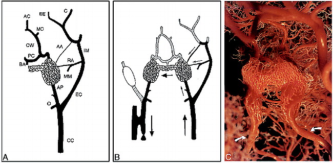
- fig 1.
Anatomic basis and features of the swine AVM model.
A, Schematic representation of the normal left carotid arterial anatomy of the swine head and neck. The carotid rete mirabile is situated at the termination of the ascending pharyngeal artery. In the swine, the internal carotid artery is very short and joins the rete to the circle of Willis. CC indicates common carotid artery; EC, external carotid artery; IM, internal maxillary artery; MM, middle meningeal artery supplying the ramus anastomoticus; RA, ramus anastomoticus; AA, arteria anastomotica; AP, ascending pharyngeal artery; O, occipital artery; BA, basilar artery; CW, circle of Willis; PC, posterior cerebral artery; MC, middle cerebral artery; AC, anterior cerebral artery; C, ciliary artery; EE, external ethmoidal artery.
B, Schematic representation of the AVM model after creation of a right carotid–jugular fistula. Arrows indicate direction of flow; that is, from the left common carotid artery to both retia mirabilia (nidus) via the three feeding arteries (left ascending pharyngeal artery, left ramus anastomoticus, and left arteria anastomotica), and retrograde down the right ascending pharyngeal artery toward the right carotid-jugular fistula. Note balloon occlusion of the right external carotid artery.
C, Detail from a plastic cast of both carotid retia mirabilia of the swine shows the complex branching pattern of this microvascular bed. The surrounding nonvascular structures have been removed. Arrows indicate left and right ascending pharyngeal arteries. (Courtesy of Marc P. Ghysels, MD, Department of Medical Imaging, University of Liege, Belgium).
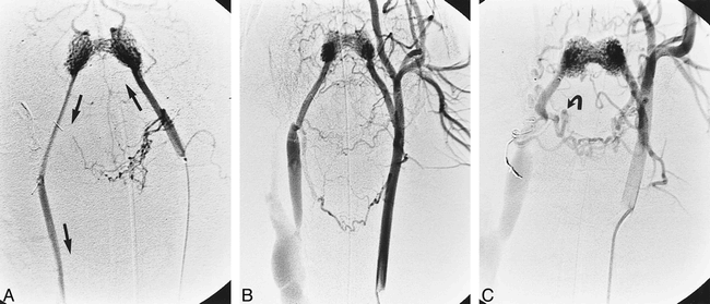
- fig 2.
A, Frontal view of a superselective left ascending pharyngeal arteriogram shows rapid shunting flow through the AVM nidus and retrograde flow in the draining vein portion of the AVM down toward the carotid-jugular fistula immediately after model construction. Straight coils were used to occlude right-sided neck branches during model construction. Arrows indicate direction of flow.
B, Frontal left common carotid arteriogram shows features of the same AVM model as in A but 2 months later. Note dilatation and some elongation of the component vessels as well as marked dilatation of the external jugular vein.
C, Frontal left common carotid arteriogram shows features of the 6-month-old AVM model. Note dilatation and elongation of its component vessels and the overall coarse pattern of nidus microvessels. In this swine, the muscular branch of the right ascending pharyngeal artery (arrow) was not occluded during model construction owing to temporary vasospasm, which prevented access. This allowed the delayed recruitment of blood via anastomoses with the namesake contralateral artery, in parallel with shunting across the AVM nidus.
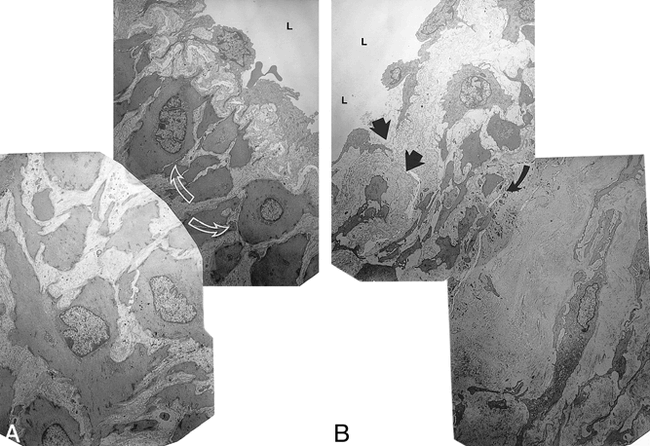
- fig 3.
Histologic and immunohistochemical features of the swine AVM models.
A, Immunohistochemical features of the nidus microvessels in the acute AVM model (ie, of normal rete mirabile microvessels) using monoclonal antibody to smooth muscle actin, followed by counterstaining with hematoxylin-eosin. Original magnification ×40.
B, Histologic section of the 2-month-old nidus vessels after staining with elastica van Gieson. Note the prominent intimal hyperplasia that occludes vessels in places and prominent disruption of the elastica. Original magnification ×16.
C, Immunohistochemical features of the nidus microvessels in the 2-month-old AVM model using monoclonal antibody to smooth muscle actin, followed by counterstaining with hematoxylin-eosin. Note medial thickening and intimal hyperplasia. Original magnification ×40.
D, Immunohistochemical features of the nidus microvessels in the 2-month-old AVM model using PC10 antibody to PCNA followed by counterstaining with hematoxylin-eosin. Note widespread presence of proliferating cells. Original magnification ×104.
E, Histologic section of the 2-month-old nidus vessels after staining with mucicarmine. Note the mucicarmine-positive ground substance among cells of hyperplastic intima. Original magnification ×40.
F, Histologic section of the 6-month-old nidus vessels after staining with elastica van Gieson. Note the multifocal destruction of the elastica layer (arrow) and the intimal hyperplasia in the form of an endoluminal cushion (arrowhead). Original magnification ×40.
G, Histologic section of the 6-month-old nidus vessels after staining with hematoxylin-eosin. Note the focal segments of mural thinning (arrow). Original magnification ×40.
- fig 4.
Electron micrographs of nidus vessels in the acute AVM model (ie, of normal rete mirabile microvessels) (A) and in the 2-month-old AVM model (B). L indicates vessel lumen. Original magnifications ×7500.
A, Note the normal and variably prominent endothelium. Smooth muscle cells (arrows) are immediately deep to the internal elastic lamina.
B, Note thinning and attenuation of the endothelium with focal breaks in the endothelial barrier (straight arrows). Underlying fibromuscular hyperplasia is noted with prominent smooth muscle and fibroblast cells, which are separated by bands of collagen and granular and fibrillar material, probably representing glycosaminoglycans. Curved arrow indicates a focus in which the internal elastic lamina is attenuated.
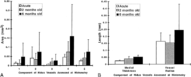
- fig 5.
A, Histogram of mean values (± SD) for five histometric parameters that characterize various areas (in mm2) of the nidus microvessels when measured in the acute, 2-month-old, and 6-month-old AVM models. The five parameters were 1) area of the vessel lumen, 2) entire area enclosed by the internal elastic lamina, 3) area of intimal hyperplasia, 4) entire area enclosed by the outer margin of media, and 5) area of media.
B, Histogram of mean values (± SD) for the medial thickness and vessel radius (in mm) of the nidus microvessels when measured in the acute, 2-month-old, and 6-month-old AVM models.


In this issue


Jump to section
Related Articles
Cited By...
- Cerebral Arteriovenous Malformation Flow Is Associated With Venous Intimal Hyperplasia
- Development of an angiogenesis animal model featuring brain arteriovenous malformation histological characteristics
- Focused post mortem dissection technique for harvest of rete mirabile in domestic swine (Sus scrofa)
- Response to Letter by Yousaf et al