
Abstract
BACKGROUND AND PURPOSE: Wada testing may provide important information for surgical planning in pediatric patients with medically refractory epilepsy, but it is often not used because of the difficulties in performing the angiographic portion of the procedure in conscious children. We reviewed our experience using propofol, a short-acting IV administered anesthetic agent, for pediatric patients undergoing Wada testing.
METHODS: In a retrospective review of Wada tests performed on patients younger than 18 years, we identified 24 cases in which propofol anesthesia was used. We reviewed the medical records of these patients, with particular reference to dose of propofol, physiological parameters during anesthesia, and adequacy of neuropsychological testing after emergence from anesthesia.
RESULTS: Patients ranged in age from 6 to 16 years (mean age, 12.5 years). Propofol induced mild reductions in blood pressure (12.4% for systolic and 13.9% for diastolic blood pressure) and heart rate (mean reduction of 4.7%), which did not require specific treatment in any patient. Recovery from anesthesia was smooth and rapid, allowing initiation of Wada testing within 15 to 25 minutes of cessation of propofol. Wada testing was successfully accomplished in all patients.
CONCLUSION: Propofol provided rapid induction of anesthesia, was administered without endotracheal intubation, and did not cause substantial changes in cardiorespiratory parameters. Propofol anesthesia allowed controlled angiography among patients as young as 6 years and did not interfere with neuropsychological testing.
In many adult patients with medically refractory epilepsy for whom surgical resection of a seizure focus is considered, the Wada test, or intracarotid amobarbital procedure (IAP), is an integral part of the preoperative evaluation (1). For those individuals with temporal lobe seizure focus, the IAP is used for lateralization of language and memory; it may also help to lateralize the seizure focus (2, 3). For patients who are candidates for extratemporal resections, the IAP is used to evaluate language function more frequently than memory (1).
Surgical management of refractory seizures in children and adolescents is becoming more common as a consequence of the recognition that poorly controlled epilepsy in this group may have significant adverse effects on psychosocial and intellectual development (4–7). The IAP is therefore being requested more frequently for pediatric patients. Performing angiography, however, presents special challenges in these patients, many of whom have IQs in the cognitively impaired to borderline intelligence range (5, 8, 9). We describe a technique for performing Wada tests in pediatric patients by use of propofol anesthesia for the angiographic portion of the procedure, which does not interfere with neuropsychological testing after Amytal injection.
Methods
We retrospectively reviewed the results obtained from patients younger than 18 years who underwent all Wada tests during a 6-year period. There were 24 cases in which anesthesia was used during the angiographic portion of the procedure; in each of these patients, propofol was IV administered. There were 15 male and nine female patients with a mean age of 12.5 years (age range, 6–16 years); the three 16-year-old patients experienced global cognitive delay. In 20 patients with epilepsy refractory to standard medical therapy, Wada testing was performed as part of a comprehensive evaluation for surgical resection of a seizure focus. Of the four other patients, two had cerebral arteriovenous malformations, one had a left frontal parenchymal hematoma secondary to hemorrhage from a cavernoma, and one had a left frontal opercular tumor. In these cases, preoperative localization of language function was necessary for surgical planning. All patients underwent baseline neuropsychological testing before the IAP to familiarize them with the procedure and to determine the appropriate level of difficulty for the IAP test items (7–10). Informed consent for the procedure was obtained from a parent or guardian in each case.
In each case, anesthesia was administered by an anesthesiologist. Continuous noninvasive monitoring of blood pressure, pulse rate, respiratory rate, and peripheral oxygen saturation was performed throughout each procedure. Supplemental oxygen was administered either by use of a facemask or nasal prongs.
Because several anesthesiologists were involved in these cases, there was some variability in the method of administration of propofol and the use of supplemental anesthetic agents. Ten (42%) patients received a combination of propofol boluses and infusion, 13 (54%) patients received propofol as an infusion only, and one patient received the drug as repeated boluses. The mean dose rate for the propofol infusion was3.15 mg/kg/hr (range, 1–6 mg/kg/hr), and the mean dose of propofol delivered as a bolus was 1.05 mg/kg (range, 0.23–2.06 mg/kg). Seven (29%) of the 24 patients received additional anesthetic agents administered concomitantly with the propofol from the beginning of the procedure. In four cases, this was nitrous oxide; one patient received IV administered fentanyl; two patients received both nitrous oxide and fentanyl.
Angiography was performed via a femoral artery puncture using 1% lidocaine for skin and subcutaneous infiltration. After selective catheterization of each internal carotid artery (ICA), biplane digital subtraction angiography of the anterior intracranial circulation was performed using nonionic contrast material diluted to 150 mg/mL iodine administered through a power injector. The volume of contrast material injected into each ICA was 6 mL, and the rate of injection was 5 or 6 mL/s. After angiography, the catheter was withdrawn to the descending aorta and was continuously flushed using heparinized saline.
After angiography, the propofol was discontinued and the patients were allowed to recover from the anesthesia. The neuropsychologist (K.P.) confirmed that the patients were at their preprocedure baseline before initiation of Wada testing. The cerebral hemisphere harboring the surgical lesion was the first to be anesthetized with amobarbital after selective catheterization of the ICA. After Amytal injection, the catheter was again withdrawn to the descending aorta and was continuously flushed using heparinized saline. The mean dose of amobarbital (at a concentration of 20 mg/mL) injected into the left ICA was 90.2 mg (range, 50–140 mg) and in the right ICA was 88.3 mg (range, 50–200 mg); the drug was injected over a 5-second interval. Effective anesthesia was confirmed by the development of contralateral facial and upper extremity plegia. In 22 of the patients after an appropriate recovery interval, the contralateral ICA was catheterized and Wada testing performed after confirmation of the catheter tip position; two patients underwent injection of only one hemisphere for evaluation of language function. One neuropsychologist (K.P.) tested all patients by using a standardized protocol to evaluate language function; memory was evaluated in appropriate cases.
In two patients, administration of amobarbital into what was ultimately determined to be the nondominant ICA induced a state of abulic mutism, possibly because of distribution of the drug into both anterior cerebral artery territories. To distinguish between mutism and bihemispheric language representation in these patients, superselective Wada testing of the ipsilateral middle cerebral artery (MCA) territory was performed. A third patient with a left frontal opercular glioma underwent superselective MCA testing for evaluation of cortical function in the territory of an MCA branch encased by tumor as part of presurgical planning. Superselective catheterization of the MCA was performed with a coaxially introduced microcatheter after systemic anticoagulation with 2000 to 4000 units of IV administered heparin.
Results
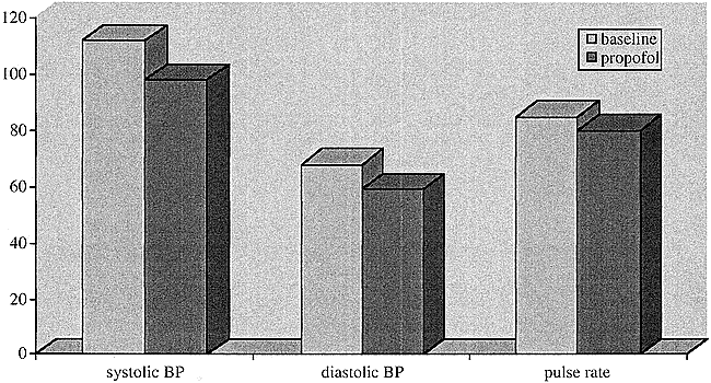
In all patients, cerebral angiography and Wada testing were successfully performed without complication. Overall, changes in physiological parameters during anesthesia were mild (Fig 1), and no specific interventions were required. The peripheral oxygen saturation ranged from 98% to 100%. In one patient, the oxygen saturation decreased to 96% after the administration of a propofol bolus but quickly returned to 100%. The respiratory rate ranged from 15 to 24 per minute, and no instances of apnea occurred. Blood pressure decreased after the administration of propofol. The mean systolic blood pressure (±SD) before the induction of anesthesia was 113 mm Hg (±9) (range, 90–130 mm Hg), which decreased to 99 mm Hg (±10) (range, 80–120) after the administration of propofol. The baseline mean diastolic blood pressure was 69 mm Hg (±8) (range, 55–85), which decreased to 59 mm Hg (±10) (range, 45–75) after the administration of propofol. The mean percentage change in systolic blood pressure was 12.4% and in diastolic blood pressure was 13.9%. The mean baseline pulse rate (±SD) was 84 (±13) per minute (range, 60–110), which decreased to 80 (±11) per minute (range, 60–100) (a 4.7% reduction).

Graphical representation of mean changes in hemodynamic parameters after the administration of propofol (n = 24)
In all patients, the emergence from anesthesia was rapid and smooth. There were no instances of agitation, nausea, or vomiting. The IAP commenced 15 to 25 minutes after discontinuing propofol. In those patients who underwent superselective MCA amobarbital injections, no complications were experienced, and in each case, unequivocal determination of language lateralization was made after the MCA amobarbital injection. Seventeen (71%) of the 24 patients underwent IAP as an outpatient procedure and were discharged home in the care of a family member after 6 hours of observation in a recovery area.
Discussion
There have been significant advances in the diagnosis and treatment of epilepsy during the last 10 years. Most patients (adults and children) with seizure disorders are successfully managed with medical therapy. Nevertheless, 10% to 20% of new patients per year with epilepsy are refractory to medical treatment (4). If a seizure focus can be lateralized and localized in these patients, then excision of that focus may eliminate the seizures or significantly reduce their frequency.
Lateralization of eloquent cortex can be achieved with cortical stimulation or Wada testing. Both methods have their limitations. Cortical stimulation, although a safe and accurate technique (4, 7), requires the implantation of subdural and/or depth electrodes or intraoperative stimulation, and only the hemisphere harboring the seizure focus is evaluated. Wada testing allows evaluation of both cerebral hemispheres. The angiographic portion of this procedure, however, may be difficult to perform in children without sedation or anesthesia, the aftereffects of which may confound the results of neuropsychological testing. At those institutions that do perform Wada testing in children and young adolescents, the patients are often coached in behavioral interventions designed to reduce anxiety and discomfort during the IAP (9–11). Such techniques, however, may not be effective in allaying anxiety and ensuring that the patient is cooperative. Children may become agitated or combative, and the procedure may have to be abandoned (7, 9, 12). In such situations, not only is no diagnostic information obtained but there is also a risk of femoral artery spasm or injury. For this reason, the IAP is not as widely used in pediatric patients, especially in children younger than 10 years (4, 12). Although functional MR imaging shows promise for the noninvasive lateralization of language (13, 14) and memory (15, 16), experience with this technique has generally been with adult patients, and it has not yet supplanted Wada testing in clinical practice.
Propofol is widely used as an anesthetic agent for pediatric patients undergoing imaging studies and some therapeutic procedures (17–21). There have been isolated reports of its use in pediatric IAPs (7, 22), but, to our knowledge, this is the largest series detailing its usefulness in this situation.
Propofol is an IV administered alkyl phenol anesthetic agent (23). It provides rapid induction of anesthesia, which can be maintained with either infusion or repeated boluses (24). It does not have analgesic properties, but this is not a requirement for cerebral angiography (when local anesthesia at the femoral puncture site is routinely used) (24).
Anesthesia with propofol does induce cardiorespiratory changes. The mean arterial pressure in children usually falls by 10% to 25% immediately after induction (25), and, in adults, reductions in systolic and diastolic blood pressure of 11% and 16%, respectively, have been observed after a bolus dose of propofol (24). Blood pressure changes are generally dose- and infusion-rate dependent (26). In pediatric patients, the pulse rate typically falls by 10% to 20% (25). These changes may persist while anesthesia is maintained with propofol (24). Because of these hemodynamic changes, it is important that patients be well-hydrated (25). Respiratory effects are characterized by hypoventilation and tend to be dose-dependent (17); apnea has been reported in 20% of children after a bolus dose of propofol (25). Hypoventilation also tends to be dose-dependent (17, 18), and hypoxemia is usually circumvented by the administration of supplemental oxygen (17, 19, 20). Although these cardiorespiratory changes are usually mild, it is recommended that propofol be administered only by a trained anesthetist (17, 25).
Recovery from propofol anesthesia is rapid and smooth. There is a very low incidence of nausea and agitation and, particularly in children, a rapid return to baseline neuropsychological function. These features make propofol an ideal anesthetic agent for pediatric patients undergoing IAP.
Our experience confirms the theoretical advantages of this technique. Femoral artery catheterization and cerebral angiography are performed with the patients under anesthesia. Because there is no degradation of digital subtraction angiography by motion artifact, anatomic information germane to surgical planning is clearly defined. This is particularly important in patients with arteriovenous malformations, for whom detailed preoperative evaluation of the angioarchitecture is critical to surgical planning. In patients with other pathologic abnormalities, knowledge of the relationship of the lesion to adjacent vascular structures may influence the surgical approach. The use of propofol does not confound the results of Wada testing, which can be initiated 15 to 25 minutes after discontinuing the drug. Although the use of propofol anesthesia does increase the length of the procedure, we think that this is more than offset by the benefits of patient comfort and safety, controlled angiography, and diagnostic neuropsychological evaluation.
A methodological flaw in our series is that seven patients received supplemental anesthetic agents, raising the possibility that within this group, propofol alone may not have provided adequate anesthesia. Based on our experience with propofol as a single anesthetic agent, we think that this is unlikely.
Conclusion
Propofol anesthesia facilitates controlled cerebral angiography in pediatric patients without compromising the results of Wada testing. It can be administered without endotracheal intubation and typically does not cause clinically significant changes in cardiorespiratory function. Because of the potential for respiratory depression, however, close monitoring of the patient by a trained anesthetist is mandatory.
Footnotes
1 This research was presented at the Annual Meeting of the American Society of Neuroradiology, San Diego, CA, May 1999.
↵2 Address reprint requests to Peter Kim Nelson, MD, Department of Radiology, Room HE208, New York University Medical Center, 560 First Avenue, New York, NY 10016.
References
- Received November 11, 1999.
- Accepted after revision February 10, 2000.
- Copyright © American Society of Neuroradiology
In this issue


Jump to section
Related Articles
Cited By...
- No citing articles found.