
Article Figures & Data
Figures
- fig 1.
A, BOLD fMR activation from a finger-tapping paradigm coregistered to an axial T1-weighted image (500/14 [TR/TE]) in patient 9. Pathologic analysis revealed a right parietal lobe glioblastoma multiforme. The areas in yellow correspond to an R value of 0.65. The areas in red correspond to an R value of 0.50. There is robust activation in the left motor cortex. The activation in the right motor cortex is barely perceptible (arrows).
B, A graph of the volume of activation for different R values for patient 9. For all R values that reveal activation, the volume of activation is greater on the side opposite the tumor.
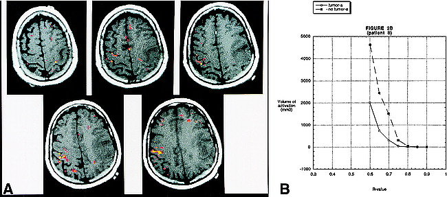
- fig 2.
A, BOLD fMR activation from a finger-tapping paradigm coregistered to an axial T1-weighted image (500/14 [TR/TE]) in patient 8. Pathologic analysis revealed a left frontoparietal glioblastoma multiforme. The areas in yellow correspond to an R value of 0.72. The areas in red correspond to an R value of 0.67. There is robust activation in the right motor cortex. It is interesting to note that the activation is seen along the most anterior and posterior aspects of the precentral gyrus. This is the location of the cortical gray matter. The central portion of the gyrus (which contains the white matter tracts) does not reveal activation. On the right side, activation is appreciated in the postcentral gyrus, which most likely represents the sensory cortex. Activation is also identified in the left motor cortex; however, the volume is much less than on the side without the tumor. The activation on the left side is seen in the most superior and medial part of the precentral gyrus—the area relatively spared by the glioma.
B, A graph of the volume of activation for different R values for patient 8. As in figure 1, for all R values that reveal activation, the volume of activation is greater on the side opposite the tumor. The absolute volume of activation and the pattern of activation, however, differ from patient 9.
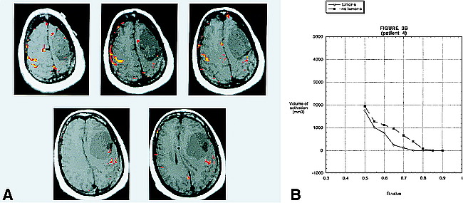
- fig 3.
A, BOLD fMR activation from a finger-tapping paradigm coregistered to an axial T1-weighted image (500/14 [TR/TE]) in patient 4. Pathologic analysis revealed a left frontal lobe meningioma. There is prominent activation of the motor cortex on the side contralateral to the tumor, at R values of 0.70 (yellow) that allows for accurate delineation of the primary motor cortex. At these R values, there is very little activation on the side with the tumor. At R values of 0.55 (red), however, the volume of activation in the primary motor cortex of the side with the tumor approaches the activation on the contralateral side. This occurs before the onset of “noise” in areas of the brain not related to the neural control of motor function. This example emphasizes the need to evaluate each patient individually at multiple R values. Areas of activation located outside the brain over the right frontal convexity probably represent cortical venous drainage.
B, A graph of the volume of activation for different R values for patient 4. There is a large difference in the volume of activation between the two sides for R values between 0.65 and 0.75. At 0.50, however, the volume of activation on the two sides is almost the same.


Tables

Quantitative data from functional and anatomic MR images in patients with tumors in or adjacent to the motor cortex

In this issue


Jump to section
Related Articles
Cited By...
- A measure of vascular reactivity to overcome neurovascular uncoupling in functional imaging of brain tumors: initial results
- Resting-State Functional Connectivity of the Middle Frontal Gyrus Can Predict Language Lateralization in Patients with Brain Tumors
- Local Glioma Cells Are Associated with Vascular Dysregulation
- Anatomic Location of Tumor Predicts the Accuracy of Motor Function Localization in Diffuse Lower-Grade Gliomas Involving the Hand Knob Area
- Presurgical Assessment of the Sensorimotor Cortex Using Resting-State fMRI
- Multimodality Brain Tumor Imaging: MR Imaging, PET, and PET/MR Imaging
- Cortical Activation Through Passive-Motion Functional MRI
- Comparison of Hypothesis- and a Novel Hybrid Data/Hypothesis-Driven Method of Functional MR Imaging Analysis in Patients with Brain Gliomas
- The Evolution of Clinical Functional Imaging during the Past 2 Decades and Its Current Impact on Neurosurgical Planning
- The Blood Oxygen Level-Dependent Functional MR Imaging Signal Can Be Used to Identify Brain Tumors and Distinguish Them from Normal Tissue
- The technology of MRI -- the next 10 years?
- Regional Impairment of Cerebrovascular Reactivity and BOLD Signal in Adults After Stroke
- Role of the healthy hemisphere in recovery after resection of the supplementary motor area
- Motor functional MRI for pre-operative and intraoperative neurosurgical guidance
- Increase in focal concentration of deoxyhaemoglobin during neuronal activity in cerebral ischaemic patients
- Reliability of Functional MR Imaging with Word-Generation Tasks for Mapping Broca's Area