Advertisement

Main menu

  • Home
  • Content
    • Current Issue
    • Accepted Manuscripts
    • Article Preview
    • Past Issue Archive
    • AJNR Case Collection
    • Case of the Week Archive
    • Classic Case Archive
    • Case of the Month Archive
  • Special Collections
    • Spinal CSF Leak Articles (Jan 2020-June 2024)
    • 2024 AJNR Journal Awards
    • Most Impactful AJNR Articles
  • Multimedia
    • AJNR Podcast
    • AJNR Scantastics
    • Video Articles
  • For Authors
    • Submit a Manuscript
    • Author Policies
    • Fast publishing of Accepted Manuscripts
    • Graphical Abstract Preparation
    • Manuscript Submission Guidelines
    • Imaging Protocol Submission
    • Submit a Case for the Case Collection
  • About Us
    • About AJNR
    • Editorial Board
  • More
    • Become a Reviewer/Academy of Reviewers
    • Subscribers
    • Permissions
    • Alerts
    • Feedback
    • Advertisers
    • ASNR Home
  • Other Publications
    • ajnr

User menu

  • Alerts
  • Log in

Search

  • Advanced search
American Journal of Neuroradiology

American Journal of Neuroradiology

ASHNR American Society of Functional Neuroradiology ASHNR American Society of Pediatric Neuroradiology ASSR
  • Alerts
  • Log in

Advanced Search

  • Home
  • Content
    • Current Issue
    • Accepted Manuscripts
    • Article Preview
    • Past Issue Archive
    • AJNR Case Collection
    • Case of the Week Archive
    • Classic Case Archive
    • Case of the Month Archive
  • Special Collections
    • Spinal CSF Leak Articles (Jan 2020-June 2024)
    • 2024 AJNR Journal Awards
    • Most Impactful AJNR Articles
  • Multimedia
    • AJNR Podcast
    • AJNR Scantastics
    • Video Articles
  • For Authors
    • Submit a Manuscript
    • Author Policies
    • Fast publishing of Accepted Manuscripts
    • Graphical Abstract Preparation
    • Manuscript Submission Guidelines
    • Imaging Protocol Submission
    • Submit a Case for the Case Collection
  • About Us
    • About AJNR
    • Editorial Board
  • More
    • Become a Reviewer/Academy of Reviewers
    • Subscribers
    • Permissions
    • Alerts
    • Feedback
    • Advertisers
    • ASNR Home
  • Follow AJNR on Twitter
  • Visit AJNR on Facebook
  • Follow AJNR on Instagram
  • Join AJNR on LinkedIn
  • RSS Feeds

Welcome to the new AJNR, Updated Hall of Fame, and more. Read the full announcements.


AJNR is seeking candidates for the position of Associate Section Editor, AJNR Case Collection. Read the full announcement.


Research ArticleBRAIN

Radiolabeled Leucocyte Imaging in Diffuse Granulomatous Involvement of the Meninges in Wegener's Granulomatosis: Scintigraphic Findings and Their Role in Monitoring Treatment Response to Specific Immunotherapy (Humanized Monoclonal Antilymphocyte Antibodies)

Joseph M. Murphy, Kottekkattu K. Balan, Andoni Toms, Beatriz Gomez-Anson and Martin Lockwood
American Journal of Neuroradiology September 2000, 21 (8) 1460-1465;
Joseph M. Murphy
  • Find this author on Google Scholar
  • Find this author on PubMed
  • Search for this author on this site
Kottekkattu K. Balan
  • Find this author on Google Scholar
  • Find this author on PubMed
  • Search for this author on this site
Andoni Toms
  • Find this author on Google Scholar
  • Find this author on PubMed
  • Search for this author on this site
Beatriz Gomez-Anson
  • Find this author on Google Scholar
  • Find this author on PubMed
  • Search for this author on this site
Martin Lockwood
  • Find this author on Google Scholar
  • Find this author on PubMed
  • Search for this author on this site
  • Article
  • Figures & Data
  • Info & Metrics
  • Responses
  • References
  • PDF
Loading

Article Figures & Data

Figures

  • Tables
  • fig 1.
    • Download figure
    • Open in new tab
    • Download powerpoint
    fig 1.

    A, Posterior view at 24 hours from a 111In-labeled leucocyte examination shows a linear midline area of increased uptake (arrow).

    B, Coronal contrast-enhanced T1-weighted (500/9/2) SE MR image shows dural thickening and enhancement in the falx (short wide arrow), tentorium cerebelli (curved arrows), and overlying the brain convexity (long thin arrow). Note how the leucocyte examination underestimates the extent of dural involvement.

    C, Posterior view at 24 hours from a 111In-labeled leucocyte examination shows normal intracranial appearance after treatment with anti-CD52.

  • fig 2.
    • Download figure
    • Open in new tab
    • Download powerpoint
    fig 2.

    A, Posterior view at 24 hours from a 99mTc-HMPAO-labeled leucocyte examination shows abnormal uptake in the midline (open arrow), overlying the brain convexity on the left side (solid arrow), and in the tentorium cerebelli bilaterally (arrowheads).

    B, Coronal contrast-enhanced T1-weighted (500/9/2) SE MR image shows dural thickening and enhancement in the falx (short wide arrow), overlying the brain convexity on the left side (long thin arrow), and in the tentorium cerebelli (curved arrows).

    C, Normal intracranial appearance after treatment with anti-CD52.
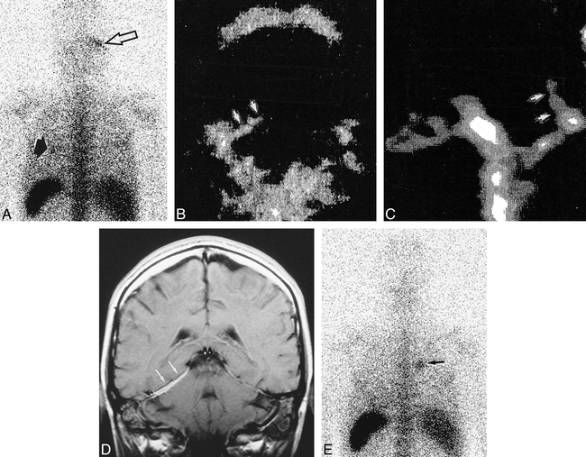
  • fig 3.
    • Download figure
    • Open in new tab
    • Download powerpoint
    fig 3.

    A, Posterior view at 24 hours from a 111In-labeled leucocyte examination shows increased uptake in the right tentorium cerebelli (arrow). A focus of increased uptake is also visible in the left lung (arrowhead), which correlated with a cavitating nodule on a high-resolution CT scan.

    B and C, Coronal (B) and sagittal (C) images from a 99mTc-HMPAO-labeled leucocyte SPECT examination show increased uptake in the right tentorium cerebelli (arrows).

    D, Coronal contrast-enhanced T1-weighted (500/9/2) SE MR image shows thickening and enhancement of the right tentorium cerebelli (arrows).

    E, Normal intracranial appearance after treatment with anti-CD52. Note focus of increased uptake in the right lung (arrow).

Tables

  • Figures
  • Table1

    Imaging findings in five patients with Wegener's granulomatosis

PreviousNext
Back to top

In this issue

American Journal of Neuroradiology
Vol. 21, Issue 8
1 Sep 2000
  • Table of Contents
  • Index by author
Advertisement
Print
Download PDF
Email Article

Thank you for your interest in spreading the word on American Journal of Neuroradiology.

NOTE: We only request your email address so that the person you are recommending the page to knows that you wanted them to see it, and that it is not junk mail. We do not capture any email address.

Enter multiple addresses on separate lines or separate them with commas.
Radiolabeled Leucocyte Imaging in Diffuse Granulomatous Involvement of the Meninges in Wegener's Granulomatosis: Scintigraphic Findings and Their Role in Monitoring Treatment Response to Specific Immunotherapy (Humanized Monoclonal Antilymphocyte Antibod…
(Your Name) has sent you a message from American Journal of Neuroradiology
(Your Name) thought you would like to see the American Journal of Neuroradiology web site.
CAPTCHA
This question is for testing whether or not you are a human visitor and to prevent automated spam submissions.
Cite this article
Joseph M. Murphy, Kottekkattu K. Balan, Andoni Toms, Beatriz Gomez-Anson, Martin Lockwood
Radiolabeled Leucocyte Imaging in Diffuse Granulomatous Involvement of the Meninges in Wegener's Granulomatosis: Scintigraphic Findings and Their Role in Monitoring Treatment Response to Specific Immunotherapy (Humanized Monoclonal Antilymphocyte Antibodies)
American Journal of Neuroradiology Sep 2000, 21 (8) 1460-1465;

Citation Manager Formats

  • BibTeX
  • Bookends
  • EasyBib
  • EndNote (tagged)
  • EndNote 8 (xml)
  • Medlars
  • Mendeley
  • Papers
  • RefWorks Tagged
  • Ref Manager
  • RIS
  • Zotero
0 Responses
Respond to this article
Share
Bookmark this article
Radiolabeled Leucocyte Imaging in Diffuse Granulomatous Involvement of the Meninges in Wegener's Granulomatosis: Scintigraphic Findings and Their Role in Monitoring Treatment Response to Specific Immunotherapy (Humanized Monoclonal Antilymphocyte Antibodies)
Joseph M. Murphy, Kottekkattu K. Balan, Andoni Toms, Beatriz Gomez-Anson, Martin Lockwood
American Journal of Neuroradiology Sep 2000, 21 (8) 1460-1465;
del.icio.us logo Twitter logo Facebook logo Mendeley logo
  • Tweet Widget
  • Facebook Like
  • Google Plus One

Jump to section

  • Article
    • Abstract
    • Methods
    • Results
    • Discussion
    • Conclusion
    • Footnotes
    • References
  • Figures & Data
  • Info & Metrics
  • Responses
  • References
  • PDF

Related Articles

  • No related articles found.
  • PubMed
  • Google Scholar

Cited By...

  • No citing articles found.
  • Crossref
  • Google Scholar

This article has not yet been cited by articles in journals that are participating in Crossref Cited-by Linking.

More in this TOC Section

  • Evaluating the Effects of White Matter Multiple Sclerosis Lesions on the Volume Estimation of 6 Brain Tissue Segmentation Methods
  • White Matter Alterations in the Brains of Patients with Active, Remitted, and Cured Cushing Syndrome: A DTI Study
  • Enhanced Axonal Metabolism during Early Natalizumab Treatment in Relapsing-Remitting Multiple Sclerosis
Show more Brain

Similar Articles

Advertisement

Indexed Content

  • Current Issue
  • Accepted Manuscripts
  • Article Preview
  • Past Issues
  • Editorials
  • Editors Choice
  • Fellow Journal Club
  • Letters to the Editor

Cases

  • Case Collection
  • Archive - Case of the Week
  • Archive - Case of the Month
  • Archive - Classic Case

Special Collections

  • Special Collections

Resources

  • News and Updates
  • Turn around Times
  • Submit a Manuscript
  • Author Policies
  • Manuscript Submission Guidelines
  • Evidence-Based Medicine Level Guide
  • Publishing Checklists
  • Graphical Abstract Preparation
  • Imaging Protocol Submission
  • Submit a Case
  • Become a Reviewer/Academy of Reviewers
  • Get Peer Review Credit from Publons

Multimedia

  • AJNR Podcast
  • AJNR SCANtastic
  • Video Articles

About Us

  • About AJNR
  • Editorial Board
  • Not an AJNR Subscriber? Join Now
  • Alerts
  • Feedback
  • Advertise with us
  • Librarian Resources
  • Permissions
  • Terms and Conditions

American Society of Neuroradiology

  • Not an ASNR Member? Join Now

© 2025 by the American Society of Neuroradiology All rights, including for text and data mining, AI training, and similar technologies, are reserved.
Print ISSN: 0195-6108 Online ISSN: 1936-959X

Powered by HighWire