Advertisement

Main menu

  • Home
  • Content
    • Current Issue
    • Accepted Manuscripts
    • Article Preview
    • Past Issue Archive
    • AJNR Case Collection
    • Case of the Week Archive
    • Classic Case Archive
    • Case of the Month Archive
  • Special Collections
    • Spinal CSF Leak Articles (Jan 2020-June 2024)
    • 2024 AJNR Journal Awards
    • Most Impactful AJNR Articles
  • Multimedia
    • AJNR Podcast
    • AJNR Scantastics
    • Video Articles
  • For Authors
    • Submit a Manuscript
    • Author Policies
    • Fast publishing of Accepted Manuscripts
    • Graphical Abstract Preparation
    • Manuscript Submission Guidelines
    • Imaging Protocol Submission
    • Submit a Case for the Case Collection
  • About Us
    • About AJNR
    • Editorial Board
  • More
    • Become a Reviewer/Academy of Reviewers
    • Subscribers
    • Permissions
    • Alerts
    • Feedback
    • Advertisers
    • ASNR Home
  • Other Publications
    • ajnr

User menu

  • Alerts
  • Log in

Search

  • Advanced search
American Journal of Neuroradiology

American Journal of Neuroradiology

ASHNR American Society of Functional Neuroradiology ASHNR American Society of Pediatric Neuroradiology ASSR
  • Alerts
  • Log in

Advanced Search

  • Home
  • Content
    • Current Issue
    • Accepted Manuscripts
    • Article Preview
    • Past Issue Archive
    • AJNR Case Collection
    • Case of the Week Archive
    • Classic Case Archive
    • Case of the Month Archive
  • Special Collections
    • Spinal CSF Leak Articles (Jan 2020-June 2024)
    • 2024 AJNR Journal Awards
    • Most Impactful AJNR Articles
  • Multimedia
    • AJNR Podcast
    • AJNR Scantastics
    • Video Articles
  • For Authors
    • Submit a Manuscript
    • Author Policies
    • Fast publishing of Accepted Manuscripts
    • Graphical Abstract Preparation
    • Manuscript Submission Guidelines
    • Imaging Protocol Submission
    • Submit a Case for the Case Collection
  • About Us
    • About AJNR
    • Editorial Board
  • More
    • Become a Reviewer/Academy of Reviewers
    • Subscribers
    • Permissions
    • Alerts
    • Feedback
    • Advertisers
    • ASNR Home
  • Follow AJNR on Twitter
  • Visit AJNR on Facebook
  • Follow AJNR on Instagram
  • Join AJNR on LinkedIn
  • RSS Feeds

Welcome to the new AJNR, Updated Hall of Fame, and more. Read the full announcements.


AJNR is seeking candidates for the position of Associate Section Editor, AJNR Case Collection. Read the full announcement.


Research ArticlePEDIATRICS

Leigh Syndrome: Serial MR Imaging and Clinical Follow-up

Junko Arii and Yuzo Tanabe
American Journal of Neuroradiology September 2000, 21 (8) 1502-1509;
Junko Arii
  • Find this author on Google Scholar
  • Find this author on PubMed
  • Search for this author on this site
Yuzo Tanabe
  • Find this author on Google Scholar
  • Find this author on PubMed
  • Search for this author on this site
  • Article
  • Figures & Data
  • Info & Metrics
  • Responses
  • References
  • PDF
Loading

Article Figures & Data

Figures

  • Tables
  • fig 1.
    • Download figure
    • Open in new tab
    • Download powerpoint
    fig 1.

    Patient 1: group BG lesion pattern. Axial T2-weighted image (2000/80/2) at the age of 7 months during an acute strokelike episode shows a hyperintense lesion in the right parietotemporal region (arrowheads)
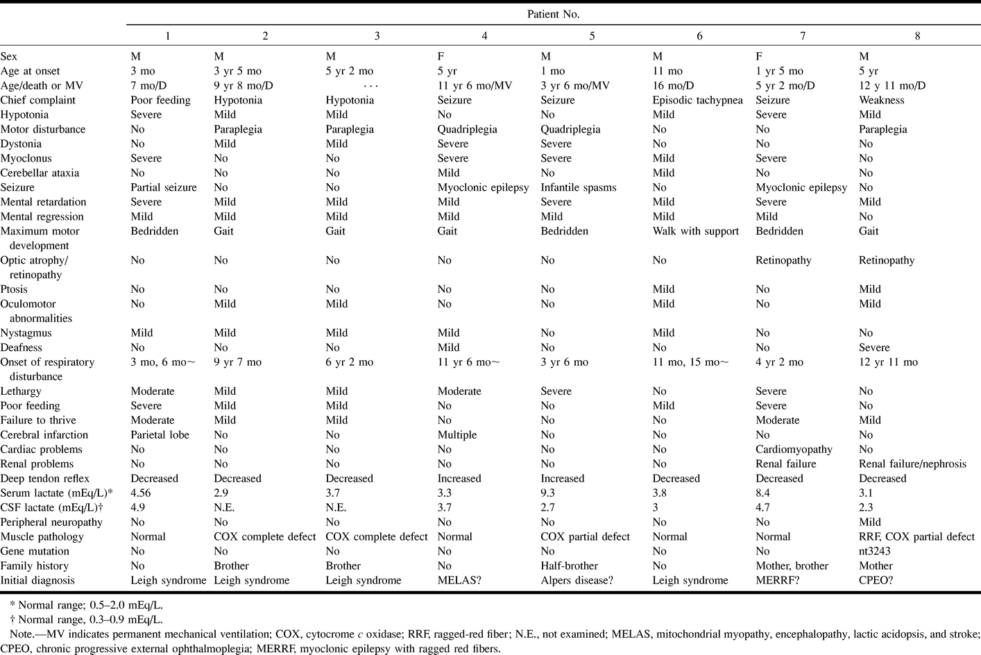
  • fig 2.
    • Download figure
    • Open in new tab
    • Download powerpoint
    fig 2.

    Patient 2: group BG lesion pattern.

    A, Axial T2-weighted image (2000/80/2) at the age of 3 years shows increased signal intensity in the bilateral putamina and caudate heads, but no detectable brain stem lesion. Clinical findings included mental retardation, muscle weakness, and failure to thrive.

    B and C, Axial (B) and sagittal (C) T2-weighted images (2000/80/2) at the age of 9 years show static and atrophic lesions in the bilateral putamina and progressive lesions in the cerebral white matter, corpus callosum, and medial medulla oblongata (arrow, C). Clinical findings included irregular breathing, lethargy, and inability to feed.

  • fig 3.
    • Download figure
    • Open in new tab
    • Download powerpoint
    fig 3.

    Patient 6: group BS lesion pattern.

    A–C, Axial T2-weighted images (2000/80/2) at the age of 11 months show lesions in the substantia nigra (arrows, B) and periaqueductal region (arrowheads, B), and subtle increased signal intensity within the medullary reticular formation (arrow, C) but not in the basal ganglia. Clinically, the patient had episodic unexplained tachypnea.

  • fig 4.
    • Download figure
    • Open in new tab
    • Download powerpoint
    fig 4.

    Patient 8: group WM lesion pattern.

    A and B, Coronal (A) and axial (B) T2-weighted images (2000/80/2) at the age of 12 years show symmetrical hyperintensity in the cerebral white matter (arrows, A), thalamus, basal ganglia, and medullary tegmentum (arrowheads, B). Clinically, the patient had acute respiratory failure.
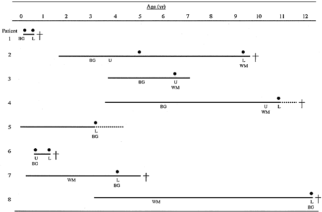
  • fig 5.
    • Download figure
    • Open in new tab
    • Download powerpoint
    fig 5.

    Brain stem lesions on MR images and abnormal respiration. Patient numbers correspond to those in Table 1. L indicates lower brain stem lesion; U, upper brain stem lesion; BG, basal ganglia lesion; WM, cerebral white matter lesion; solid line, duration of the disease; solid circle, acute respiratory failure; dotted line, permanent mechanical ventilation; cross, death

Tables

  • Figures
  • TABLE 1:
    • View popup
    • Download powerpoint
    TABLE 1:

    Clinical features and results of investigations in Leigh syndrome

  • TABLE 2:
    • View popup
    • Download powerpoint
    TABLE 2:

    MR imaging findings in 41 patients with Leigh syndrome reported in the literature

PreviousNext
Back to top

In this issue

American Journal of Neuroradiology
Vol. 21, Issue 8
1 Sep 2000
  • Table of Contents
  • Index by author
Advertisement
Print
Download PDF
Email Article

Thank you for your interest in spreading the word on American Journal of Neuroradiology.

NOTE: We only request your email address so that the person you are recommending the page to knows that you wanted them to see it, and that it is not junk mail. We do not capture any email address.

Enter multiple addresses on separate lines or separate them with commas.
Leigh Syndrome: Serial MR Imaging and Clinical Follow-up
(Your Name) has sent you a message from American Journal of Neuroradiology
(Your Name) thought you would like to see the American Journal of Neuroradiology web site.
CAPTCHA
This question is for testing whether or not you are a human visitor and to prevent automated spam submissions.
Cite this article
Junko Arii, Yuzo Tanabe
Leigh Syndrome: Serial MR Imaging and Clinical Follow-up
American Journal of Neuroradiology Sep 2000, 21 (8) 1502-1509;

Citation Manager Formats

  • BibTeX
  • Bookends
  • EasyBib
  • EndNote (tagged)
  • EndNote 8 (xml)
  • Medlars
  • Mendeley
  • Papers
  • RefWorks Tagged
  • Ref Manager
  • RIS
  • Zotero
0 Responses
Respond to this article
Share
Bookmark this article
Leigh Syndrome: Serial MR Imaging and Clinical Follow-up
Junko Arii, Yuzo Tanabe
American Journal of Neuroradiology Sep 2000, 21 (8) 1502-1509;
del.icio.us logo Twitter logo Facebook logo Mendeley logo
  • Tweet Widget
  • Facebook Like
  • Google Plus One

Jump to section

  • Article
    • Abstract
    • Methods
    • Results
    • Discussion
    • Conclusion
    • Acknowledgments
    • References
  • Figures & Data
  • Info & Metrics
  • Responses
  • References
  • PDF

Related Articles

  • No related articles found.
  • PubMed
  • Google Scholar

Cited By...

  • Maltodextrin administration ameliorates brain pathology in a mouse model of mitochondrial disease
  • Defined neuronal populations drive fatal phenotype in Leigh Syndrome
  • Dentate Update: Imaging Features of Entities That Affect the Dentate Nucleus
  • mTOR Inhibition Alleviates Mitochondrial Disease in a Mouse Model of Leigh Syndrome
  • Complex I deficiency due to loss of Ndufs4 in the brain results in progressive encephalopathy resembling Leigh syndrome
  • Crossref
  • Google Scholar

This article has not yet been cited by articles in journals that are participating in Crossref Cited-by Linking.

More in this TOC Section

  • Comparison of Image Quality and Radiation Dose in Pediatric Temporal Bone CT Using Photon-Counting Detector CT and Energy-Integrating Detector CT
  • SyMRI & MR Fingerprinting in Brainstem Myelination
  • Axenfeld-Rieger Syndrome: Neuroimaging Findings
Show more Pediatrics

Similar Articles

Advertisement

Indexed Content

  • Current Issue
  • Accepted Manuscripts
  • Article Preview
  • Past Issues
  • Editorials
  • Editors Choice
  • Fellow Journal Club
  • Letters to the Editor

Cases

  • Case Collection
  • Archive - Case of the Week
  • Archive - Case of the Month
  • Archive - Classic Case

Special Collections

  • Special Collections

Resources

  • News and Updates
  • Turn around Times
  • Submit a Manuscript
  • Author Policies
  • Manuscript Submission Guidelines
  • Evidence-Based Medicine Level Guide
  • Publishing Checklists
  • Graphical Abstract Preparation
  • Imaging Protocol Submission
  • Submit a Case
  • Become a Reviewer/Academy of Reviewers
  • Get Peer Review Credit from Publons

Multimedia

  • AJNR Podcast
  • AJNR SCANtastic
  • Video Articles

About Us

  • About AJNR
  • Editorial Board
  • Not an AJNR Subscriber? Join Now
  • Alerts
  • Feedback
  • Advertise with us
  • Librarian Resources
  • Permissions
  • Terms and Conditions

American Society of Neuroradiology

  • Not an ASNR Member? Join Now

© 2025 by the American Society of Neuroradiology All rights, including for text and data mining, AI training, and similar technologies, are reserved.
Print ISSN: 0195-6108 Online ISSN: 1936-959X

Powered by HighWire