
Article Figures & Data
Figures
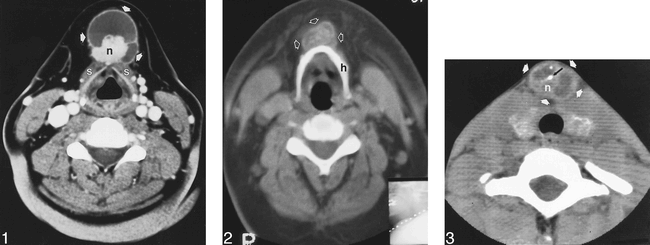
- fig 1.
Papillary thyroid carcinoma of the thyroglossal duct. Contrast-enhanced CT at the level of the thyroid cartilage reveals a cystic mass in the anterior neck (arrows), inseparable from the strap muscles (s), with septation and a lobular mural nodule (n).
- fig 2.
Papillary thyroid carcinoma of the thyroglossal duct. Unenhanced CT at the level of the hyoid bone reveals a cystic mass (arrows) anterior to the hyoid bone (h) with irregular calcifications throughout the mass.
- fig 3.
Papillary thyroid carcinoma of the thyroglossal duct. Unenhanced CT at the level of the thyroid gland reveals a complex cystic mass (white arrows) with septations, a mural nodule (n), and calcifications (black arrow)
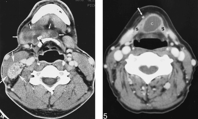
- fig 4.
Papillary thyroid carcinoma of the thyroglossal duct. Contrast-enhanced CT at the level of the hyoid bone reveals a cystic and solid mass (small arrows) extending from the anterior hyoid bone to the right lateral neck. Calcifications are identified within the mass (black arrow). A large, heterogeneous mass with calcifications in the posterior right neck (large arrows) represents metastatic disease to a cervical lymph node.
- fig 5.
Benign thyroglossal duct cyst. Contrast-enhanced CT below the level of the hyoid bone reveals a cystic mass (c) inseparable from the strap muscles (s). The mass has a thin, smooth rim, and the contents are of fluid density. No calcifications are identified within the mass. The anterior jugular vein (arrow) is displaced by the mass
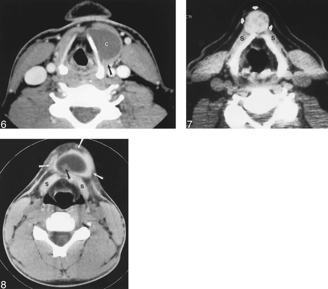
- fig 6.
Previously infected thyroglossal duct cyst. Contrast-enhanced CT through the larynx reveals a cyst (c) adjacent to the left thyroid cartilage. The cyst contents are higher than fluid density. The posterior wall of the mass is thick (arrow), but no nodules are noted. The cyst contains no calcifications. The patient had multiple episodes of anterior neck pain prior to this examination.
- fig 7.
Ectopic thyroid gland. Unenhanced CT at the level of the thyroid cartilage reveals a heterogeneous, dense, midline mass (arrows) intimately associated with the strap muscles (s). No cystic component is identified.
- fig 8.
Dermoid tumor. Unenhanced CT below the level of the hyoid bone reveals a cystic midline mass (white arrows) anterior to the strap muscles (s). A mural nodule (black arrow) is noted within the mass. The center of the mass measured −20 HU, indicating fat content


