
Article Figures & Data
Figures
- fig 1.
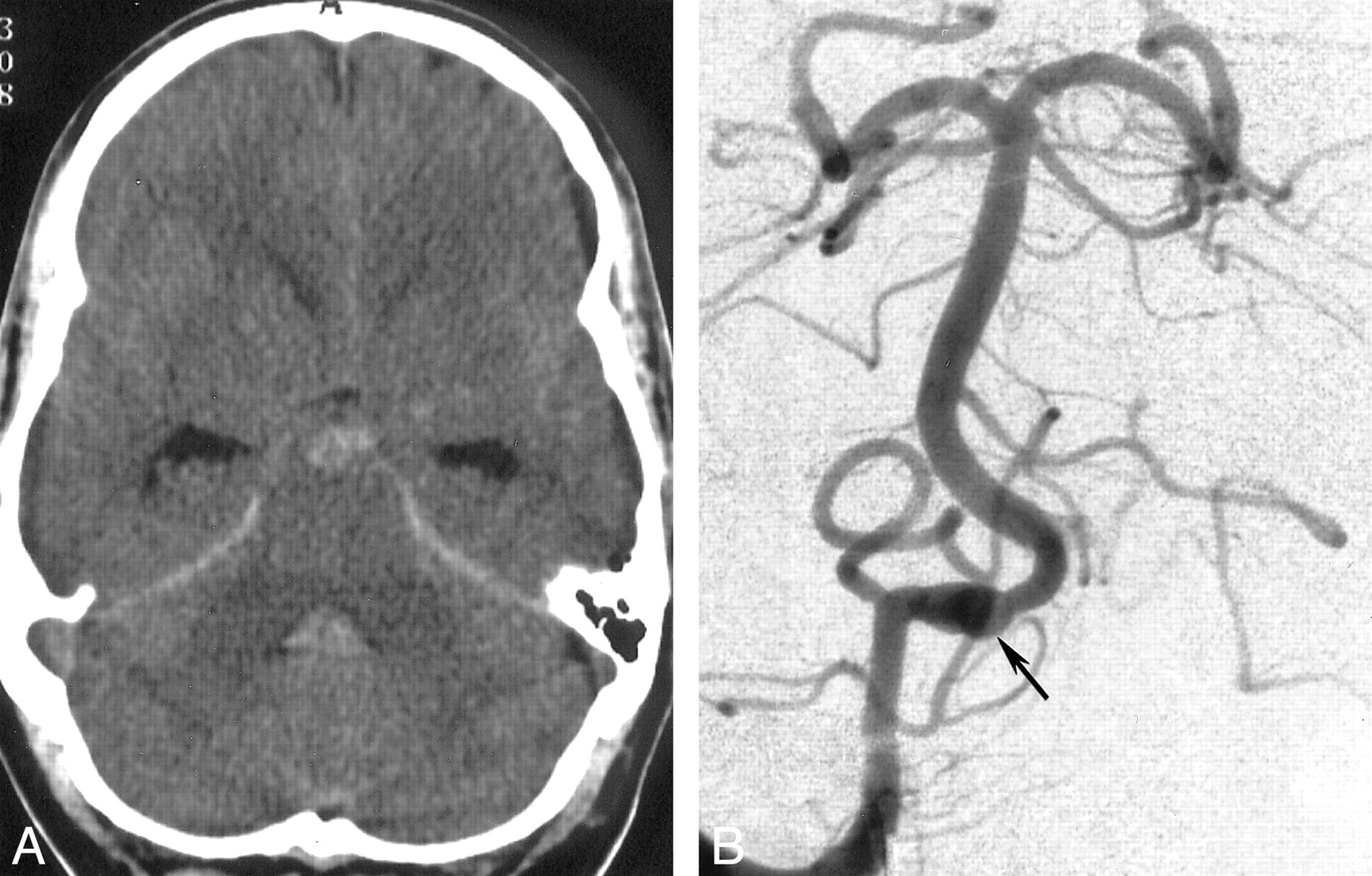
Initial images.
A, Transverse CT scan obtained on the day of admission. Blood is present within the prepontine subarachnoid space and fourth ventricle.
B, Anteroposterior arteriogram obtained with a right vertebral artery injection. Fusiform dilatation of the distal vertebral artery, distal to the posterior inferior cerebellar artery origin, indicates intracranial dissection (arrow).
- fig 2.
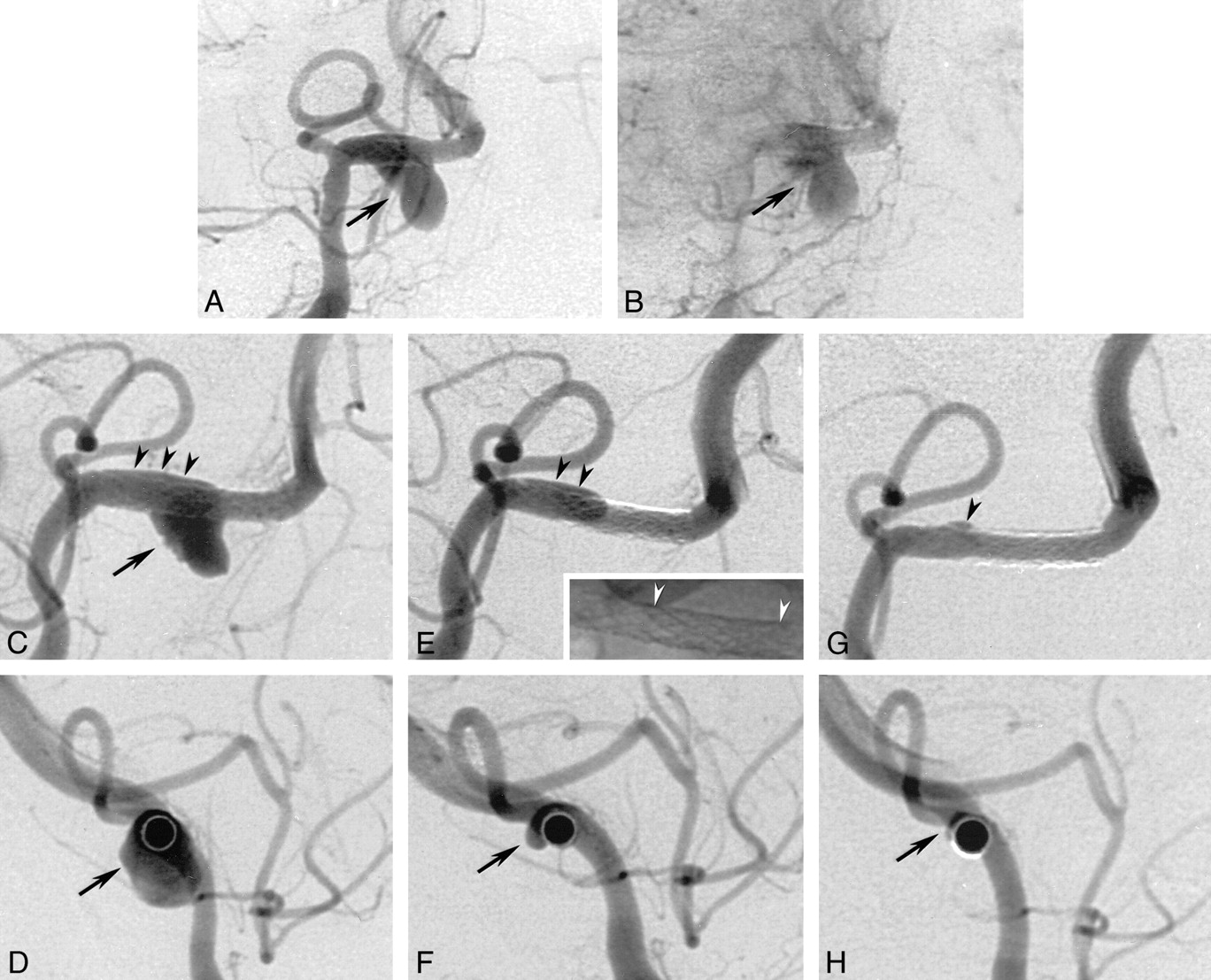
Follow-up anteroposterior arteriograms obtained with a right vertebral artery injection 5 wk after bleeding.
A, Image shows aneurysmal growth (arrow) of approximately 8 mm.
B, Image shows a further slight increase (arrow) in the size of the dome, 5 d later.
- fig 3.
Arteriograms obtained after stent placement.
A and B, Anteroposterior projection arteriograms obtained immediately after stent placement. After deployment of two overlapping stents (S670, 3/12 and 3/18 mm) distal to the posterior inferior cerebellar artery origin, the aneurysmal neck is fully covered by the portion with reduced porosity. The vertebral artery is patent, and the aneurysm still shows filling; however, a minimal delay in the washout of the intraaneurysmal filling is observed (arrow).
C, Right anteroposterior-oblique arteriogram obtained 3 d after stent placement. The overlapping stents are patent, but the aneurysmal dome shows some small filling defects adjacent to the aneurysmal wall (arrow) consistent with the beginning of intraaneurysmal thrombosis. A small amount of contrast medium is seen in the space between the stents and the proximal vessel wall where the stents are not completely adjacent to the vessel wall (arrowheads).
D, Lateral orthogonal projection of the course of the artery, obtained 3 d after stent placement, more clearly shows the circumferential extent of the pseudoaneurysm (arrow) during the post–stent-placement stage
E, Right anteroposterior-oblique arteriogram obtained 4 wk later. The two stents are patent, whereas the aneurysm shows subtotal occlusion. The filling of the extra space is now diminished in its extension (black arrowheads) and corresponds to the decreased size of the pseudoaneurysm in F. This probably does not represent the original arterial wall but rather the dissecting membrane that is the initial part of the developed pseudoaneurysm. Miniature: nonsubstracted image of the two stents shows the overlapping portion between the white arrowheads.
F, Lateral orthogonal projection of the course of the stented artery, obtained 4 wk after stent placement, more clearly shows the circumferential extent of the pseudoaneurysm (arrow) during the regression stage.
G, Right anteroposterior-oblique arteriogram obtained 3 mo after stent placement shows complete occlusion of the pseudoaneurysm, with minimal extravasation in the area of the previously dissected arterial wall (arrowhead). A patent right vertebral artery with no notable intimal hyperplasia is depicted.
H, Lateral orthogonal projection of the course of the stented artery, obtained 3 mo after stent placement, more clearly shows the total occlusion of the pseudoaneurysm (arrow).
- fig 4.
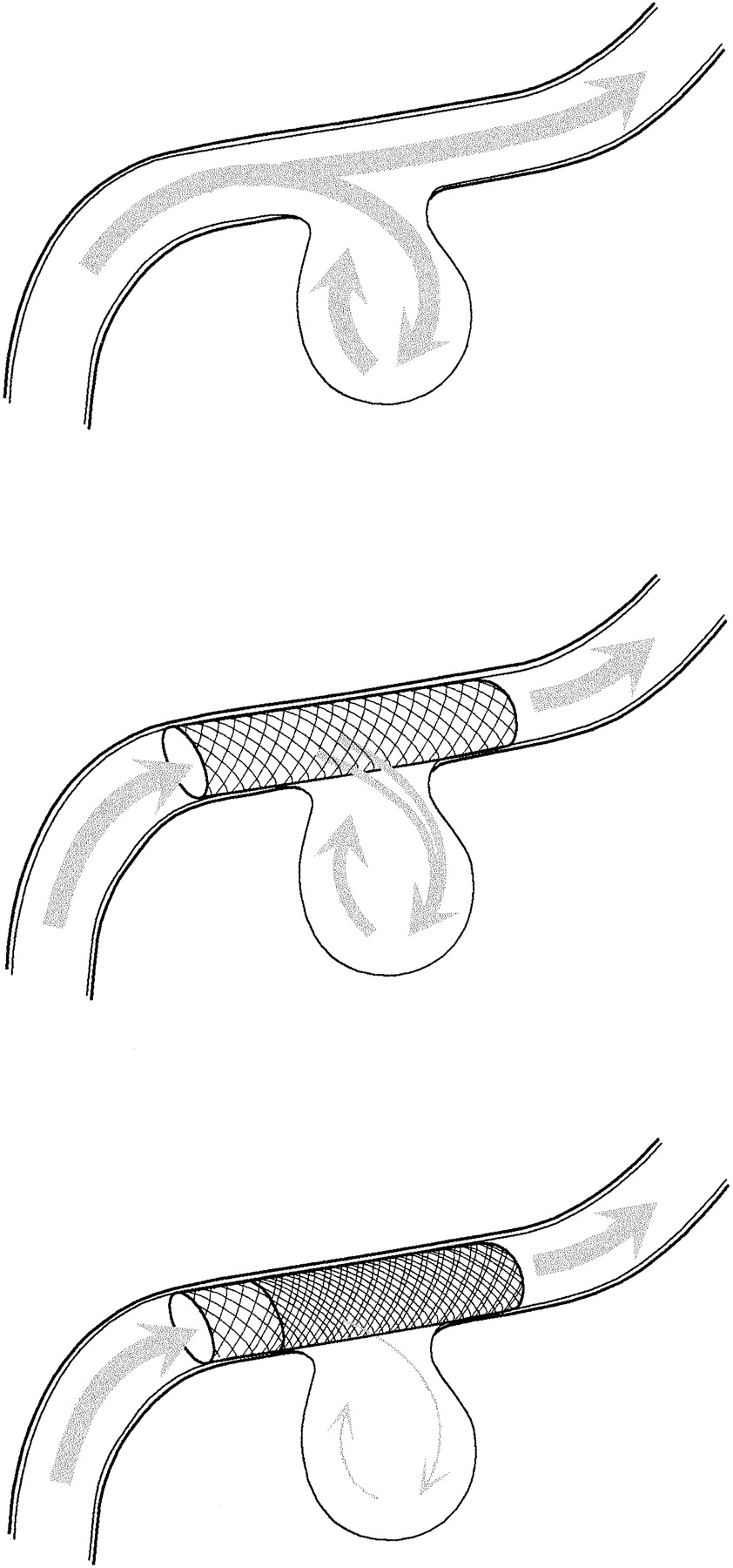
Illustration of the reduction of the intra-aneurysmal flow caused by decreased porosity due to double stent placement


In this issue


Jump to section
Related Articles
Cited By...
- Vascular angular remodeling by kissing-Y stenting in wide necked intracranial bifurcation aneurysms
- Endovascular treatment of acute intracranial vertebral artery dissection: long-term follow-up results of internal trapping and reconstructive treatment using coils and stents
- Multiple overlapping stents as monotherapy in the treatment of 'blister' pseudoaneurysms arising from the supraclinoid internal carotid artery: a single institution series and review of the literature
- Using Leo Plus stent as flow diverter and endoluminal remodeling in endovascular treatment of intracranial fusiform aneurysms
- A Flow-Diverting Stent Is Not a Pressure-Diverting Stent
- Flow Diverters Can Occlude Aneurysms and Preserve Arterial Branches: A New Experimental Model
- Stent-Assisted Coiling of Intracranial Bifurcation Aneurysms Leads to Immediate and Delayed Intracranial Vascular Angle Remodeling
- Incidence and Risk Factors of Recurrence After Endovascular Treatment of Intracranial Vertebrobasilar Dissecting Aneurysms
- Clinical and Angiographic Follow-Up of Stent-Only Therapy for Acute Intracranial Vertebrobasilar Dissecting Aneurysms