Advertisement

Main menu

  • Home
  • Content
    • Current Issue
    • Accepted Manuscripts
    • Article Preview
    • Past Issue Archive
    • AJNR Case Collection
    • Case of the Week Archive
    • Classic Case Archive
    • Case of the Month Archive
  • Special Collections
    • Spinal CSF Leak Articles (Jan 2020-June 2024)
    • 2024 AJNR Journal Awards
    • Most Impactful AJNR Articles
  • Multimedia
    • AJNR Podcast
    • AJNR Scantastics
    • Video Articles
  • For Authors
    • Submit a Manuscript
    • Author Policies
    • Fast publishing of Accepted Manuscripts
    • Graphical Abstract Preparation
    • Manuscript Submission Guidelines
    • Imaging Protocol Submission
    • Submit a Case for the Case Collection
  • About Us
    • About AJNR
    • Editorial Board
  • More
    • Become a Reviewer/Academy of Reviewers
    • Subscribers
    • Permissions
    • Alerts
    • Feedback
    • Advertisers
    • ASNR Home
  • Other Publications
    • ajnr

User menu

  • Alerts
  • Log in

Search

  • Advanced search
American Journal of Neuroradiology

American Journal of Neuroradiology

ASHNR American Society of Functional Neuroradiology ASHNR American Society of Pediatric Neuroradiology ASSR
  • Alerts
  • Log in

Advanced Search

  • Home
  • Content
    • Current Issue
    • Accepted Manuscripts
    • Article Preview
    • Past Issue Archive
    • AJNR Case Collection
    • Case of the Week Archive
    • Classic Case Archive
    • Case of the Month Archive
  • Special Collections
    • Spinal CSF Leak Articles (Jan 2020-June 2024)
    • 2024 AJNR Journal Awards
    • Most Impactful AJNR Articles
  • Multimedia
    • AJNR Podcast
    • AJNR Scantastics
    • Video Articles
  • For Authors
    • Submit a Manuscript
    • Author Policies
    • Fast publishing of Accepted Manuscripts
    • Graphical Abstract Preparation
    • Manuscript Submission Guidelines
    • Imaging Protocol Submission
    • Submit a Case for the Case Collection
  • About Us
    • About AJNR
    • Editorial Board
  • More
    • Become a Reviewer/Academy of Reviewers
    • Subscribers
    • Permissions
    • Alerts
    • Feedback
    • Advertisers
    • ASNR Home
  • Follow AJNR on Twitter
  • Visit AJNR on Facebook
  • Follow AJNR on Instagram
  • Join AJNR on LinkedIn
  • RSS Feeds

Welcome to the new AJNR, Updated Hall of Fame, and more. Read the full announcements.


AJNR is seeking candidates for the position of Associate Section Editor, AJNR Case Collection. Read the full announcement.


Research ArticleSPINE

Functional MR Imaging of the Cervical Spinal Cord by Use of Median Nerve Stimulation and Fist Clenching

Walter H. Backes, Werner H. Mess and Jan T. Wilmink
American Journal of Neuroradiology November 2001, 22 (10) 1854-1859;
Walter H. Backes
  • Find this author on Google Scholar
  • Find this author on PubMed
  • Search for this author on this site
Werner H. Mess
  • Find this author on Google Scholar
  • Find this author on PubMed
  • Search for this author on this site
Jan T. Wilmink
  • Find this author on Google Scholar
  • Find this author on PubMed
  • Search for this author on this site
  • Article
  • Figures & Data
  • Info & Metrics
  • Responses
  • References
  • PDF
Loading

Article Figures & Data

Figures

  • Tables
  • fig 1.
    • Download figure
    • Open in new tab
    • Download powerpoint
    fig 1.

    Example of a compound muscle action potential recording evoked by electrical median nerve stimulation. The small signal deflection (left) represents the compound muscle action potential measured on the flexor carpi radialis muscle, and the strong zigzag pattern (right) is caused by the strong gradient switching of the running MR imaging system
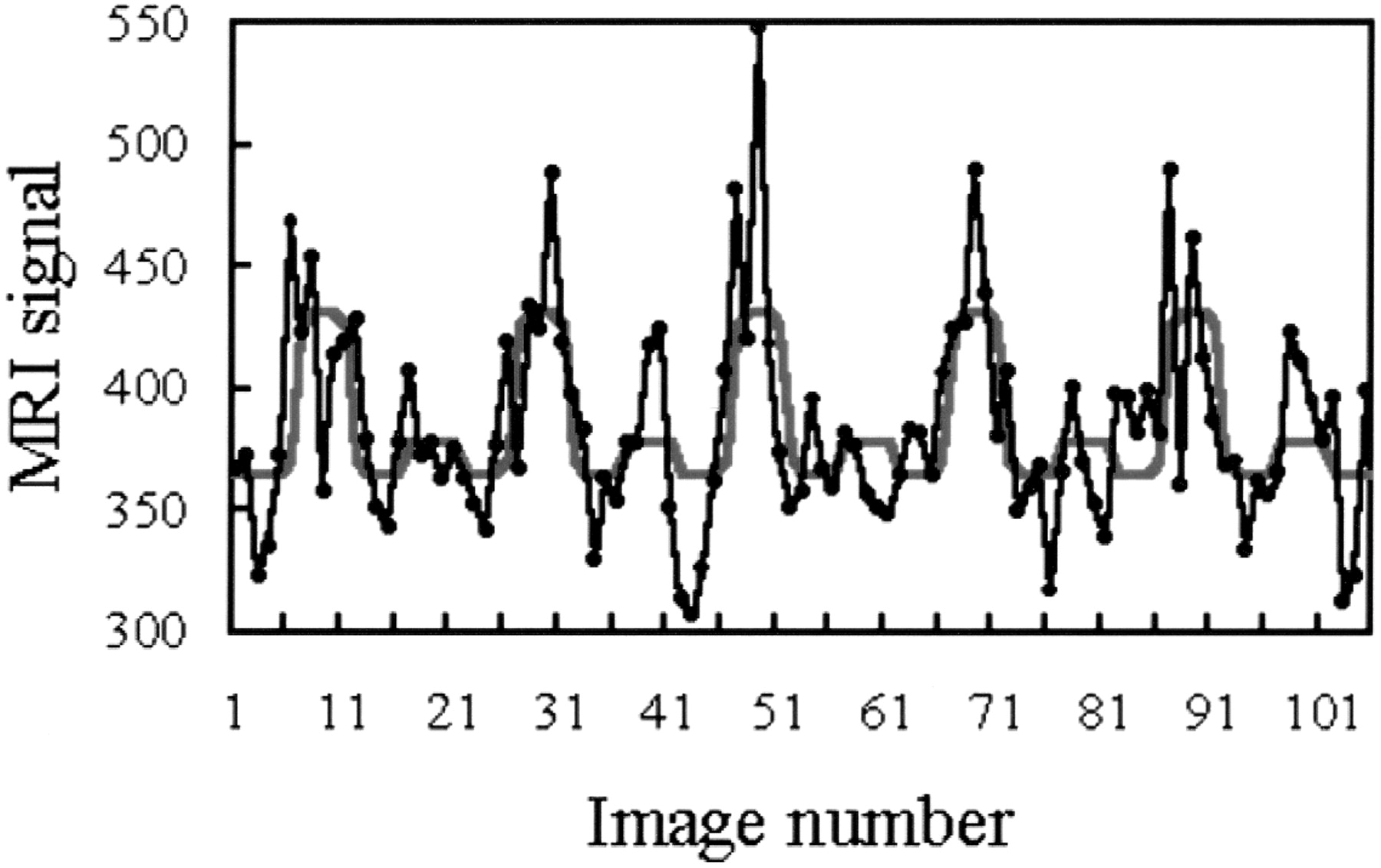
  • fig 2.
    • Download figure
    • Open in new tab
    • Download powerpoint
    fig 2.

    Example of the time course of the MR imaging signal evoked by median nerve stimulation in a spinal cord voxel. During execution, the paradigm proceeds as follows: rest, right hand stimulation, rest, left hand stimulation, and so on. In each 30-s epoch, five images were acquired. The black dots and lines represent the raw data; the red curve, the model function fitted to the responses. Note the considerable left-right differences in amplitude despite the standardized stimulus intensity
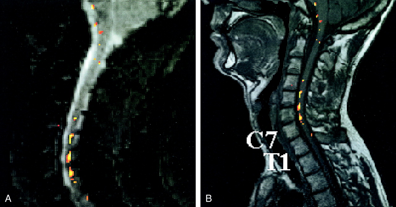
  • fig 3.
    • Download figure
    • Open in new tab
    • Download powerpoint
    fig 3.

    Example of a statistical parametric map obtained in an fMR imaging session with median nerve stimulation. Sagittal views of the statistical parametric maps of the fMR imaging responses superimposed on the first acquired T2*-weighted image (A) and the spatially corresponding anatomic T1-weighted image section (B). The voxels displayed in colors of the activation pattern analyzed with a statistical threshold of P < .01. Vertebrae C7 and T1 are indicated. The color bar indicates the statistical Z values
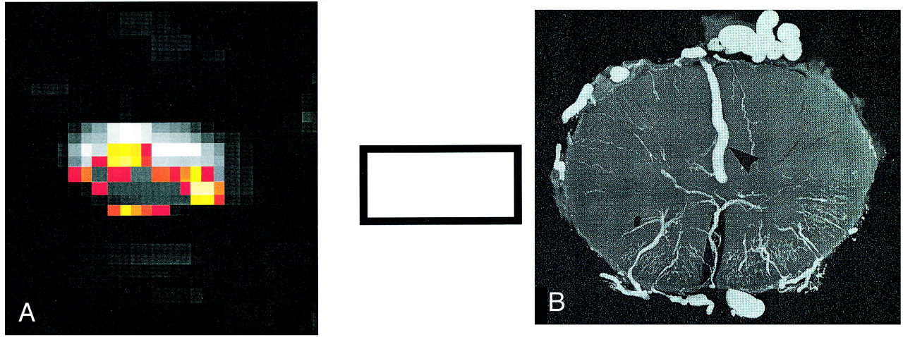
  • fig 4.
    • Download figure
    • Open in new tab
    • Download powerpoint
    fig 4.

    Transverse images in the lower cervical cord acquired during fMR imaging sessions.

    A, fMR imaging responses evoked predominantly within the cord as well as on the cord surface during the fist-clenching task.

    B, Illustration of the veins on the cord surface in a transverse cross-section. (Radiograph reproduced with permission by A.K. Thron.)
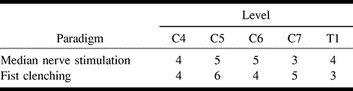
Tables

  • Figures
  • TABLE 1:
    • View popup
    • Download powerpoint
    TABLE 1:

    Subjects with fMR imaging responses

  • TABLE 2:
    • View popup
    • Download powerpoint
    TABLE 2:

    Subjects with significant fMR imaging activation at various vertebral levels

PreviousNext
Back to top

In this issue

American Journal of Neuroradiology: 22 (10)
American Journal of Neuroradiology
Vol. 22, Issue 10
1 Nov 2001
  • Table of Contents
  • Index by author
Advertisement
Print
Download PDF
Email Article

Thank you for your interest in spreading the word on American Journal of Neuroradiology.

NOTE: We only request your email address so that the person you are recommending the page to knows that you wanted them to see it, and that it is not junk mail. We do not capture any email address.

Enter multiple addresses on separate lines or separate them with commas.
Functional MR Imaging of the Cervical Spinal Cord by Use of Median Nerve Stimulation and Fist Clenching
(Your Name) has sent you a message from American Journal of Neuroradiology
(Your Name) thought you would like to see the American Journal of Neuroradiology web site.
CAPTCHA
This question is for testing whether or not you are a human visitor and to prevent automated spam submissions.
Cite this article
Walter H. Backes, Werner H. Mess, Jan T. Wilmink
Functional MR Imaging of the Cervical Spinal Cord by Use of Median Nerve Stimulation and Fist Clenching
American Journal of Neuroradiology Nov 2001, 22 (10) 1854-1859;

Citation Manager Formats

  • BibTeX
  • Bookends
  • EasyBib
  • EndNote (tagged)
  • EndNote 8 (xml)
  • Medlars
  • Mendeley
  • Papers
  • RefWorks Tagged
  • Ref Manager
  • RIS
  • Zotero
0 Responses
Respond to this article
Share
Bookmark this article
Functional MR Imaging of the Cervical Spinal Cord by Use of Median Nerve Stimulation and Fist Clenching
Walter H. Backes, Werner H. Mess, Jan T. Wilmink
American Journal of Neuroradiology Nov 2001, 22 (10) 1854-1859;
del.icio.us logo Twitter logo Facebook logo Mendeley logo
  • Tweet Widget
  • Facebook Like
  • Google Plus One

Jump to section

  • Article
    • Abstract
    • Methods
    • Results
    • Discussion
    • Conclusion
    • Footnotes
    • References
  • Figures & Data
  • Info & Metrics
  • Responses
  • References
  • PDF

Related Articles

  • No related articles found.
  • PubMed
  • Google Scholar

Cited By...

  • Spatial distribution of hand-grasp motor task activity in spinal cord functional magnetic resonance imaging
  • Spatial distribution of hand-grasp motor task activity in spinal cord functional magnetic resonance imaging
  • Functional Ultrasound Imaging of the Human Spinal Cord
  • Functional Responses in the Human Spinal Cord during Willed Motor Actions: Evidence for Side- and Rate-Dependent Activity
  • Magnetic Resonance Imaging of Neuronal Function in the Spinal Cord: Spinal fMRI
  • Crossref
  • Google Scholar

This article has not yet been cited by articles in journals that are participating in Crossref Cited-by Linking.

More in this TOC Section

  • Bern Score Validity for SIH
  • MP2RAGE 7T in MS Lesions of the Cervical Spine
  • Deep Learning for STIR Spine MRI Quality
Show more Spine

Similar Articles

Advertisement

Indexed Content

  • Current Issue
  • Accepted Manuscripts
  • Article Preview
  • Past Issues
  • Editorials
  • Editors Choice
  • Fellow Journal Club
  • Letters to the Editor

Cases

  • Case Collection
  • Archive - Case of the Week
  • Archive - Case of the Month
  • Archive - Classic Case

Special Collections

  • Special Collections

Resources

  • News and Updates
  • Turn around Times
  • Submit a Manuscript
  • Author Policies
  • Manuscript Submission Guidelines
  • Evidence-Based Medicine Level Guide
  • Publishing Checklists
  • Graphical Abstract Preparation
  • Imaging Protocol Submission
  • Submit a Case
  • Become a Reviewer/Academy of Reviewers
  • Get Peer Review Credit from Publons

Multimedia

  • AJNR Podcast
  • AJNR SCANtastic
  • Video Articles

About Us

  • About AJNR
  • Editorial Board
  • Not an AJNR Subscriber? Join Now
  • Alerts
  • Feedback
  • Advertise with us
  • Librarian Resources
  • Permissions
  • Terms and Conditions

American Society of Neuroradiology

  • Not an ASNR Member? Join Now

© 2025 by the American Society of Neuroradiology All rights, including for text and data mining, AI training, and similar technologies, are reserved.
Print ISSN: 0195-6108 Online ISSN: 1936-959X

Powered by HighWire