Advertisement

Main menu

  • Home
  • Content
    • Current Issue
    • Accepted Manuscripts
    • Article Preview
    • Past Issue Archive
    • AJNR Case Collection
    • Case of the Week Archive
    • Classic Case Archive
    • Case of the Month Archive
  • Special Collections
    • Spinal CSF Leak Articles (Jan 2020-June 2024)
    • 2024 AJNR Journal Awards
    • Most Impactful AJNR Articles
  • Multimedia
    • AJNR Podcast
    • AJNR Scantastics
    • Video Articles
  • For Authors
    • Submit a Manuscript
    • Author Policies
    • Fast publishing of Accepted Manuscripts
    • Graphical Abstract Preparation
    • Manuscript Submission Guidelines
    • Imaging Protocol Submission
    • Submit a Case for the Case Collection
  • About Us
    • About AJNR
    • Editorial Board
  • More
    • Become a Reviewer/Academy of Reviewers
    • Subscribers
    • Permissions
    • Alerts
    • Feedback
    • Advertisers
    • ASNR Home
  • Other Publications
    • ajnr

User menu

  • Alerts
  • Log in

Search

  • Advanced search
American Journal of Neuroradiology

American Journal of Neuroradiology

ASHNR American Society of Functional Neuroradiology ASHNR American Society of Pediatric Neuroradiology ASSR
  • Alerts
  • Log in

Advanced Search

  • Home
  • Content
    • Current Issue
    • Accepted Manuscripts
    • Article Preview
    • Past Issue Archive
    • AJNR Case Collection
    • Case of the Week Archive
    • Classic Case Archive
    • Case of the Month Archive
  • Special Collections
    • Spinal CSF Leak Articles (Jan 2020-June 2024)
    • 2024 AJNR Journal Awards
    • Most Impactful AJNR Articles
  • Multimedia
    • AJNR Podcast
    • AJNR Scantastics
    • Video Articles
  • For Authors
    • Submit a Manuscript
    • Author Policies
    • Fast publishing of Accepted Manuscripts
    • Graphical Abstract Preparation
    • Manuscript Submission Guidelines
    • Imaging Protocol Submission
    • Submit a Case for the Case Collection
  • About Us
    • About AJNR
    • Editorial Board
  • More
    • Become a Reviewer/Academy of Reviewers
    • Subscribers
    • Permissions
    • Alerts
    • Feedback
    • Advertisers
    • ASNR Home
  • Follow AJNR on Twitter
  • Visit AJNR on Facebook
  • Follow AJNR on Instagram
  • Join AJNR on LinkedIn
  • RSS Feeds

Welcome to the new AJNR, Updated Hall of Fame, and more. Read the full announcements.


AJNR is seeking candidates for the position of Associate Section Editor, AJNR Case Collection. Read the full announcement.


Research ArticleHEAD AND NECK

Normal Laryngeal CT Findings after Supracricoid Partial Laryngectomy

N. Bely-Toueg, P. Halimi, O. Laccourreye, F. Laskri, D. Brasnu and G. Frija
American Journal of Neuroradiology November 2001, 22 (10) 1872-1880;
N. Bely-Toueg
  • Find this author on Google Scholar
  • Find this author on PubMed
  • Search for this author on this site
P. Halimi
  • Find this author on Google Scholar
  • Find this author on PubMed
  • Search for this author on this site
O. Laccourreye
  • Find this author on Google Scholar
  • Find this author on PubMed
  • Search for this author on this site
F. Laskri
  • Find this author on Google Scholar
  • Find this author on PubMed
  • Search for this author on this site
D. Brasnu
  • Find this author on Google Scholar
  • Find this author on PubMed
  • Search for this author on this site
G. Frija
  • Find this author on Google Scholar
  • Find this author on PubMed
  • Search for this author on this site
  • Article
  • Figures & Data
  • Info & Metrics
  • Responses
  • References
  • PDF
Loading

Article Figures & Data

Figures

  • Figure1
    • Download figure
    • Open in new tab
    • Download powerpoint

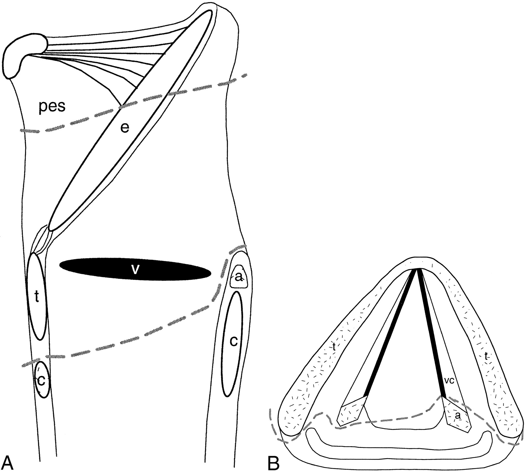
    fig. 1. A and B, Sagittal and axial drawings of structures resected during CHP (dashed lines). pes indicates preepiglottic space; e, epiglottis; a, arytenoid; c, cricoid cartilage; v, ventricle; t, thyroid cartilage; vc, vocal cords

  • Figure2
    • Download figure
    • Open in new tab
    • Download powerpoint

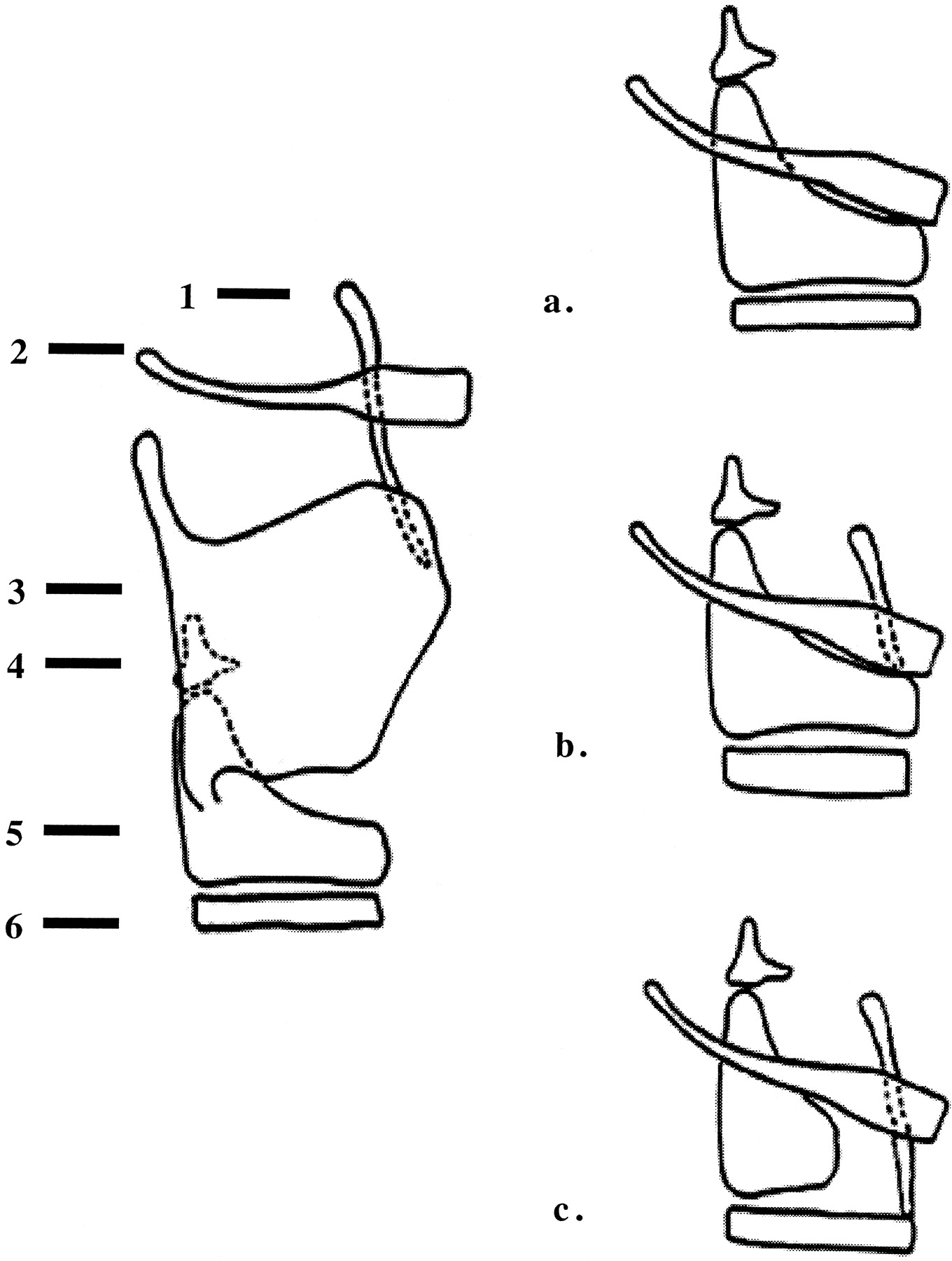
    fig. 2. Types of reconstruction with SCPL. 1 indicates epiglottis; 2, hyoid bone; 3, thyroid cartilage; 4, arytenoid cartilage; 5, cricoid cartilage; 6, tracheal ring.

    A, CHP.

    B, CHEP.

    C, TCHEP.

  • Figure3
    • Download figure
    • Open in new tab
    • Download powerpoint

    fig. 3. A and B, Sagittal and axial drawings of structures resected during CHEP (dashed lines).

    Note.—pes indicates preepiglottis space; e, epiglottis; a, arytenoid; c, cricoid cartilage; v, ventricle; t, thyroid cartilage; vc, vocal cords.

  • Figure4
    • Download figure
    • Open in new tab
    • Download powerpoint

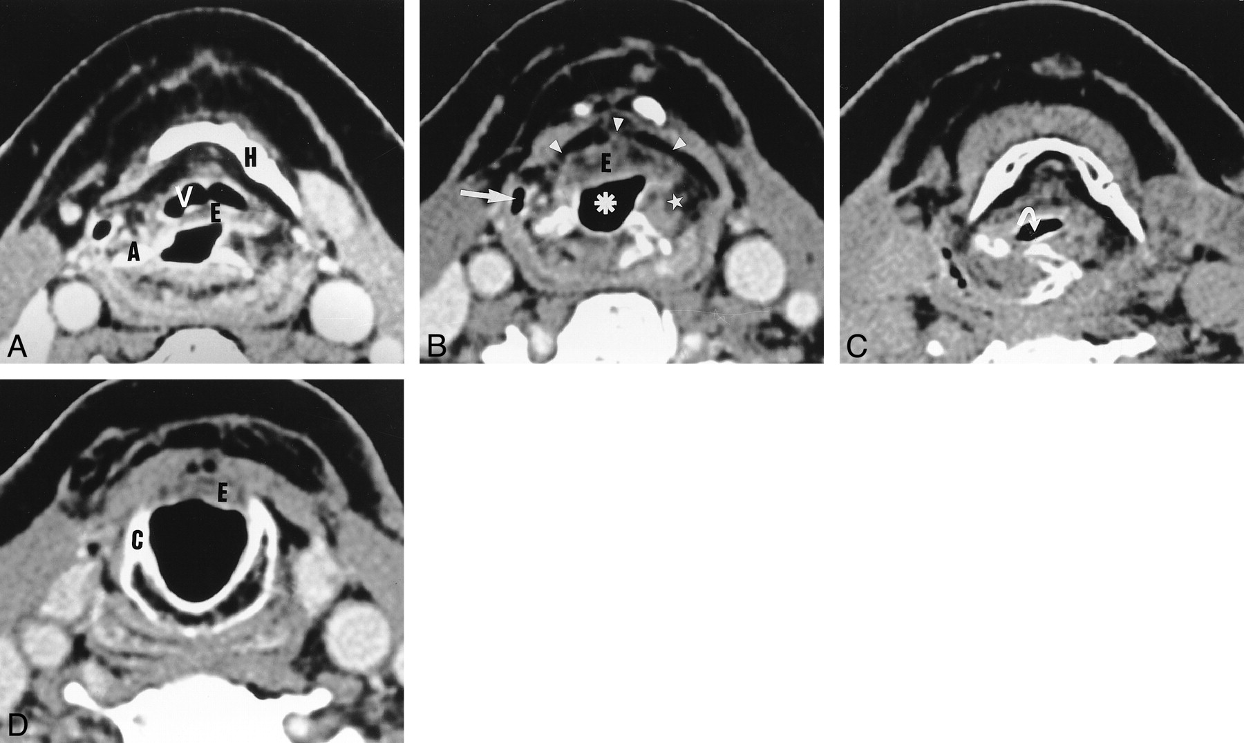
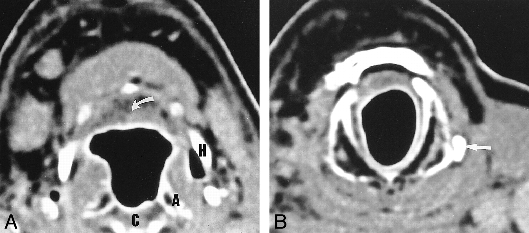
    fig. 4. Axial CT scans obtained after SCPL with CHEP.

    A, The vallecula (V) and epiglottis (E) are depicted at the level of the hyoid bone (H).

    B, Below the level of the hyoid bone, the neovestibule (asterisk) is limited anteriorly by the epiglottis (E) and laterally by the NAFs (star). The lateral recess of the hypopharynx (arrow) and the preepiglottic space (arrowheads) are depicted.

    C, During phonation, the neoglottis is oval and transversally oriented (arrow).

    D, At the level of the cricoid cartilage (C), the subglottic lumen is empty and outlined by a thin mucosa. The inferior border of the epiglottis (E) cannot be distinguished.

  • Figure5
    • Download figure
    • Open in new tab
    • Download powerpoint

    fig. 5. Axial CT scans obtained after SCPL with CHEP.

    A, Hyoid bone (H), epiglottis (E), and neoaryepiglottic fold (star).

    B, Slightly lower image shows that the left arytenoid has been resected and replaced by an exessive soft tissue (star). The right arytenoid (A) is depicted.

    C, At the level of the cricoid cartilage, the excessive soft tissue is visible in the subglottic lumen (arrow). A slightly thickened aspect of the mucosa covering the cricoid cartilage is depicted (arrowhead).

  • Figure6
    • Download figure
    • Open in new tab
    • Download powerpoint

    fig. 6. Axial CT scans obtained after SCPL with CHEP below the level of the hyoid.

    A, The lateral recess of the hypopharynx is virtual (arrow).

    B, During the Valsalva maneuver, the recess unfolds completely. Its medial limit is the neoaryepiglottic fold, which is thickened and slightly heterogeneous.

  • Figure7
    • Download figure
    • Open in new tab
    • Download powerpoint

    fig. 7. Axial CT scans obtained after SCPL with CHP.

    A, Soft tissue (arrow) corresponding to the base of the tongue is depicted behind the hyoid bone (H) instead of the epiglottis and valleculae. The pexis projects the arytenoids (A), upper border of the cricoid cartilage (C), and hyoid bone (H) to the same level.

    B, At the level of the cricoid cartilage, the mucosa is thin and regular. The left inferior horn of the thyroid cartilage has been spared (arrow). The right horn is not visible.

  • Figure8
    • Download figure
    • Open in new tab
    • Download powerpoint

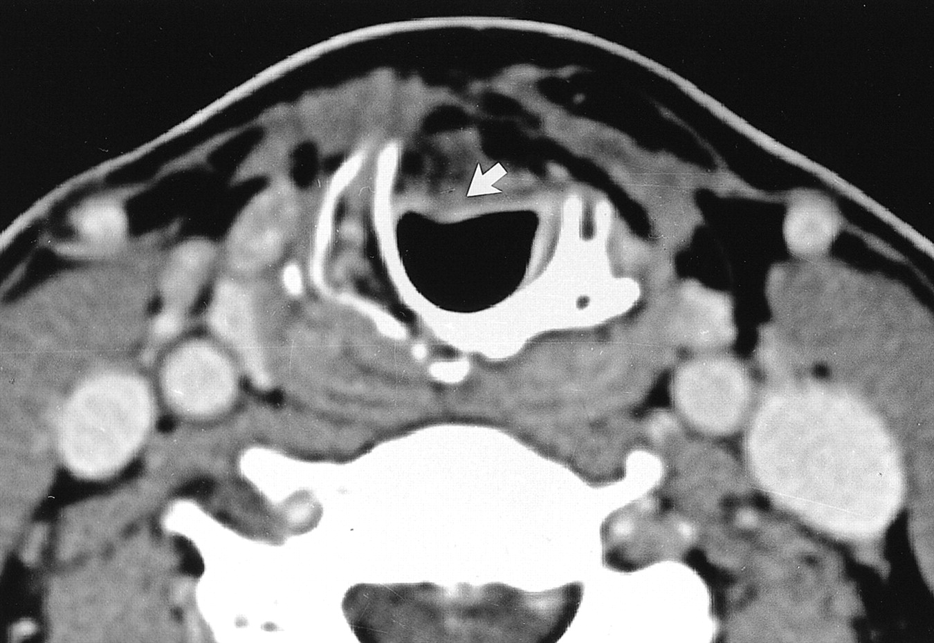
    fig. 8. Axial CT scan obtained after SCPL with TCHEP. The anterior arch of the cricoid cartilage has been resected. Therefore, the remaining epiglottis (arrow) is anchored to the first two tracheal rings

  • Figure9
    • Download figure
    • Open in new tab
    • Download powerpoint

    fig. 9. Axial CT scans of recurrrent disease in a patient who underwent SCPL with CHEP 2 y before.

    A, Image depicts strong enhancement (star) of the right NAF compared with that of the left. This finding is very suggestive of recurrence.

    B, The enhancement follows the lateral wall of the neovestibule (arrowheads). C, The enhancement proceeds down to the cricoid cartilage mucosa, which is thicker than usual (straight arrow), with the extralaryngeal spread of tumor (curved arrow).

  • Figure10
    • Download figure
    • Open in new tab
    • Download powerpoint

    fig. 10. Axial CT scans of recurrence 3 y after SCPL with CHP.

    A, Enhancement of the right lateral wall of the neovestibule (arrows).

    B, Enhancement spreading downward to the submucosa of the cricoid cartilage (arrow). C, Enhancement extending upward to the right side of the base of the tongue (arrow).

  • Figure11
    • Download figure
    • Open in new tab
    • Download powerpoint

    fig. 11. Axial CT scans in a patient who underwent SCPL with CHEP 5 y before. At nasofibroscopy, the patient had an anterior cervical cutaneous fistula with an arytenoid that was only slightly thickened.

    A, Massive extralaryngeal recurrence around the cricohyoidopexy (arrows).

    B, Recurrence spreading downward to the pretracheal tissue (stars).

  • Figure12
    • Download figure
    • Open in new tab
    • Download powerpoint

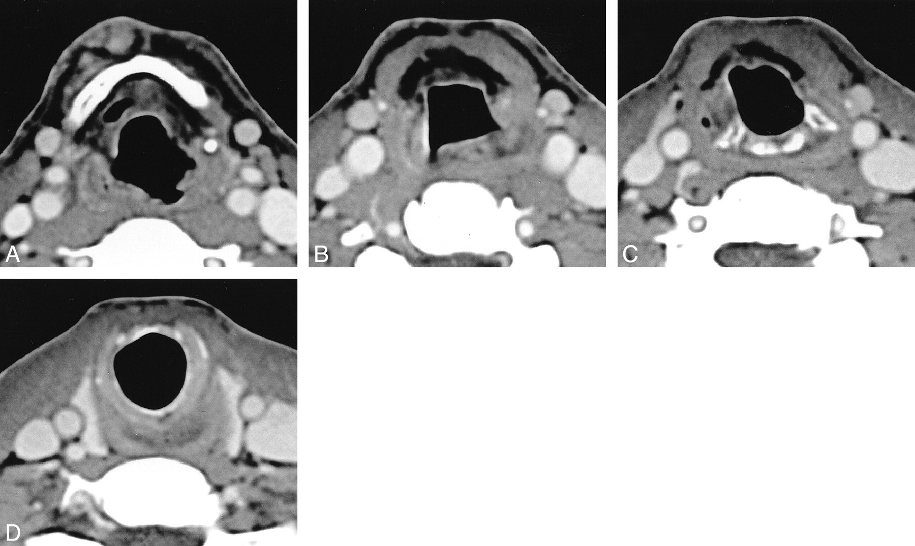
    fig. 12. Axial CT scans obtained 6 mo after SCPL with CHEP in a patient with severe swallowing disorders. The distance between the lower limit of the hyoid bone (in A) and the lower limit of the cricoid cartilage (in D) is more than 3.5 cm. This finding implies failure of the pexis, which was confirmed at surgery

PreviousNext
Back to top

In this issue

American Journal of Neuroradiology: 22 (10)
American Journal of Neuroradiology
Vol. 22, Issue 10
1 Nov 2001
  • Table of Contents
  • Index by author
Advertisement
Print
Download PDF
Email Article

Thank you for your interest in spreading the word on American Journal of Neuroradiology.

NOTE: We only request your email address so that the person you are recommending the page to knows that you wanted them to see it, and that it is not junk mail. We do not capture any email address.

Enter multiple addresses on separate lines or separate them with commas.
Normal Laryngeal CT Findings after Supracricoid Partial Laryngectomy
(Your Name) has sent you a message from American Journal of Neuroradiology
(Your Name) thought you would like to see the American Journal of Neuroradiology web site.
CAPTCHA
This question is for testing whether or not you are a human visitor and to prevent automated spam submissions.
Cite this article
N. Bely-Toueg, P. Halimi, O. Laccourreye, F. Laskri, D. Brasnu, G. Frija
Normal Laryngeal CT Findings after Supracricoid Partial Laryngectomy
American Journal of Neuroradiology Nov 2001, 22 (10) 1872-1880;

Citation Manager Formats

  • BibTeX
  • Bookends
  • EasyBib
  • EndNote (tagged)
  • EndNote 8 (xml)
  • Medlars
  • Mendeley
  • Papers
  • RefWorks Tagged
  • Ref Manager
  • RIS
  • Zotero
0 Responses
Respond to this article
Share
Bookmark this article
Normal Laryngeal CT Findings after Supracricoid Partial Laryngectomy
N. Bely-Toueg, P. Halimi, O. Laccourreye, F. Laskri, D. Brasnu, G. Frija
American Journal of Neuroradiology Nov 2001, 22 (10) 1872-1880;
del.icio.us logo Twitter logo Facebook logo Mendeley logo
  • Tweet Widget
  • Facebook Like
  • Google Plus One

Jump to section

  • Article
    • Abstract
    • Methods
    • Results
    • Discussion
    • Footnotes
    • References
  • Figures & Data
  • Info & Metrics
  • Responses
  • References
  • PDF

Related Articles

  • No related articles found.
  • PubMed
  • Google Scholar

Cited By...

  • Failure of OPHL type IIb due to undiagnosed Eagle syndrome
  • Crossref
  • Google Scholar

This article has not yet been cited by articles in journals that are participating in Crossref Cited-by Linking.

More in this TOC Section

  • Correlation of Apparent Diffusion Coefficient at 3T with Prognostic Parameters of Retinoblastoma
  • Parathyroid Lesions: Characterization with Dual-Phase Arterial and Venous Enhanced CT of the Neck
  • MR Diagnosis of Facial Neuritis: Diagnostic Performance of Contrast-Enhanced 3D-FLAIR Technique Compared with Contrast-Enhanced 3D-T1-Fast-Field Echo with Fat Suppression
Show more Head and Neck

Similar Articles

Advertisement

Indexed Content

  • Current Issue
  • Accepted Manuscripts
  • Article Preview
  • Past Issues
  • Editorials
  • Editors Choice
  • Fellow Journal Club
  • Letters to the Editor

Cases

  • Case Collection
  • Archive - Case of the Week
  • Archive - Case of the Month
  • Archive - Classic Case

Special Collections

  • Special Collections

Resources

  • News and Updates
  • Turn around Times
  • Submit a Manuscript
  • Author Policies
  • Manuscript Submission Guidelines
  • Evidence-Based Medicine Level Guide
  • Publishing Checklists
  • Graphical Abstract Preparation
  • Imaging Protocol Submission
  • Submit a Case
  • Become a Reviewer/Academy of Reviewers
  • Get Peer Review Credit from Publons

Multimedia

  • AJNR Podcast
  • AJNR SCANtastic
  • Video Articles

About Us

  • About AJNR
  • Editorial Board
  • Not an AJNR Subscriber? Join Now
  • Alerts
  • Feedback
  • Advertise with us
  • Librarian Resources
  • Permissions
  • Terms and Conditions

American Society of Neuroradiology

  • Not an ASNR Member? Join Now

© 2025 by the American Society of Neuroradiology All rights, including for text and data mining, AI training, and similar technologies, are reserved.
Print ISSN: 0195-6108 Online ISSN: 1936-959X

Powered by HighWire