Advertisement

Main menu

  • Home
  • Content
    • Current Issue
    • Accepted Manuscripts
    • Article Preview
    • Past Issue Archive
    • Video Articles
    • AJNR Case Collection
    • Case of the Week Archive
    • Case of the Month Archive
    • Classic Case Archive
  • Special Collections
    • AJNR Awards
    • Low-Field MRI
    • Alzheimer Disease
    • ASNR Foundation Special Collection
    • Photon-Counting CT
    • View All
  • Multimedia
    • AJNR Podcasts
    • AJNR SCANtastic
    • Trainee Corner
    • MRI Safety Corner
    • Imaging Protocols
  • For Authors
    • Submit a Manuscript
    • Submit a Video Article
    • Submit an eLetter to the Editor/Response
    • Manuscript Submission Guidelines
    • Statistical Tips
    • Fast Publishing of Accepted Manuscripts
    • Graphical Abstract Preparation
    • Imaging Protocol Submission
    • Author Policies
  • About Us
    • About AJNR
    • Editorial Board
    • Editorial Board Alumni
  • More
    • Become a Reviewer/Academy of Reviewers
    • Subscribers
    • Permissions
    • Alerts
    • Feedback
    • Advertisers
    • ASNR Home

User menu

  • Alerts
  • Log in

Search

  • Advanced search
American Journal of Neuroradiology

American Journal of Neuroradiology

ASHNR American Society of Functional Neuroradiology ASHNR American Society of Pediatric Neuroradiology ASSR
  • Alerts
  • Log in

Advanced Search

  • Home
  • Content
    • Current Issue
    • Accepted Manuscripts
    • Article Preview
    • Past Issue Archive
    • Video Articles
    • AJNR Case Collection
    • Case of the Week Archive
    • Case of the Month Archive
    • Classic Case Archive
  • Special Collections
    • AJNR Awards
    • Low-Field MRI
    • Alzheimer Disease
    • ASNR Foundation Special Collection
    • Photon-Counting CT
    • View All
  • Multimedia
    • AJNR Podcasts
    • AJNR SCANtastic
    • Trainee Corner
    • MRI Safety Corner
    • Imaging Protocols
  • For Authors
    • Submit a Manuscript
    • Submit a Video Article
    • Submit an eLetter to the Editor/Response
    • Manuscript Submission Guidelines
    • Statistical Tips
    • Fast Publishing of Accepted Manuscripts
    • Graphical Abstract Preparation
    • Imaging Protocol Submission
    • Author Policies
  • About Us
    • About AJNR
    • Editorial Board
    • Editorial Board Alumni
  • More
    • Become a Reviewer/Academy of Reviewers
    • Subscribers
    • Permissions
    • Alerts
    • Feedback
    • Advertisers
    • ASNR Home
  • Follow AJNR on Twitter
  • Visit AJNR on Facebook
  • Follow AJNR on Instagram
  • Join AJNR on LinkedIn
  • RSS Feeds

AJNR Awards, New Junior Editors, and more. Read the latest AJNR updates

  • Getting new auth cookie, if you see this message a lot, tell someone!
  • Getting new auth cookie, if you see this message a lot, tell someone!
Research ArticleBRAIN

Blood Volume of Gliomas Determined by Double-Echo Dynamic Perfusion-Weighted MR Imaging: A Preliminary Study

Hidemasa Uematsu, Masayuki Maeda, Norihiro Sadato, Tsuyoshi Matsuda, Yoshiyuki Ishimori, Yoshio Koshimoto, Hirohiko Kimura, Hiroki Yamada, Yasutaka Kawamura, Yoshiharu Yonekura and Harumi Itoh
American Journal of Neuroradiology November 2001, 22 (10) 1915-1919;
Hidemasa Uematsu
  • Find this author on Google Scholar
  • Find this author on PubMed
  • Search for this author on this site
Masayuki Maeda
  • Find this author on Google Scholar
  • Find this author on PubMed
  • Search for this author on this site
Norihiro Sadato
  • Find this author on Google Scholar
  • Find this author on PubMed
  • Search for this author on this site
Tsuyoshi Matsuda
  • Find this author on Google Scholar
  • Find this author on PubMed
  • Search for this author on this site
Yoshiyuki Ishimori
  • Find this author on Google Scholar
  • Find this author on PubMed
  • Search for this author on this site
Yoshio Koshimoto
  • Find this author on Google Scholar
  • Find this author on PubMed
  • Search for this author on this site
Hirohiko Kimura
  • Find this author on Google Scholar
  • Find this author on PubMed
  • Search for this author on this site
Hiroki Yamada
  • Find this author on Google Scholar
  • Find this author on PubMed
  • Search for this author on this site
Yasutaka Kawamura
  • Find this author on Google Scholar
  • Find this author on PubMed
  • Search for this author on this site
Yoshiharu Yonekura
  • Find this author on Google Scholar
  • Find this author on PubMed
  • Search for this author on this site
Harumi Itoh
  • Find this author on Google Scholar
  • Find this author on PubMed
  • Search for this author on this site
  • Article
  • Figures & Data
  • Info & Metrics
  • Responses
  • References
  • PDF
Loading

Article Figures & Data

Figures

  • Tables
  • fig 1.
    • Download figure
    • Open in new tab
    • Download powerpoint
    fig 1.

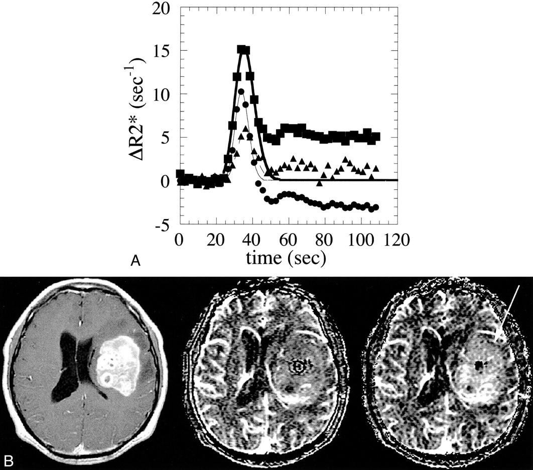
    Case 2: 54-year-old man with a GBM.

    A, Time course of the ΔR2* values of the GBM and normal white matter. The ΔR2*T1C values of the GBM are represented by solid squares and its gamma-fitted curve by a thick line; ΔR2*T1U is represented by solid circles and its gamma-fitted curve a thin line. The ΔR2*T1C values of normal white matter are represented by solid triangles and its gamma-fitted curve by a dashed line.

    B, A stereotactic biopsy was performed before the MR study. A transaxial section of a contrast-enhanced T1-weighted (333/10/3) spin-echo image is shown on the left, the corresponding VT1U map is in the middle, and the VT1C map is on the right. The GBM shows nonhomogeneously dense contrast enhancement in the left frontotemporal lobe. Note that blood volume of the tumor is much more prominent on the VT1C map (arrow) than on the VT1U map. The qualitative analysis was rated as 1 in this case. The VT1U/VT1C ratio is 0.41.
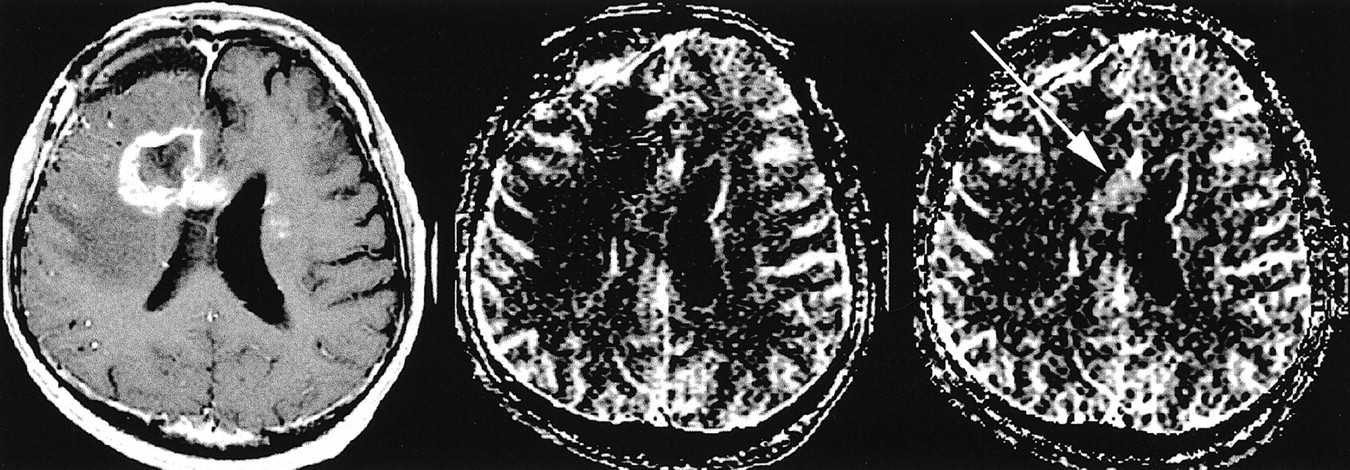
  • fig 2.
    • Download figure
    • Open in new tab
    • Download powerpoint
    fig 2.

    Case 5: 37-year-old man with a GBM who had previously undergone surgical resection, irradiation, and chemotherapy for a GBM of the right frontal lobe. Recently, a recurrent tumor was found on follow-up CT studies. A transaxial section of a contrast-enhanced T1-weighted spin-echo MR image (333/10/3) is shown on the left, a corresponding VT1U map is in the middle, and the VT1C map is on the right. The recurrent GBM shows dense contrast enhancement in the right frontal lobe, extending to the corpus callosum. Small, enhanced foci are also depicted in the left frontal lobe. Note that the lesion in the corpus callosum is depicted as an area of hypervascular blood volume on the VT1C map (arrow), and the blood volume of this lesion is similar to that of gray matter on the VT1U map, indicating the tumor blood volume is underestimated on the VT1U map. The qualitative analysis was rated as 1 in this case. The VT1U/VT1C ratio is 0.64

Tables

  • Figures
  • Table1

    Results of quantitative and qualitative analysis of blood volume in GBMs

PreviousNext
Back to top

In this issue

American Journal of Neuroradiology: 22 (10)
American Journal of Neuroradiology
Vol. 22, Issue 10
1 Nov 2001
  • Table of Contents
  • Index by author
Advertisement
Print
Download PDF
Email Article

Thank you for your interest in spreading the word on American Journal of Neuroradiology.

NOTE: We only request your email address so that the person you are recommending the page to knows that you wanted them to see it, and that it is not junk mail. We do not capture any email address.

Enter multiple addresses on separate lines or separate them with commas.
Blood Volume of Gliomas Determined by Double-Echo Dynamic Perfusion-Weighted MR Imaging: A Preliminary Study
(Your Name) has sent you a message from American Journal of Neuroradiology
(Your Name) thought you would like to see the American Journal of Neuroradiology web site.
CAPTCHA
This question is for testing whether or not you are a human visitor and to prevent automated spam submissions.
Cite this article
Hidemasa Uematsu, Masayuki Maeda, Norihiro Sadato, Tsuyoshi Matsuda, Yoshiyuki Ishimori, Yoshio Koshimoto, Hirohiko Kimura, Hiroki Yamada, Yasutaka Kawamura, Yoshiharu Yonekura, Harumi Itoh
Blood Volume of Gliomas Determined by Double-Echo Dynamic Perfusion-Weighted MR Imaging: A Preliminary Study
American Journal of Neuroradiology Nov 2001, 22 (10) 1915-1919;

Citation Manager Formats

  • BibTeX
  • Bookends
  • EasyBib
  • EndNote (tagged)
  • EndNote 8 (xml)
  • Medlars
  • Mendeley
  • Papers
  • RefWorks Tagged
  • Ref Manager
  • RIS
  • Zotero
0 Responses
Respond to this article
Share
Bookmark this article
Blood Volume of Gliomas Determined by Double-Echo Dynamic Perfusion-Weighted MR Imaging: A Preliminary Study
Hidemasa Uematsu, Masayuki Maeda, Norihiro Sadato, Tsuyoshi Matsuda, Yoshiyuki Ishimori, Yoshio Koshimoto, Hirohiko Kimura, Hiroki Yamada, Yasutaka Kawamura, Yoshiharu Yonekura, Harumi Itoh
American Journal of Neuroradiology Nov 2001, 22 (10) 1915-1919;
del.icio.us logo Twitter logo Facebook logo Mendeley logo
  • Tweet Widget
  • Facebook Like
  • Google Plus One

Jump to section

  • Article
    • Abstract
    • Methods
    • Results
    • Discussion
    • Conclusion
    • Acknowledgments
    • Footnotes
    • References
  • Figures & Data
  • Info & Metrics
  • Responses
  • References
  • PDF

Related Articles

  • No related articles found.
  • PubMed
  • Google Scholar

Cited By...

  • Optimization of Acquisition and Analysis Methods for Clinical Dynamic Susceptibility Contrast MRI Using a Population-Based Digital Reference Object
  • Improving Perfusion Measurement in DSC-MR Imaging with Multiecho Information for Arterial Input Function Determination
  • On the Use of DSC-MRI for Measuring Vascular Permeability
  • The Effect of Pulse Sequence Parameters and Contrast Agent Dose on Percentage Signal Recovery in DSC-MRI: Implications for Clinical Applications
  • The Role of Preload and Leakage Correction in Gadolinium-Based Cerebral Blood Volume Estimation Determined by Comparison with MION as a Criterion Standard
  • Percentage Signal Recovery Derived from MR Dynamic Susceptibility Contrast Imaging Is Useful to Differentiate Common Enhancing Malignant Lesions of the Brain
  • Relative Cerebral Blood Volume Values to Differentiate High-Grade Glioma Recurrence from Posttreatment Radiation Effect: Direct Correlation between Image-Guided Tissue Histopathology and Localized Dynamic Susceptibility-Weighted Contrast-Enhanced Perfusion MR Imaging Measurements
  • Crossref
  • Google Scholar

This article has not yet been cited by articles in journals that are participating in Crossref Cited-by Linking.

More in this TOC Section

  • Optimal MRI Sequence for Identifying Occlusion Location in Acute Stroke: Which Value of Time-Resolved Contrast-Enhanced MRA?
  • Evaluating the Effects of White Matter Multiple Sclerosis Lesions on the Volume Estimation of 6 Brain Tissue Segmentation Methods
  • Quiet PROPELLER MRI Techniques Match the Quality of Conventional PROPELLER Brain Imaging Techniques
Show more Brain

Similar Articles

Advertisement

Indexed Content

  • Current Issue
  • Accepted Manuscripts
  • Article Preview
  • Past Issues
  • Editorials
  • Editor's Choice
  • Fellows' Journal Club
  • Letters to the Editor
  • Video Articles

Cases

  • Case Collection
  • Archive - Case of the Week
  • Archive - Case of the Month
  • Archive - Classic Case

More from AJNR

  • Trainee Corner
  • Imaging Protocols
  • MRI Safety Corner
  • Book Reviews

Multimedia

  • AJNR Podcasts
  • AJNR Scantastics

Resources

  • Turnaround Time
  • Submit a Manuscript
  • Submit a Video Article
  • Submit an eLetter to the Editor/Response
  • Manuscript Submission Guidelines
  • Statistical Tips
  • Fast Publishing of Accepted Manuscripts
  • Graphical Abstract Preparation
  • Imaging Protocol Submission
  • Evidence-Based Medicine Level Guide
  • Publishing Checklists
  • Author Policies
  • Become a Reviewer/Academy of Reviewers
  • News and Updates

About Us

  • About AJNR
  • Editorial Board
  • Editorial Board Alumni
  • Alerts
  • Permissions
  • Not an AJNR Subscriber? Join Now
  • Advertise with Us
  • Librarian Resources
  • Feedback
  • Terms and Conditions
  • AJNR Editorial Board Alumni

American Society of Neuroradiology

  • Not an ASNR Member? Join Now

© 2025 by the American Society of Neuroradiology All rights, including for text and data mining, AI training, and similar technologies, are reserved.
Print ISSN: 0195-6108 Online ISSN: 1936-959X

Powered by HighWire