Advertisement

Main menu

  • Home
  • Content
    • Current Issue
    • Accepted Manuscripts
    • Article Preview
    • Past Issue Archive
    • AJNR Case Collection
    • Case of the Week Archive
    • Classic Case Archive
    • Case of the Month Archive
  • Special Collections
    • Spinal CSF Leak Articles (Jan 2020-June 2024)
    • 2024 AJNR Journal Awards
    • Most Impactful AJNR Articles
  • Multimedia
    • AJNR Podcast
    • AJNR Scantastics
    • Video Articles
  • For Authors
    • Submit a Manuscript
    • Author Policies
    • Fast publishing of Accepted Manuscripts
    • Graphical Abstract Preparation
    • Manuscript Submission Guidelines
    • Imaging Protocol Submission
    • Submit a Case for the Case Collection
  • About Us
    • About AJNR
    • Editorial Board
  • More
    • Become a Reviewer/Academy of Reviewers
    • Subscribers
    • Permissions
    • Alerts
    • Feedback
    • Advertisers
    • ASNR Home
  • Other Publications
    • ajnr

User menu

  • Alerts
  • Log in

Search

  • Advanced search
American Journal of Neuroradiology

American Journal of Neuroradiology

ASHNR American Society of Functional Neuroradiology ASHNR American Society of Pediatric Neuroradiology ASSR
  • Alerts
  • Log in

Advanced Search

  • Home
  • Content
    • Current Issue
    • Accepted Manuscripts
    • Article Preview
    • Past Issue Archive
    • AJNR Case Collection
    • Case of the Week Archive
    • Classic Case Archive
    • Case of the Month Archive
  • Special Collections
    • Spinal CSF Leak Articles (Jan 2020-June 2024)
    • 2024 AJNR Journal Awards
    • Most Impactful AJNR Articles
  • Multimedia
    • AJNR Podcast
    • AJNR Scantastics
    • Video Articles
  • For Authors
    • Submit a Manuscript
    • Author Policies
    • Fast publishing of Accepted Manuscripts
    • Graphical Abstract Preparation
    • Manuscript Submission Guidelines
    • Imaging Protocol Submission
    • Submit a Case for the Case Collection
  • About Us
    • About AJNR
    • Editorial Board
  • More
    • Become a Reviewer/Academy of Reviewers
    • Subscribers
    • Permissions
    • Alerts
    • Feedback
    • Advertisers
    • ASNR Home
  • Follow AJNR on Twitter
  • Visit AJNR on Facebook
  • Follow AJNR on Instagram
  • Join AJNR on LinkedIn
  • RSS Feeds

Welcome to the new AJNR, Updated Hall of Fame, and more. Read the full announcements.


AJNR is seeking candidates for the position of Associate Section Editor, AJNR Case Collection. Read the full announcement.


Research ArticleBRAIN

MR Imaging, Single-photon Emission CT, and Neurocognitive Performance after Mild Traumatic Brain Injury

Paul A.M. Hofman, Sven Z. Stapert, Marinus J.P.G. van Kroonenburgh, Jelle Jolles, Jelle de Kruijk and Jan T. Wilmink
American Journal of Neuroradiology March 2001, 22 (3) 441-449;
Paul A.M. Hofman
  • Find this author on Google Scholar
  • Find this author on PubMed
  • Search for this author on this site
Sven Z. Stapert
  • Find this author on Google Scholar
  • Find this author on PubMed
  • Search for this author on this site
Marinus J.P.G. van Kroonenburgh
  • Find this author on Google Scholar
  • Find this author on PubMed
  • Search for this author on this site
Jelle Jolles
  • Find this author on Google Scholar
  • Find this author on PubMed
  • Search for this author on this site
Jelle de Kruijk
  • Find this author on Google Scholar
  • Find this author on PubMed
  • Search for this author on this site
Jan T. Wilmink
  • Find this author on Google Scholar
  • Find this author on PubMed
  • Search for this author on this site
  • Article
  • Figures & Data
  • Info & Metrics
  • Responses
  • References
  • PDF
Loading

Article Figures & Data

Figures

  • Tables
  • fig 1.
    • Download figure
    • Open in new tab
    • Download powerpoint
    fig 1.

    A and B, FLAIR images of a 36-year-old man who fell from a bicycle. Contusions are seen in the left temporal lobe as well as in the left frontal lobe. Note also the right frontal extracerebral hemorrhage
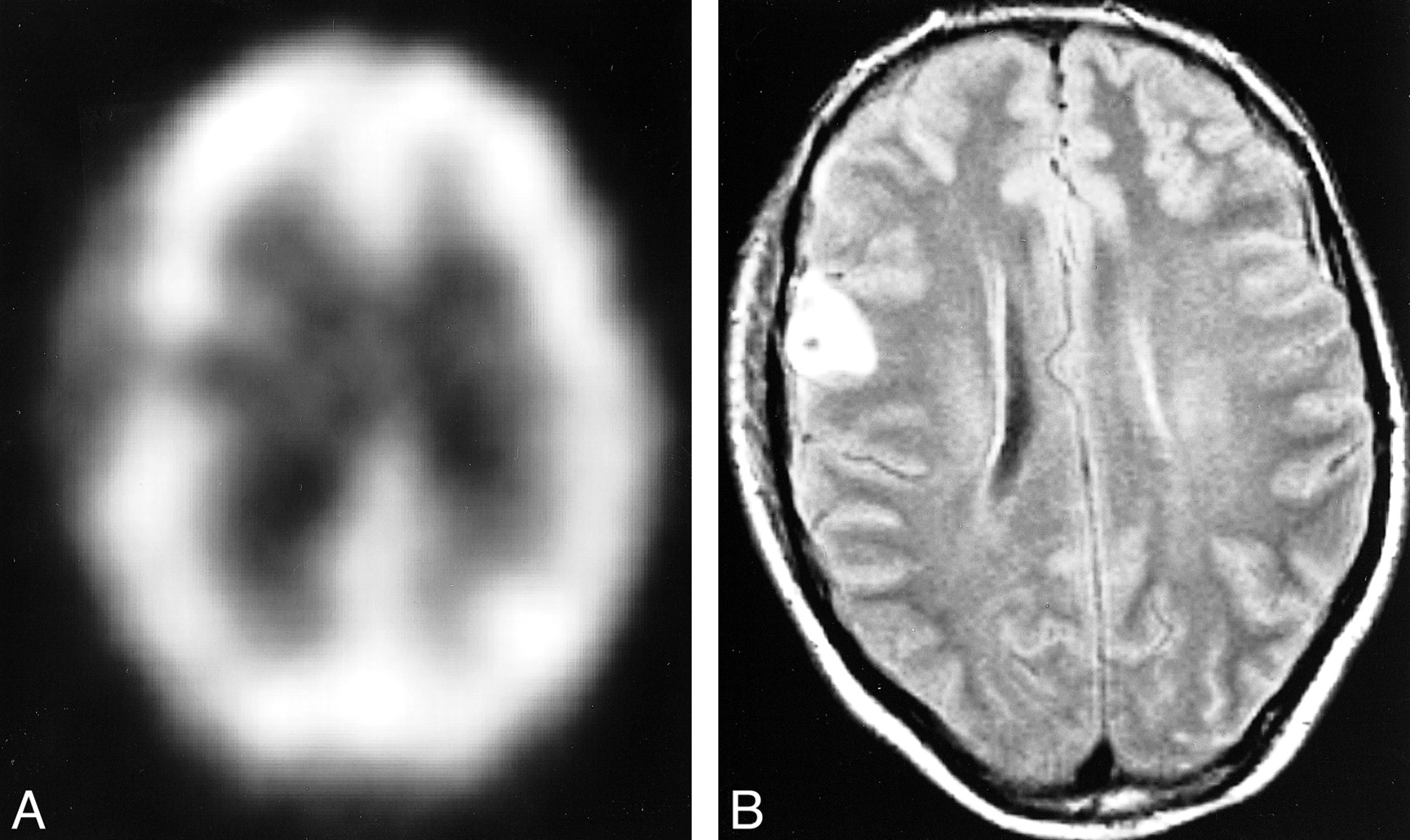
  • fig 2.
    • Download figure
    • Open in new tab
    • Download powerpoint
    fig 2.

    Images obtained from a 23-year-old man who was involved in a car accident.

    A, SPECT image shows a left frontal perfusion deficit.

    B, FLAIR image shows no lesions at this location.

    C, Owing to diffuse axonal injury, T2*-weighted image shows two deep hemorrhagic lesions.

  • fig 3.
    • Download figure
    • Open in new tab
    • Download powerpoint
    fig 3.

    Images obtained from a 37-year-old bicyclist.

    A, SPECT image shows a right frontal perfusion deficit.

    B, Corresponding FLAIR image shows a hemorrhagic contusion.
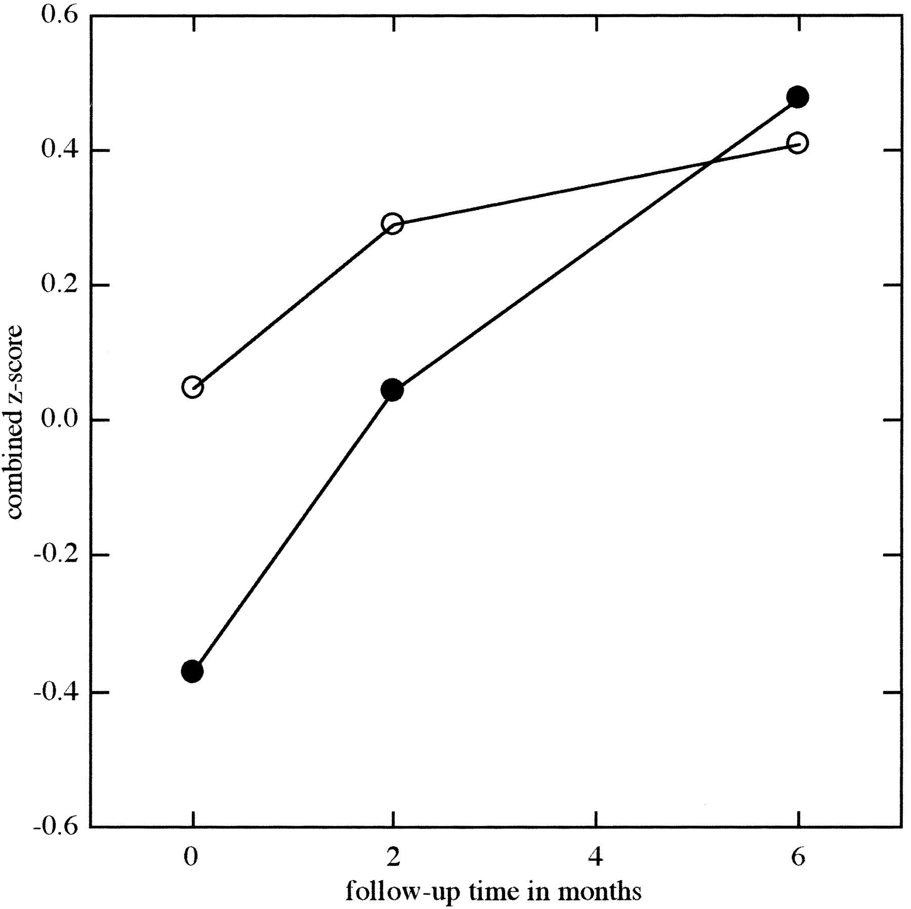
  • fig 4.
    • Download figure
    • Open in new tab
    • Download powerpoint
    fig 4.

    Combined z scores of the patients with normal (○) and abnormal (•) MR findings over the 6-month follow-up period

Tables

  • Figures
  • TABLE 1:
    • View popup
    • Download powerpoint
    TABLE 1:

    Demographic data, trauma characteristics, imaging, and neurocognitive results of the mTBI patients

  • TABLE 2:
    • View popup
    • Download powerpoint
    TABLE 2:

    Independent sample t test on demographic variables and injury severity characteristics for patients with normal and abnormal MR findings

  • TABLE 3:
    • View popup
    • Download powerpoint
    TABLE 3:

    Mean z score and SD (between brackets) of the neu~rocognitive examination (NCE) of patients with normal and ab~normal MR findings

  • TABLE 4:
    • View popup
    • Download powerpoint
    TABLE 4:

    Mean number of posttraumatic complaints, as rated on a 28-item questionnaire

PreviousNext
Back to top

In this issue

American Journal of Neuroradiology
Vol. 22, Issue 3
1 Mar 2001
  • Table of Contents
  • Index by author
Advertisement
Print
Download PDF
Email Article

Thank you for your interest in spreading the word on American Journal of Neuroradiology.

NOTE: We only request your email address so that the person you are recommending the page to knows that you wanted them to see it, and that it is not junk mail. We do not capture any email address.

Enter multiple addresses on separate lines or separate them with commas.
MR Imaging, Single-photon Emission CT, and Neurocognitive Performance after Mild Traumatic Brain Injury
(Your Name) has sent you a message from American Journal of Neuroradiology
(Your Name) thought you would like to see the American Journal of Neuroradiology web site.
CAPTCHA
This question is for testing whether or not you are a human visitor and to prevent automated spam submissions.
Cite this article
Paul A.M. Hofman, Sven Z. Stapert, Marinus J.P.G. van Kroonenburgh, Jelle Jolles, Jelle de Kruijk, Jan T. Wilmink
MR Imaging, Single-photon Emission CT, and Neurocognitive Performance after Mild Traumatic Brain Injury
American Journal of Neuroradiology Mar 2001, 22 (3) 441-449;

Citation Manager Formats

  • BibTeX
  • Bookends
  • EasyBib
  • EndNote (tagged)
  • EndNote 8 (xml)
  • Medlars
  • Mendeley
  • Papers
  • RefWorks Tagged
  • Ref Manager
  • RIS
  • Zotero
0 Responses
Respond to this article
Share
Bookmark this article
MR Imaging, Single-photon Emission CT, and Neurocognitive Performance after Mild Traumatic Brain Injury
Paul A.M. Hofman, Sven Z. Stapert, Marinus J.P.G. van Kroonenburgh, Jelle Jolles, Jelle de Kruijk, Jan T. Wilmink
American Journal of Neuroradiology Mar 2001, 22 (3) 441-449;
del.icio.us logo Twitter logo Facebook logo Mendeley logo
  • Tweet Widget
  • Facebook Like
  • Google Plus One

Jump to section

  • Article
    • Abstract
    • Methods
    • Results
    • Discussion
    • Conclusion
    • Footnotes
    • References
  • Figures & Data
  • Info & Metrics
  • Responses
  • References
  • PDF

Related Articles

  • No related articles found.
  • PubMed
  • Google Scholar

Cited By...

  • Biomarkers of Traumatic Brain Injury: Temporal Changes in Body Fluids
  • Concussion is confusing us all
  • A prospective study of gray matter abnormalities in mild traumatic brain injury
  • What evidence exists for new strategies or technologies in the diagnosis of sports concussion and assessment of recovery?
  • Decreased Fractional Anisotropy Evaluated Using Tract-Based Spatial Statistics and Correlated with Cognitive Dysfunction in Patients with Mild Traumatic Brain Injury in the Chronic Stage
  • Mild traumatic brain injury and Postconcussion Syndrome: a neuropsychological perspective
  • Contributions of neuroimaging, balance testing, electrophysiology and blood markers to the assessment of sport-related concussion
  • HEAD INJURY FOR NEUROLOGISTS
  • Crossref
  • Google Scholar

This article has not yet been cited by articles in journals that are participating in Crossref Cited-by Linking.

More in this TOC Section

  • SWI or T2*: Which MRI Sequence to Use in the Detection of Cerebral Microbleeds? The Karolinska Imaging Dementia Study
  • Progression of Microstructural Damage in Spinocerebellar Ataxia Type 2: A Longitudinal DTI Study
  • Usefulness of Quantitative Susceptibility Mapping for the Diagnosis of Parkinson Disease
Show more Brain

Similar Articles

Advertisement

Indexed Content

  • Current Issue
  • Accepted Manuscripts
  • Article Preview
  • Past Issues
  • Editorials
  • Editors Choice
  • Fellow Journal Club
  • Letters to the Editor

Cases

  • Case Collection
  • Archive - Case of the Week
  • Archive - Case of the Month
  • Archive - Classic Case

Special Collections

  • Special Collections

Resources

  • News and Updates
  • Turn around Times
  • Submit a Manuscript
  • Author Policies
  • Manuscript Submission Guidelines
  • Evidence-Based Medicine Level Guide
  • Publishing Checklists
  • Graphical Abstract Preparation
  • Imaging Protocol Submission
  • Submit a Case
  • Become a Reviewer/Academy of Reviewers
  • Get Peer Review Credit from Publons

Multimedia

  • AJNR Podcast
  • AJNR SCANtastic
  • Video Articles

About Us

  • About AJNR
  • Editorial Board
  • Not an AJNR Subscriber? Join Now
  • Alerts
  • Feedback
  • Advertise with us
  • Librarian Resources
  • Permissions
  • Terms and Conditions

American Society of Neuroradiology

  • Not an ASNR Member? Join Now

© 2025 by the American Society of Neuroradiology All rights, including for text and data mining, AI training, and similar technologies, are reserved.
Print ISSN: 0195-6108 Online ISSN: 1936-959X

Powered by HighWire