Advertisement

Main menu

  • Home
  • Content
    • Current Issue
    • Accepted Manuscripts
    • Article Preview
    • Past Issue Archive
    • Video Articles
    • AJNR Case Collection
    • Case of the Week Archive
    • Case of the Month Archive
    • Classic Case Archive
  • Special Collections
    • AJNR Awards
    • Low-Field MRI
    • Alzheimer Disease
    • ASNR Foundation Special Collection
    • Photon-Counting CT
    • View All
  • Multimedia
    • AJNR Podcasts
    • AJNR SCANtastic
    • Trainee Corner
    • MRI Safety Corner
    • Imaging Protocols
  • For Authors
    • Submit a Manuscript
    • Submit a Video Article
    • Submit an eLetter to the Editor/Response
    • Manuscript Submission Guidelines
    • Statistical Tips
    • Fast Publishing of Accepted Manuscripts
    • Graphical Abstract Preparation
    • Imaging Protocol Submission
    • Author Policies
  • About Us
    • About AJNR
    • Editorial Board
    • Editorial Board Alumni
  • More
    • Become a Reviewer/Academy of Reviewers
    • Subscribers
    • Permissions
    • Alerts
    • Feedback
    • Advertisers
    • ASNR Home

User menu

  • Alerts
  • Log in

Search

  • Advanced search
American Journal of Neuroradiology

American Journal of Neuroradiology

ASHNR American Society of Functional Neuroradiology ASHNR American Society of Pediatric Neuroradiology ASSR
  • Alerts
  • Log in

Advanced Search

  • Home
  • Content
    • Current Issue
    • Accepted Manuscripts
    • Article Preview
    • Past Issue Archive
    • Video Articles
    • AJNR Case Collection
    • Case of the Week Archive
    • Case of the Month Archive
    • Classic Case Archive
  • Special Collections
    • AJNR Awards
    • Low-Field MRI
    • Alzheimer Disease
    • ASNR Foundation Special Collection
    • Photon-Counting CT
    • View All
  • Multimedia
    • AJNR Podcasts
    • AJNR SCANtastic
    • Trainee Corner
    • MRI Safety Corner
    • Imaging Protocols
  • For Authors
    • Submit a Manuscript
    • Submit a Video Article
    • Submit an eLetter to the Editor/Response
    • Manuscript Submission Guidelines
    • Statistical Tips
    • Fast Publishing of Accepted Manuscripts
    • Graphical Abstract Preparation
    • Imaging Protocol Submission
    • Author Policies
  • About Us
    • About AJNR
    • Editorial Board
    • Editorial Board Alumni
  • More
    • Become a Reviewer/Academy of Reviewers
    • Subscribers
    • Permissions
    • Alerts
    • Feedback
    • Advertisers
    • ASNR Home
  • Follow AJNR on Twitter
  • Visit AJNR on Facebook
  • Follow AJNR on Instagram
  • Join AJNR on LinkedIn
  • RSS Feeds

AJNR Awards, New Junior Editors, and more. Read the latest AJNR updates

Research ArticleBRAIN

Diffusion Anisotropy of the Internal Capsule and the Corona Radiata in Association with Stroke and Tumors as Measured by Diffusion-weighted MR Imaging

Shuichi Higano, Jianhui Zhong, David A. Shrier, Dean K. Shibata, Yukinori Takase, Henry Wang and Yuji Numaguchi
American Journal of Neuroradiology March 2001, 22 (3) 456-463;
Shuichi Higano
  • Find this author on Google Scholar
  • Find this author on PubMed
  • Search for this author on this site
Jianhui Zhong
  • Find this author on Google Scholar
  • Find this author on PubMed
  • Search for this author on this site
David A. Shrier
  • Find this author on Google Scholar
  • Find this author on PubMed
  • Search for this author on this site
Dean K. Shibata
  • Find this author on Google Scholar
  • Find this author on PubMed
  • Search for this author on this site
Yukinori Takase
  • Find this author on Google Scholar
  • Find this author on PubMed
  • Search for this author on this site
Henry Wang
  • Find this author on Google Scholar
  • Find this author on PubMed
  • Search for this author on this site
Yuji Numaguchi
  • Find this author on Google Scholar
  • Find this author on PubMed
  • Search for this author on this site
  • Article
  • Figures & Data
  • Info & Metrics
  • Responses
  • References
  • PDF
Loading

Article Figures & Data

Figures

  • Tables
  • fig 1.
    • Download figure
    • Open in new tab
    • Download powerpoint
    fig 1.

    Screen of defining ROI. The DAV image (upper left), the ASD map image (lower left), and the Dz image (lower right) were displayed on the screen, and the location of the defined ROI were shown on the three images simultaneously

  • fig 2.
    • Download figure
    • Open in new tab
    • Download powerpoint
    fig 2.

    Mean and SD of AI(ASD) of the internal capsule or the corona radiata involved by various types of lesions. Analysis of variance, F = 39.6; P < .0001; Post hoc test (Scheffe); *, P = .05 as compared with the control group; **, P < .0001 as compared with both control and uninvolved groups

  • fig 3.
    • Download figure
    • Open in new tab
    • Download powerpoint
    fig 3.

    Acute cerebral infarction of the right corona radiata with moderate-severe left hemiparesis.

    A, T2-weighted echo-planar image (b = 0) shows an oval hyperintense lesion involving the right corona radiata.

    B, Apparent diffusion coefficient image shows a decreased diffusion coefficient in the lesion.

    C, In the ASD image, the diffusion anisotropy is also reduced.

    D, T2-weighted echo-planar image (10000/98 [TR/TEeff]) that includes the internal capsule shows no involvement of the right internal capsule, although high intensity spotty lesions were noted in the bilateral lentiform nuclei.

    E, Apparent diffusion coefficient image of the same section as that shown in D shows no definite abnormality in the internal capsule.

    F, ASD image shows decreased diffusion anisotropy in the posterior limb of the right internal capsule as compared with the left side.

  • fig 4.
    • Download figure
    • Open in new tab
    • Download powerpoint
    fig 4.

    Glioblastoma involving the right corona radiata and basal ganglia with moderate-severe hemiparesis.

    A, T2-weighted echo-planar image shows an irregular mass with marked perifocal edema involving the right corona radiata.

    B, Apparent diffusion coefficient image shows an irregular mass with marked perifocal edema involving the right corona radiata.

    C, In the ASD image, the diffusion anisotropy of the right corona radiata is hardly noted.

  • fig 5.
    • Download figure
    • Open in new tab
    • Download powerpoint
    fig 5.

    Mean and SD of AI(ASD) of the internal capsule or the corona radiata for each degree of hemiparesis. Analysis of variance, F = 17.0; P < .0001; Post hoc test (Bonferroni); *, P < .0001 as compared with every other group

  • fig 6.
    • Download figure
    • Open in new tab
    • Download powerpoint
    fig 6.

    Comparison of AI(ASD) value (mean and SD) of normal-appearing internal capsule and corona radiata between the patients with entirely intact corticospinal tract (Intact) and those having some involved segments of the corticospinal tract (Involved)

Tables

  • Figures
  • TABLE 1:
    • View popup
    • Download powerpoint
    TABLE 1:

    Relationship between the degree of hemiparesis and the type of pathologic abnormality
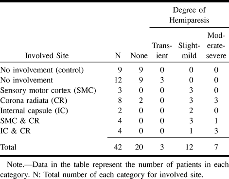
  • TABLE 2:
    • View popup
    • Download powerpoint
    TABLE 2:

    Relationship between the involved site of the cortico~spinal pathway and the degree of hemiparesis

PreviousNext
Back to top

In this issue

American Journal of Neuroradiology
Vol. 22, Issue 3
1 Mar 2001
  • Table of Contents
  • Index by author
Advertisement
Print
Download PDF
Email Article

Thank you for your interest in spreading the word on American Journal of Neuroradiology.

NOTE: We only request your email address so that the person you are recommending the page to knows that you wanted them to see it, and that it is not junk mail. We do not capture any email address.

Enter multiple addresses on separate lines or separate them with commas.
Diffusion Anisotropy of the Internal Capsule and the Corona Radiata in Association with Stroke and Tumors as Measured by Diffusion-weighted MR Imaging
(Your Name) has sent you a message from American Journal of Neuroradiology
(Your Name) thought you would like to see the American Journal of Neuroradiology web site.
CAPTCHA
This question is for testing whether or not you are a human visitor and to prevent automated spam submissions.
Cite this article
Shuichi Higano, Jianhui Zhong, David A. Shrier, Dean K. Shibata, Yukinori Takase, Henry Wang, Yuji Numaguchi
Diffusion Anisotropy of the Internal Capsule and the Corona Radiata in Association with Stroke and Tumors as Measured by Diffusion-weighted MR Imaging
American Journal of Neuroradiology Mar 2001, 22 (3) 456-463;

Citation Manager Formats

  • BibTeX
  • Bookends
  • EasyBib
  • EndNote (tagged)
  • EndNote 8 (xml)
  • Medlars
  • Mendeley
  • Papers
  • RefWorks Tagged
  • Ref Manager
  • RIS
  • Zotero
0 Responses
Respond to this article
Share
Bookmark this article
Diffusion Anisotropy of the Internal Capsule and the Corona Radiata in Association with Stroke and Tumors as Measured by Diffusion-weighted MR Imaging
Shuichi Higano, Jianhui Zhong, David A. Shrier, Dean K. Shibata, Yukinori Takase, Henry Wang, Yuji Numaguchi
American Journal of Neuroradiology Mar 2001, 22 (3) 456-463;
del.icio.us logo Twitter logo Facebook logo Mendeley logo
  • Tweet Widget
  • Facebook Like
  • Google Plus One

Jump to section

  • Article
    • Abstract
    • Methods
    • Results
    • Discussion
    • Conclusion
    • Footnotes
    • References
  • Figures & Data
  • Info & Metrics
  • Responses
  • References
  • PDF

Related Articles

  • No related articles found.
  • PubMed
  • Google Scholar

Cited By...

  • No citing articles found.
  • Crossref
  • Google Scholar

This article has not yet been cited by articles in journals that are participating in Crossref Cited-by Linking.

More in this TOC Section

  • Multimodal CT Provides Improved Performance for Lacunar Infarct Detection
  • Optimal MRI Sequence for Identifying Occlusion Location in Acute Stroke: Which Value of Time-Resolved Contrast-Enhanced MRA?
  • Evaluating the Effects of White Matter Multiple Sclerosis Lesions on the Volume Estimation of 6 Brain Tissue Segmentation Methods
Show more Brain

Similar Articles

Advertisement

Indexed Content

  • Current Issue
  • Accepted Manuscripts
  • Article Preview
  • Past Issues
  • Editorials
  • Editor's Choice
  • Fellows' Journal Club
  • Letters to the Editor
  • Video Articles

Cases

  • Case Collection
  • Archive - Case of the Week
  • Archive - Case of the Month
  • Archive - Classic Case

More from AJNR

  • Trainee Corner
  • Imaging Protocols
  • MRI Safety Corner
  • Book Reviews

Multimedia

  • AJNR Podcasts
  • AJNR Scantastics

Resources

  • Turnaround Time
  • Submit a Manuscript
  • Submit a Video Article
  • Submit an eLetter to the Editor/Response
  • Manuscript Submission Guidelines
  • Statistical Tips
  • Fast Publishing of Accepted Manuscripts
  • Graphical Abstract Preparation
  • Imaging Protocol Submission
  • Evidence-Based Medicine Level Guide
  • Publishing Checklists
  • Author Policies
  • Become a Reviewer/Academy of Reviewers
  • News and Updates

About Us

  • About AJNR
  • Editorial Board
  • Editorial Board Alumni
  • Alerts
  • Permissions
  • Not an AJNR Subscriber? Join Now
  • Advertise with Us
  • Librarian Resources
  • Feedback
  • Terms and Conditions
  • AJNR Editorial Board Alumni

American Society of Neuroradiology

  • Not an ASNR Member? Join Now

© 2025 by the American Society of Neuroradiology All rights, including for text and data mining, AI training, and similar technologies, are reserved.
Print ISSN: 0195-6108 Online ISSN: 1936-959X

Powered by HighWire