Advertisement

Main menu

  • Home
  • Content
    • Current Issue
    • Accepted Manuscripts
    • Article Preview
    • Past Issue Archive
    • AJNR Case Collection
    • Case of the Week Archive
    • Classic Case Archive
    • Case of the Month Archive
  • Special Collections
    • Spinal CSF Leak Articles (Jan 2020-June 2024)
    • 2024 AJNR Journal Awards
    • Most Impactful AJNR Articles
  • Multimedia
    • AJNR Podcast
    • AJNR Scantastics
    • Video Articles
  • For Authors
    • Submit a Manuscript
    • Author Policies
    • Fast publishing of Accepted Manuscripts
    • Graphical Abstract Preparation
    • Manuscript Submission Guidelines
    • Imaging Protocol Submission
    • Submit a Case for the Case Collection
  • About Us
    • About AJNR
    • Editorial Board
  • More
    • Become a Reviewer/Academy of Reviewers
    • Subscribers
    • Permissions
    • Alerts
    • Feedback
    • Advertisers
    • ASNR Home
  • Other Publications
    • ajnr

User menu

  • Alerts
  • Log in

Search

  • Advanced search
American Journal of Neuroradiology

American Journal of Neuroradiology

ASHNR American Society of Functional Neuroradiology ASHNR American Society of Pediatric Neuroradiology ASSR
  • Alerts
  • Log in

Advanced Search

  • Home
  • Content
    • Current Issue
    • Accepted Manuscripts
    • Article Preview
    • Past Issue Archive
    • AJNR Case Collection
    • Case of the Week Archive
    • Classic Case Archive
    • Case of the Month Archive
  • Special Collections
    • Spinal CSF Leak Articles (Jan 2020-June 2024)
    • 2024 AJNR Journal Awards
    • Most Impactful AJNR Articles
  • Multimedia
    • AJNR Podcast
    • AJNR Scantastics
    • Video Articles
  • For Authors
    • Submit a Manuscript
    • Author Policies
    • Fast publishing of Accepted Manuscripts
    • Graphical Abstract Preparation
    • Manuscript Submission Guidelines
    • Imaging Protocol Submission
    • Submit a Case for the Case Collection
  • About Us
    • About AJNR
    • Editorial Board
  • More
    • Become a Reviewer/Academy of Reviewers
    • Subscribers
    • Permissions
    • Alerts
    • Feedback
    • Advertisers
    • ASNR Home
  • Follow AJNR on Twitter
  • Visit AJNR on Facebook
  • Follow AJNR on Instagram
  • Join AJNR on LinkedIn
  • RSS Feeds

Welcome to the new AJNR, Updated Hall of Fame, and more. Read the full announcements.


AJNR is seeking candidates for the position of Associate Section Editor, AJNR Case Collection. Read the full announcement.


Research ArticleBRAIN

Chordoid Glioma: A Neoplasm Unique to the Hypothalamus and Anterior Third Ventricle

Martin G. Pomper, Theodore J. Passe, Peter C. Burger, Bernd W. Scheithauer and Daniel J. Brat
American Journal of Neuroradiology March 2001, 22 (3) 464-469;
Martin G. Pomper
  • Find this author on Google Scholar
  • Find this author on PubMed
  • Search for this author on this site
Theodore J. Passe
  • Find this author on Google Scholar
  • Find this author on PubMed
  • Search for this author on this site
Peter C. Burger
  • Find this author on Google Scholar
  • Find this author on PubMed
  • Search for this author on this site
Bernd W. Scheithauer
  • Find this author on Google Scholar
  • Find this author on PubMed
  • Search for this author on this site
Daniel J. Brat
  • Find this author on Google Scholar
  • Find this author on PubMed
  • Search for this author on this site
  • Article
  • Figures & Data
  • Info & Metrics
  • Responses
  • References
  • PDF
Loading

Article Figures & Data

Figures

  • Tables
  • fig 1.
    • Download figure
    • Open in new tab
    • Download powerpoint
    fig 1.

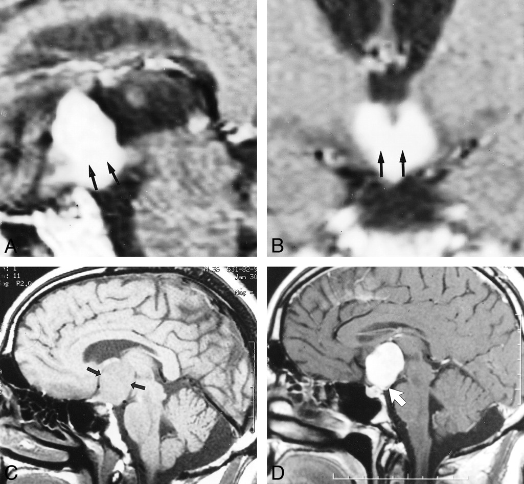
    Representative appearance of chordoid glioma on MR image. Note the location of the mass in the hypothalamus/third ventricle region, dense enhancement, and greatest dimension in the superoinferior orientation.

    A, Sagittal contrast-enhanced spin-echo MR image (500/15) of Patient 1, a 59-year-old man. Note hypothalamic involvement (arrow).

    B, Coronal contrast-enhanced spin-echo MR image (500/15) of Patient 1, a 59-year-old man. Note hypothalamic involvement (arrow).

    C, Unenhanced sagittal spin-echo MR image (500/14) of Patient 3, a 36-year-old man. Arrows demarcate the suprasellar mass.

    D, Contrast-enhanced sagittal spin-echo MR image (500/14) of Patient 3, a 36-year-old man. Note the posterior displacement of the infundibulum (arrow).

  • fig 2.
    • Download figure
    • Open in new tab
    • Download powerpoint
    fig 2.

    CT scans of Patient 2, a 41-year-old woman with chordoid glioma.

    A, Unenhanced axial CT scan reveals a hyperdense mass (arrow) that enhances uniformly in the suprasellar region.

    B, Contrast-enhanced axial CT scan reveals a hyperdense mass that enhances uniformly (arrow) in the suprasellar region.

  • fig 3.
    • Download figure
    • Open in new tab
    • Download powerpoint
    fig 3.

    Images of Patient 5, a 35-year-old woman with chordoid glioma.

    A, Axial T1-weighted spin-echo MR image (450/20) reveals an isointense hypothalamic/third ventricular mass (arrows).

    B, Axial fluid-attenuated inversion recovery MR image (8912/142; inversion time, 2200 ms) reveals an iso- to slightly hyperintense hypothalamic/third ventricular mass (large arrow). Note vasogenic edema within the basal ganglia (small arrows), posterior limbs of the internal capsules, and lateral geniculate ganglia of the thalami (arrowheads).

    C, Axial fluid-attenuated inversion recovery MR image (8912/142; inversion time, 2200 ms) obtained at a level approximately 1.5 cm inferior to that shown in B. Note splaying of the cerebral peduncles (large arrows) and bilateral involvement of the optic tracts (small arrows).

  • fig 4.
    • Download figure
    • Open in new tab
    • Download powerpoint
    fig 4.

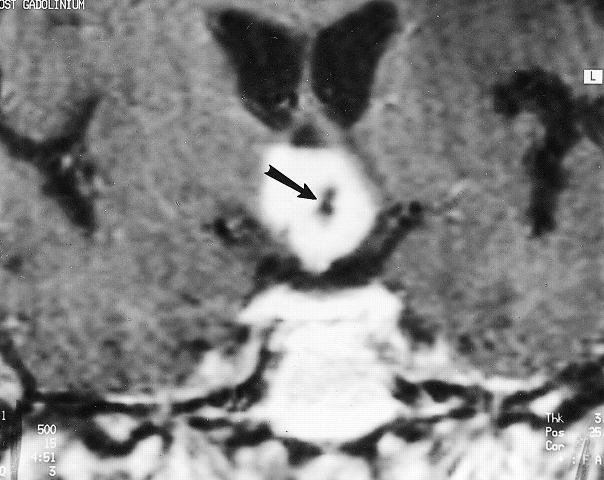
    Image of Patient 1, a 59-year-old man with chordoid glioma. Contrast-enhanced coronal T1-weighted spin-echo MR image (500/15) reveals a densely enhancing hypothalamic/third ventricular mass with a central nonenhancing component representing a small cyst or necrosis (arrow)

  • fig 5.
    • Download figure
    • Open in new tab
    • Download powerpoint
    fig 5.

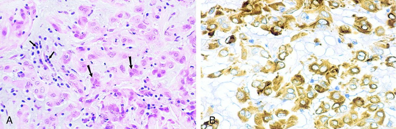
    Photomicrographs of chordoid glioma (Patient 5).

    A, Hematoxylin and eosin stain of chordoid glioma reveals cords and clusters of eosinophilic epithelioid tumor cells (large arrows) dispersed within a slightly bluish mucinous matrix. A lymphoplasmacytic infiltrate is consistently present (small arrows).

    B, Glial fibrillary acidic protein stain of chordoid glioma tumor cells shows strong, diffuse, cytoplasmic immunoreactivity.

Tables

  • Figures
  • Table1

    Patient characteristics and imaging findings

PreviousNext
Back to top

In this issue

American Journal of Neuroradiology
Vol. 22, Issue 3
1 Mar 2001
  • Table of Contents
  • Index by author
Advertisement
Print
Download PDF
Email Article

Thank you for your interest in spreading the word on American Journal of Neuroradiology.

NOTE: We only request your email address so that the person you are recommending the page to knows that you wanted them to see it, and that it is not junk mail. We do not capture any email address.

Enter multiple addresses on separate lines or separate them with commas.
Chordoid Glioma: A Neoplasm Unique to the Hypothalamus and Anterior Third Ventricle
(Your Name) has sent you a message from American Journal of Neuroradiology
(Your Name) thought you would like to see the American Journal of Neuroradiology web site.
CAPTCHA
This question is for testing whether or not you are a human visitor and to prevent automated spam submissions.
Cite this article
Martin G. Pomper, Theodore J. Passe, Peter C. Burger, Bernd W. Scheithauer, Daniel J. Brat
Chordoid Glioma: A Neoplasm Unique to the Hypothalamus and Anterior Third Ventricle
American Journal of Neuroradiology Mar 2001, 22 (3) 464-469;

Citation Manager Formats

  • BibTeX
  • Bookends
  • EasyBib
  • EndNote (tagged)
  • EndNote 8 (xml)
  • Medlars
  • Mendeley
  • Papers
  • RefWorks Tagged
  • Ref Manager
  • RIS
  • Zotero
0 Responses
Respond to this article
Share
Bookmark this article
Chordoid Glioma: A Neoplasm Unique to the Hypothalamus and Anterior Third Ventricle
Martin G. Pomper, Theodore J. Passe, Peter C. Burger, Bernd W. Scheithauer, Daniel J. Brat
American Journal of Neuroradiology Mar 2001, 22 (3) 464-469;
del.icio.us logo Twitter logo Facebook logo Mendeley logo
  • Tweet Widget
  • Facebook Like
  • Google Plus One

Jump to section

  • Article
    • Abstract
    • Methods
    • Results
    • Discussion
    • Conclusion
    • Footnotes
    • References
  • Figures & Data
  • Info & Metrics
  • Responses
  • References
  • PDF

Related Articles

  • No related articles found.
  • PubMed
  • Google Scholar

Cited By...

  • Primary hypothalamic third ventriclular Burkitt's lymphoma: a case report with emphasis on differential diagnosis
  • Crossref
  • Google Scholar

This article has not yet been cited by articles in journals that are participating in Crossref Cited-by Linking.

More in this TOC Section

  • Evaluating the Effects of White Matter Multiple Sclerosis Lesions on the Volume Estimation of 6 Brain Tissue Segmentation Methods
  • White Matter Alterations in the Brains of Patients with Active, Remitted, and Cured Cushing Syndrome: A DTI Study
  • Enhanced Axonal Metabolism during Early Natalizumab Treatment in Relapsing-Remitting Multiple Sclerosis
Show more Brain

Similar Articles

Advertisement

Indexed Content

  • Current Issue
  • Accepted Manuscripts
  • Article Preview
  • Past Issues
  • Editorials
  • Editors Choice
  • Fellow Journal Club
  • Letters to the Editor

Cases

  • Case Collection
  • Archive - Case of the Week
  • Archive - Case of the Month
  • Archive - Classic Case

Special Collections

  • Special Collections

Resources

  • News and Updates
  • Turn around Times
  • Submit a Manuscript
  • Author Policies
  • Manuscript Submission Guidelines
  • Evidence-Based Medicine Level Guide
  • Publishing Checklists
  • Graphical Abstract Preparation
  • Imaging Protocol Submission
  • Submit a Case
  • Become a Reviewer/Academy of Reviewers
  • Get Peer Review Credit from Publons

Multimedia

  • AJNR Podcast
  • AJNR SCANtastic
  • Video Articles

About Us

  • About AJNR
  • Editorial Board
  • Not an AJNR Subscriber? Join Now
  • Alerts
  • Feedback
  • Advertise with us
  • Librarian Resources
  • Permissions
  • Terms and Conditions

American Society of Neuroradiology

  • Not an ASNR Member? Join Now

© 2025 by the American Society of Neuroradiology All rights, including for text and data mining, AI training, and similar technologies, are reserved.
Print ISSN: 0195-6108 Online ISSN: 1936-959X

Powered by HighWire