
Article Figures & Data
Figures
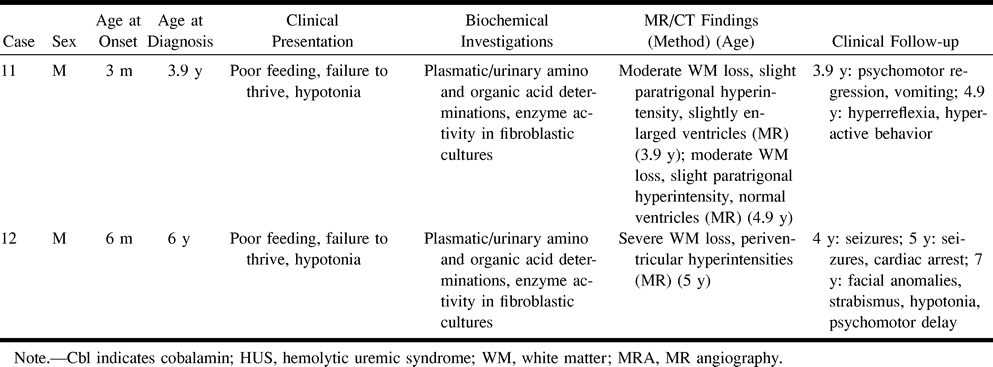
- fig 1.
Case 1.
A and B, Axial T2-weighted MR images at 2 months show the supratentorial white matter is markedly edematous and hyperintense, the U fibers are diffusely involved, the basal ganglia are spared, and the ventricular size is normal. Periencephalic CSF collections are still consistent with immaturity of CSF absorption at this age.
C and D, Axial T2-weighted MR images at 10 months show the supratentorial white matter is still abnormally hyperintense, but edema has resolved and a certain degree of white matter loss is becoming apparent, especially around the trigones of the lateral ventricles and in the parietal lobes. The basal ganglia are normal. An arachnoid cyst (asterisk) has developed adjacent to the right frontal lobe.
E and F, Axial T2-weighted MR images at 24 months show the white matter loss is now particularly severe in the paratrigonal areas, where the cortex nearly abuts on the ventricular surface, and in the parietal lobes; however, there is bulk loss throughout the whole supratentorial white matter. There is also delayed myelination, as shown by absent hypointensity in the anterior limbs of both internal capsules. The right frontal arachnoid cyst (asterisk) is essentially unchanged in size.
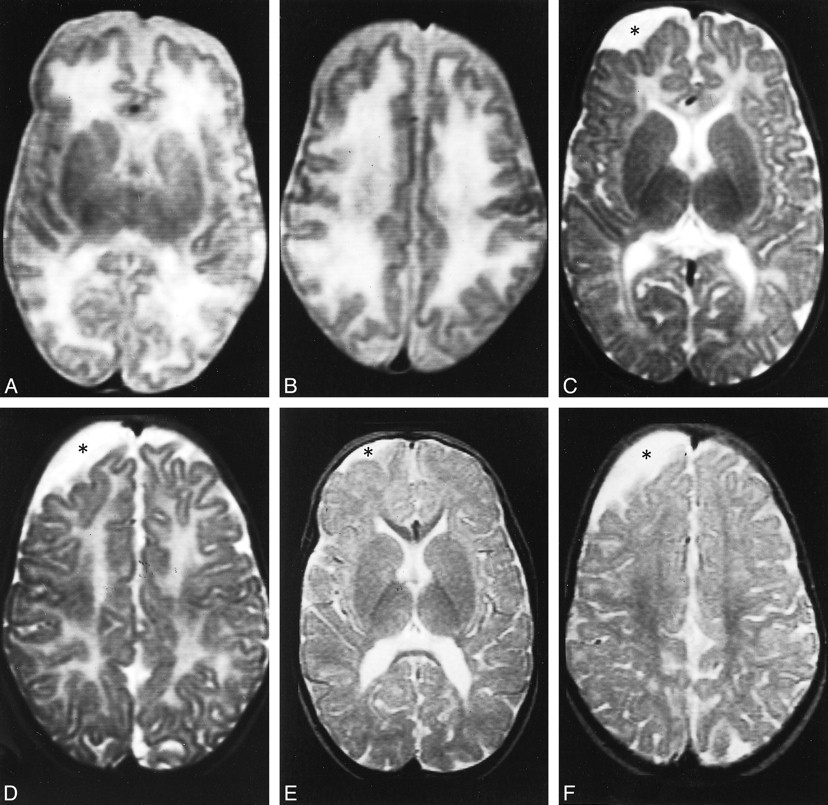
- fig 2.
Case 2.
A, Unenhanced CT scan at age 3 months shows the supratentorial white matter is probably slightly hypodense, especially in the paraventricular and subcortical regions; however, swelling is not particularly pronounced in this case. Both the ventricles and the subarachnoid spaces are slightly dilated, consistent with immaturity of CSF absorption at this age.
B–D, Axial T2-weighted (B), axial fluid-attenuated inversion-recovery (C), and sagittal T1-weighted (D) MR images at 7 years show marked and diffuse bulk loss in the supratentorial white matter; the cortex nearly abuts on the ventricular surface. Slight residual hyperintensity is seen in the periventricular regions, especially posterior to the left trigone (arrowheads, B and C). The basal ganglia are spared, and the lateral ventricles are slightly enlarged ex-vacuo. Notice marked, diffuse thinning of the corpus callosum (arrows, D).
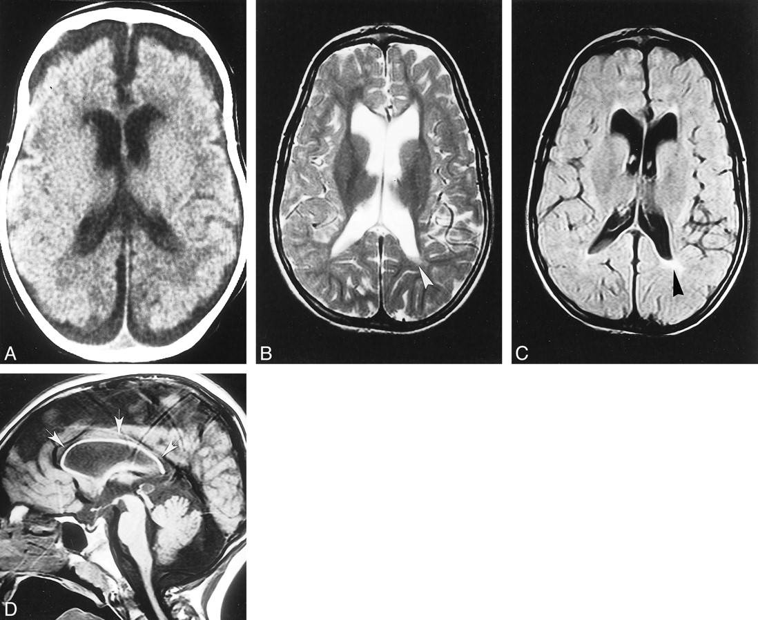
- fig 3.
Case 3. Axial T2-weighted MR image at age 3 months shows marked dilatation of the lateral ventricles. The white matter is markedly and diffusely hyperintense whereas the basal ganglia are spared.
fig 4. Case 4.
A and B, Axial proton density–weighted (A) and T2-weighted (B) MR images at 24 months show slight supratentorial white matter loss and delayed myelination with hyperintense areas in the posterior paraventricular regions (arrows). A focal area of gliosis in the nucleocapsular regions is depicted as hyperintense focus both on proton density—and T2-weighted images (arrowhead).
- fig 5.
The metabolism of MMA-HC. The disease results from impaired hepatic conversion of dietary cobalamin (Cbl) to both methylcobalamin (MeCbl) and adenosylcobalamin (AdoCbl). Complementation studies individuate three genetic subsets (CblC, CblD, and CblF) within MMA-HC, whereas other complementation groups represent biosynthetic defects that are restricted to either AdoCbl (CblA, CblB) or MeCbl (Cbl E, CblG) and that, as a consequence, represent different diseases. Absence of MeCbl and AdoCbl results in defective activity of methionine synthase (MS) and methylmalonyl-CoA mutase, respectively; the eventual biochemical picture is represented by hyperhomocysteinemia, homocystinuria, hypomethioninemia, methylmalonic acidemia, and methylmalonic aciduria. CBS indicates cystathionine β-synthetase; MTHFR, 5,10-methylenetetrahydrofolate reductase. Modified from (7) and reproduced with permission from Springer, Berlin
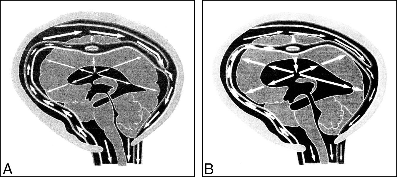
- fig 6.
Greitz and Greitz's theory on the pathogenesis of hydrocephalus.
A, Normal systole. The systolic pulse wave causes a large expansion of the arteries with a concomitant and successful dampening of the arterial pressure. The pressure is immediately transmitted to the entire subarachnoid space. After some delay, a small pressure wave is transmitted to the brain via the intracerebral arteries. This causes a slight brain expansion and a transcerebral mantle pressure gradient of normal magnitude, which keeps the ventricles patent and of normal size.
B, Hydrocephalus systole. The arterial pulsations are restricted, so little or no dampening of the arterial pressure occurs. The undamped pulse pressure is therefore transmitted into the brain, giving rise to an increased transcerebral mantle pressure gradient. Ventricular dilatation results. Reproduced from (19) with permission from Lippincott Williams & Wilkins.


Tables

Clinical and neuroradiologic findings in 12 patients with combined methylmalonic aciduria and homocystinuria

In this issue


Jump to section
Related Articles
Cited By...
- Neurodevelopmental and neuropsychiatric disorders in cobalamin C disease: a case report and review of the literature
- Analysis of 70 patients with hydrocephalus due to cobalamin C deficiency
- Clinical Reasoning: A young woman with rapid mental deterioration and leukoencephalopathy: A treatable cause
- Clinical Images - A Quarterly Column: Subacute Combined Degeneration of the Spinal Cord
- Vitamin B12 status and rate of brain volume loss in community-dwelling elderly