
Article Figures & Data
Figures
- fig 1.
Four contiguous axial view CT scans of the temporal bone, obtained from cephalad to caudad through the internal auditory canal and cochlea.
A, Note the normal location of the origin of the labyrinthine segment of the facial nerve (black arrowhead).
B, Note the normal sized basilar, second, and apical turns of the cochlea. Black arrow, normal modiolus.
C, Note the normal sized basilar, second, and apical turns of the cochlea. Black arrow, normal cochlea.
D, Note the normal sized basilar, second, and apical turns of the cochlea. Black arrow, normal basilar turn; open arrow, normal separation of the second and apical turns
- fig 2.
Cochleae in cases of facial nerve displacement were examined for presence and size of basilar, second, and apical turns.
A and B, Axial view CT scans of the temporal bone show pronounced anteromedial facial nerve migration bilaterally (black arrowheads), with pronounced widening of Bill's bar (black arrow).
C, Axial view CT scan of the temporal bone shows the right- sided hypoplastic cochlea with small first turn (black arrowhead). The left- sided cochlea was also hypoplastic and is not shown. (Same magnification as that shown in fig 1.)
D, Axial view CT scan of the temporal bone shows the right-sided hypoplastic cochlea with small second turn (black arrowhead). The left-sided cochlea was also hypoplastic and is not shown. (Same scale as that shown in fig 1.)
- fig 3.
Cochleae in cases of facial nerve displacement were examined for presence and size of basilar, second, and apical turns.
A, Axial view CT scan of the temporal bone shows mild/moderate migration of the right facial nerve (black arrowhead) and mild/moderate widening of Bill's bar (small black arrow).
B, Basilar turn of the cochlea was normal, second turn was small, and third turn was absent (large black arrow). (Same scale as that shown in fig 1.)
- fig 4.
Absence of all cochlear turns with presence of only rudimentary bud-like cavity connected to vestibule was categorized as common cavity.
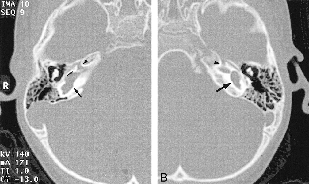
A and B, Axial view CT scans of the temporal bone show pronounced anteromedial facial nerve migration bilaterally (black arrowheads), with widening of Bill's bar (small black arrow) in a patient with a common cavity deformity bilaterally (large black arrows). (Same scale as that shown in fig 1.)
- fig 5.
CT scans of Mondini malformations.
A and B, Axial view CT scans obtained through the temporal bone show Mondini malformations of the cochlea bilaterally, with lack of separation of the second and third turns (black arrowheads).
C and D, Axial view CT scans of the same patient, obtained more cephalad, show normal origins of the facial nerves from the internal auditory canals bilaterally (black arrowheads). Note also prominent vestibules (large black arrows) and enlarged right vestibular aqueduct (small black arrow). (Same scale as that shown in fig 1.)


Tables

Facial nerve migration and non-Mondini-type malformations

In this issue


Jump to section
Related Articles
Cited By...
- No citing articles found.