
Article Figures & Data
Figures
- fig 1.
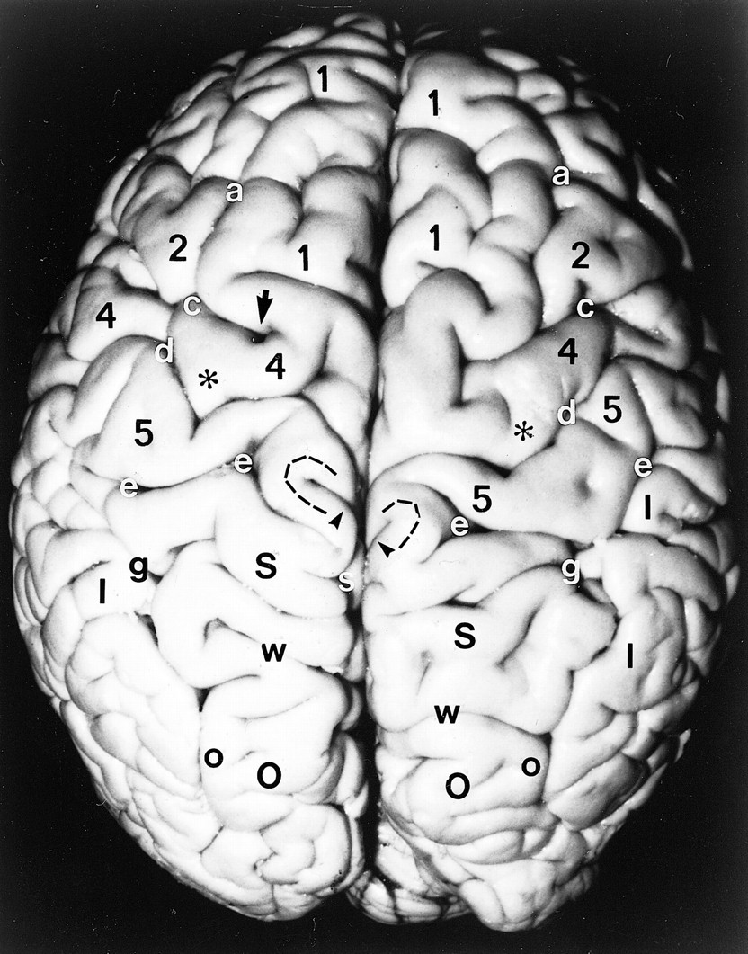
Anatomic specimen. Superior surface of the brain after removal of the pia-arachnoid and surface vasculature. Note the positions, alignments, and interrelationships among the gyri: superior frontal gyrus (1), middle frontal gyrus (2), precentral gyrus (4), postcentral gyrus (5), pars deflection (dashed curve), superior parietal lobule (large black S), inferior parietal lobule (I), and superior occipital gyrus (large O), and among the sulci: superior frontal sulcus (a), precentral sulcus (c), central sulcus (d), postcentral sulcus (e), including the medial parentheses (also e), pars marginalis (arrowheads), intraparietal sulcus (g), subparietal sulcus (small white s), parietooccipital sulcus (w), and intraoccipital sulcus (small o). Note the notch (arrow) in the precentral sulcus anterior to the hand-motor area, the sharp inscription of the central sulcus just medial to the knob (asterisks) of the hand-motor area, the medial positions of the pars marginalis, SPS and POS, the nesting of the pars deflection (dashed curve) between the pars marginalis (arrowheads) and the postcentral parenthesis (e) on each side, and the convergence of the posterior portions of the IPS toward the parasagittal line
- fig 2.
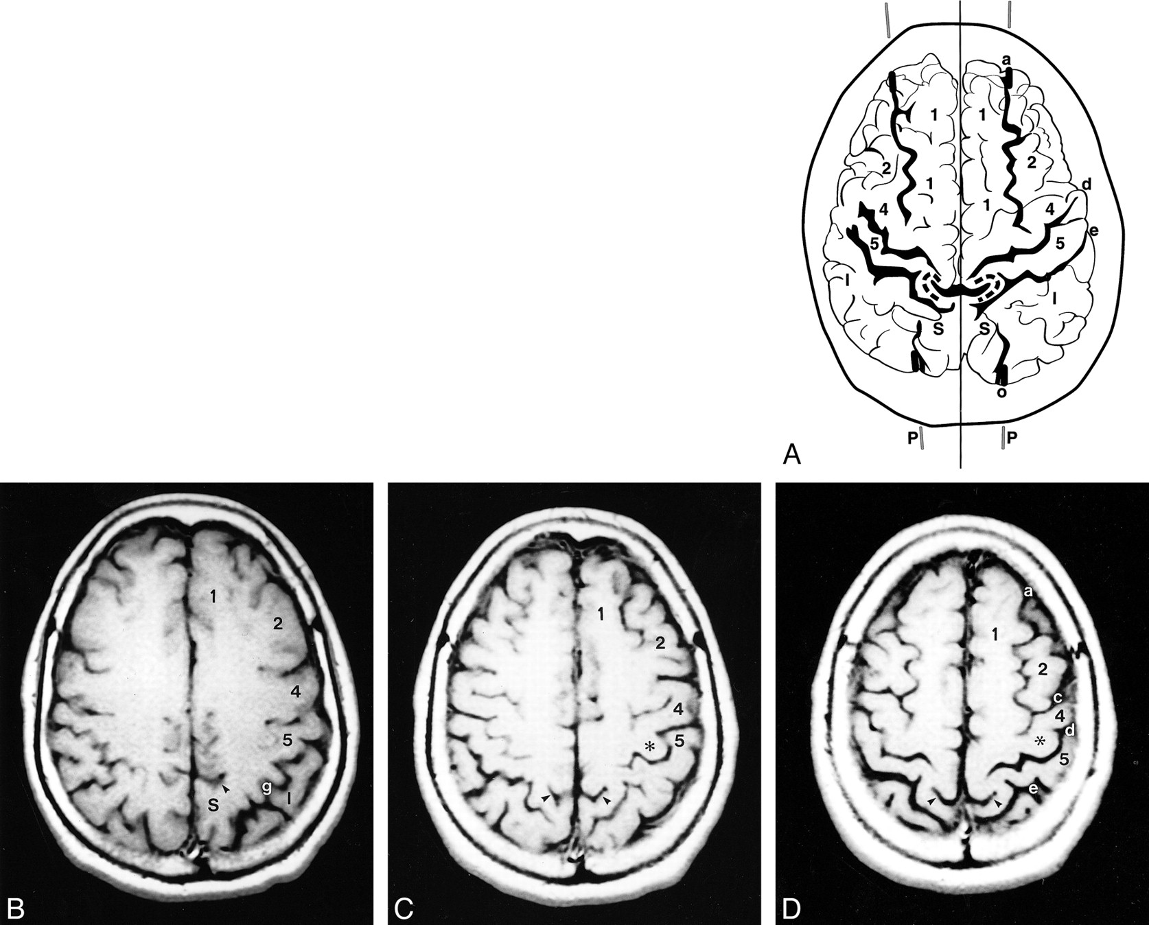
A–F, Anatomic relationships of the parasagittal line in a 92-year-old man. The composite representation (A) was traced from serial axial CT images (B–F). Labels as in figure 1. M indicates midline. In A, the curved gray lines surrounding the partes marginales (arrowheads) represent the pars deflections and correspond to the dashed curved lines seen around the partes marginales in figure 1. In the supraventricular sections, the parasagittal line (paramedian lines, P) demarcate the deepest extensions of the medial sulci and separate them from the deepest extensions of the lateral sulci. In the upper sections, multiple transverse sulci show short sharp deflections, which align along the parasagittal line. Note specifically the alignment of the SFS (a) and IOS (o) along the parasagittal line and their alignment with the notches in the precentral sulci (arrows), with the medial aspects of the knobs (asterisks) of the hand-motor areas, and with the inscriptions in the central sulci (d) just medial to the hand-motor areas. The partes marginales (arrowheads), pars deflections (gray pencil lines), and postcentral parentheses lie medial to the parasagittal line. In this composite tracing, the inferiorly situated occipital sulci superimpose upon the superior parietal lobules (S)
- fig 3.
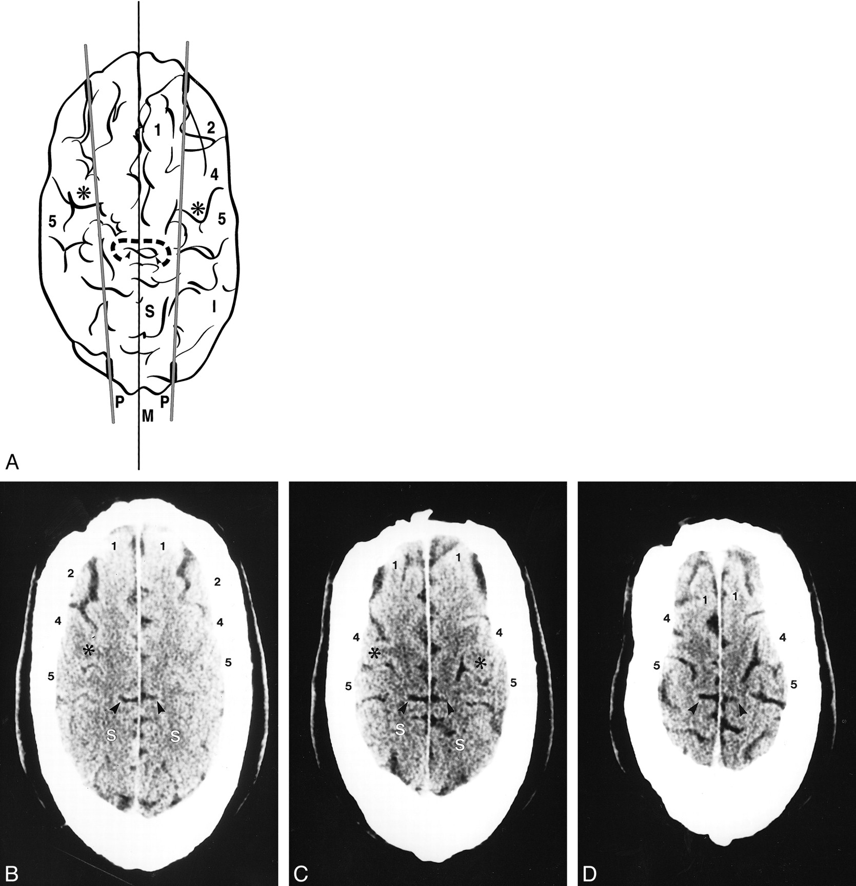
A–D, Anatomic relationships of the parasagittal line on MR images. The composite representation (A) was traced from serial axial MR images (B–D). Labels as in figures 1 and 2. Despite the differences in the scan angles and technology, MR images display the same anatomic relationships and the same alignments as CT scans do. Because the scan angle used for MR imaging is more horizontal, the partes marginales and adjacent sulci project further posteriorly on the serial axial images
- fig 4.
Parasagittal line. Limited anatomic diversity is seen on composite tracings from four different subjects. Labels as in figures 1 and 2.
A, 27-year-old man. The anatomic alignment is stereotypical.
B, 42-year-old woman. The parasagittal line traverses the midpoint of a double arc epsilon-shaped hand-motor area (small asterisks) on the right. On the left, the medial edge of the single ω-shaped hand-motor area (large asterisk) lies medial to the parasagittal line, while the postcentral parenthesis (open arrow) lies on the line.
C, 13-year-old boy. Unusually deep extension of the right pars marginalis (emphasized in black, arrowheads) displaces the right pars deflection onto the parasagittal line. The precentral notches (solid black circles) and hand-motor areas (asterisks) align well with the parasagittal line. The arrows outside the brain contour at 5 o'clock and 7 o'clock indicate notches in the brain contour along the IOS, corresponding to the bony occipital ridges (see fig 5).
D, 81-year-old woman. Very deep invagination of the partes marginales displace the pars deflections onto the parasagittal line bilaterally
- fig 5.
Molding of the occipital bone in a 76-year-old man. Normal scalloping of the inner table of the skull by the gyri situated to each side of the IPS-IOS commonly leaves small vertically oriented bony ridges (arrowheads) aligned along the parasagittal line. I = inferior parietal lobule, large O = superior occipital gyrus, S = superior parietal lobule, a = superior frontal sulcus, g = intraparietal sulcus, small o = intraoccipital sulcus, w = parietooccipital sulcus
- fig 6.
Sagittal synostosis in a 44-year-old man who had had bilateral strip craniectomies in the remote past.
A, Composite tracing.
B–D, Three of the serial axial CT sections from which the composite (A) was traced. Calvarial elongation and transverse compression have not altered the fundamental relationships of the parasagittal line. Labels as in figures 1 and 2
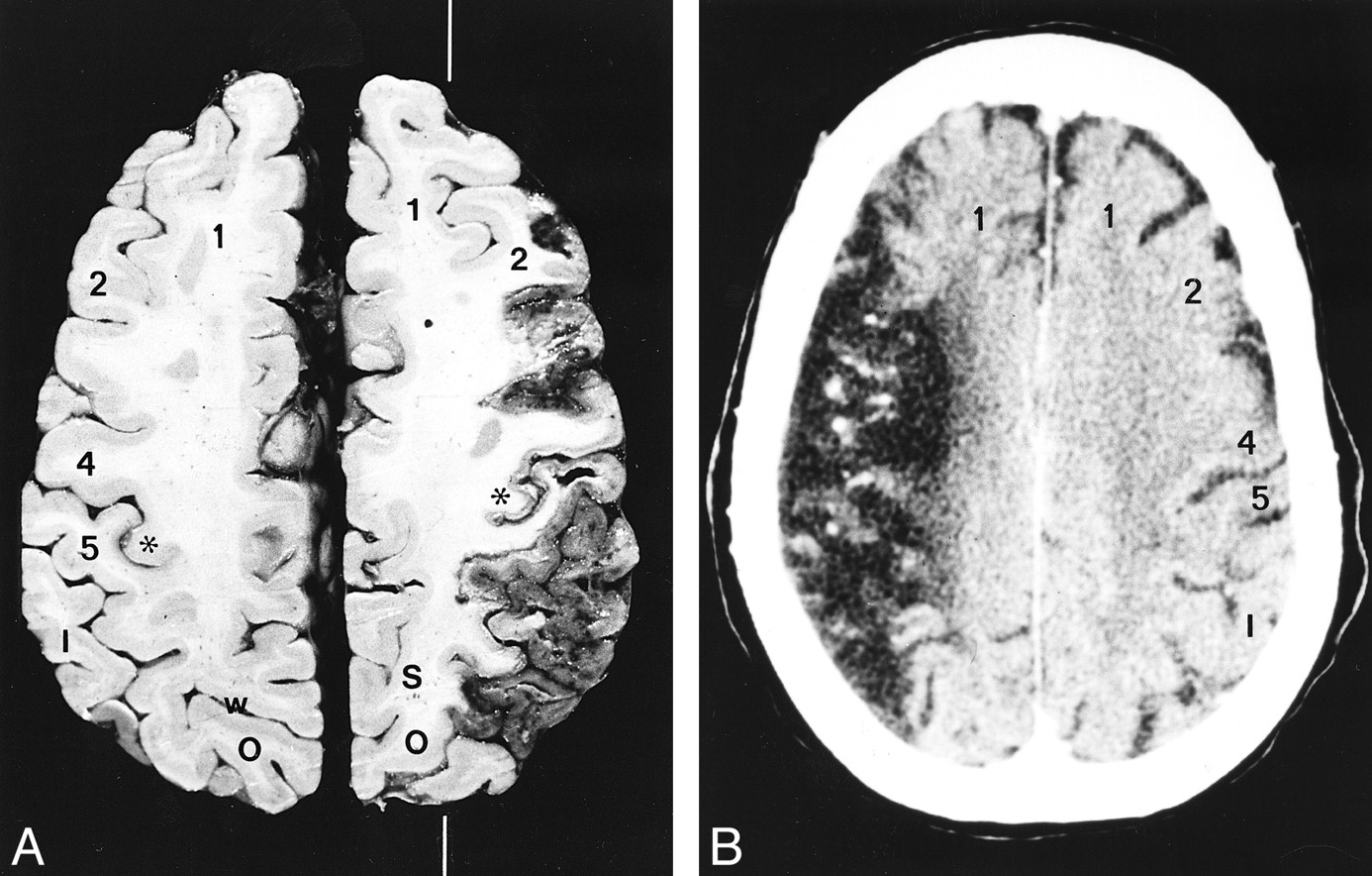
- fig 7.
The parasagittal line and cerebral infarction. Middle cerebral artery infarctions in two subjects.
A, Anatomic section. The white line indicates the position of the parasagittal line. (Case courtesy of Drs. Anne Osborne and Tessa Hedley-Whyte.)
B, Noncontrast CT scan. In the supraventricular sections, the medial borders of infarctions tend to align along the parasagittal line


Tables
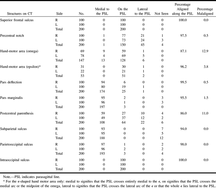
 TABLE 1:
Positions of anatomic structures with respect to the parasagittal line on CT (n = 200 hemipheres)
- TABLE 2:
Positions of anatomic structures with respect to the parasagittal line on MR (n = 200 hemispheres)


