
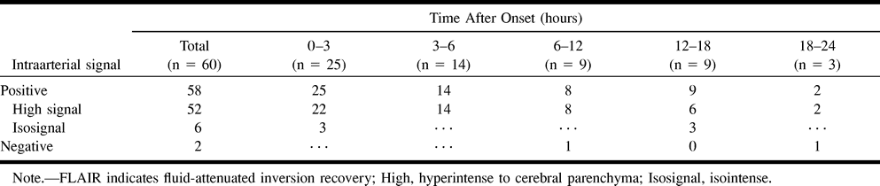
Article Figures & Data
Figures
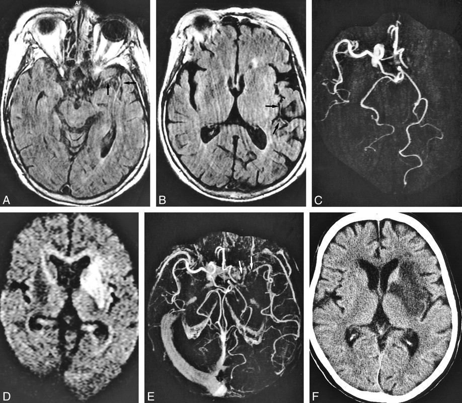
- fig 1.
A 78-year-old woman who underwent imaging 35 minutes after onset of right hemiparesis and aphasia.
A and B, FLAIR images (8000/10/1 [TR/TE/excitation]), TI = 2000) show contiguous intraarterial signal, which is hyperintense to cerebral parenchyma in M1, M2, and M3 segments of the left middle cerebral artery (arrows).
C, MR angiogram (32/6.8, flip angle = 15 degrees) shows corresponding lack of TOF effect in M1, M2, and M3 segments of the left middle cerebral artery.
D, Diffusion-weighted image (500/123/1, b = 1200) shows faintly hyperintense lesion localized in the area fed by left lateral lenticulostriate artery (arrow); however, there is no change in the hemispheric territory of the left middle cerebral artery.
E, Postcontrast MR angiogram (32/6.8, flip angle = 15 degrees) shows intraluminal enhancement in the left middle cerebral artery except for in the distal potion of the M1 segment. Arrow indicates complete obstruction in distal portion of left M1 segment.
F, T2-weighted image (4500/96/1) obtained 7 days after onset confirmed final infarction is in the entire territory of the left middle cerebral artery. Note the diffusion-weighted lesion is still smaller than the area of intraarterial signal distribution. Intraarterial signal consists of not only complete obstruction but also slow flow. The area of final infarct corresponds to the area of intraarterial signal distribution.
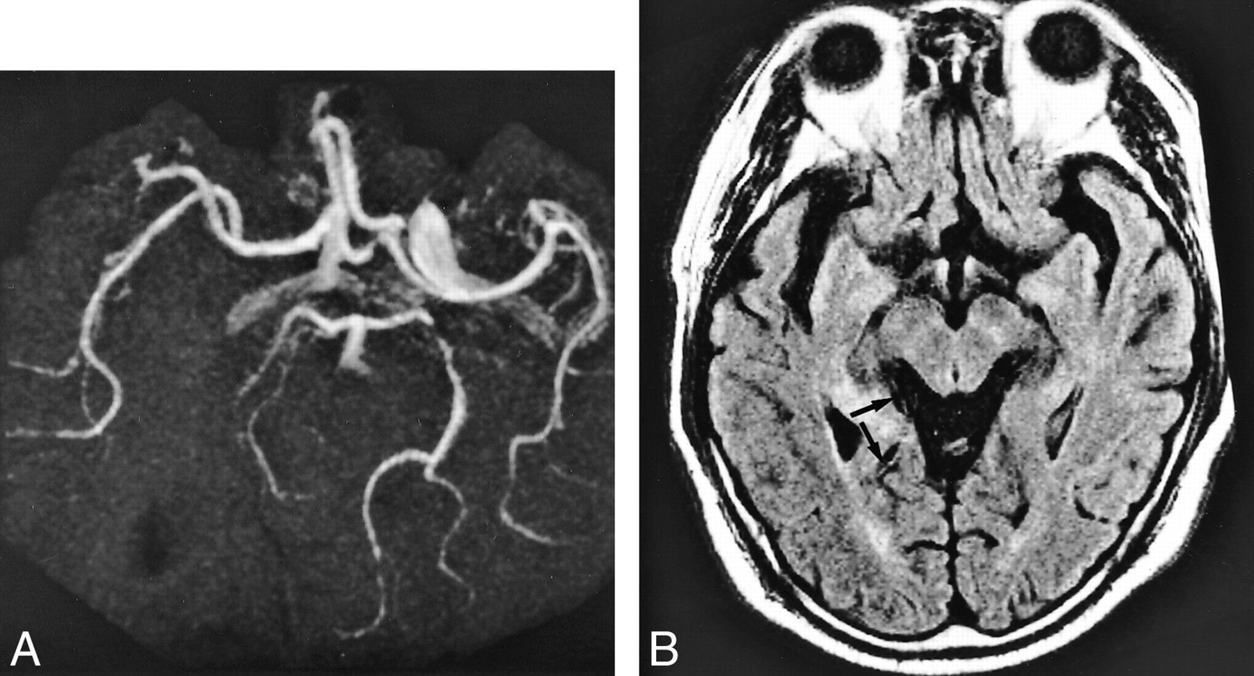
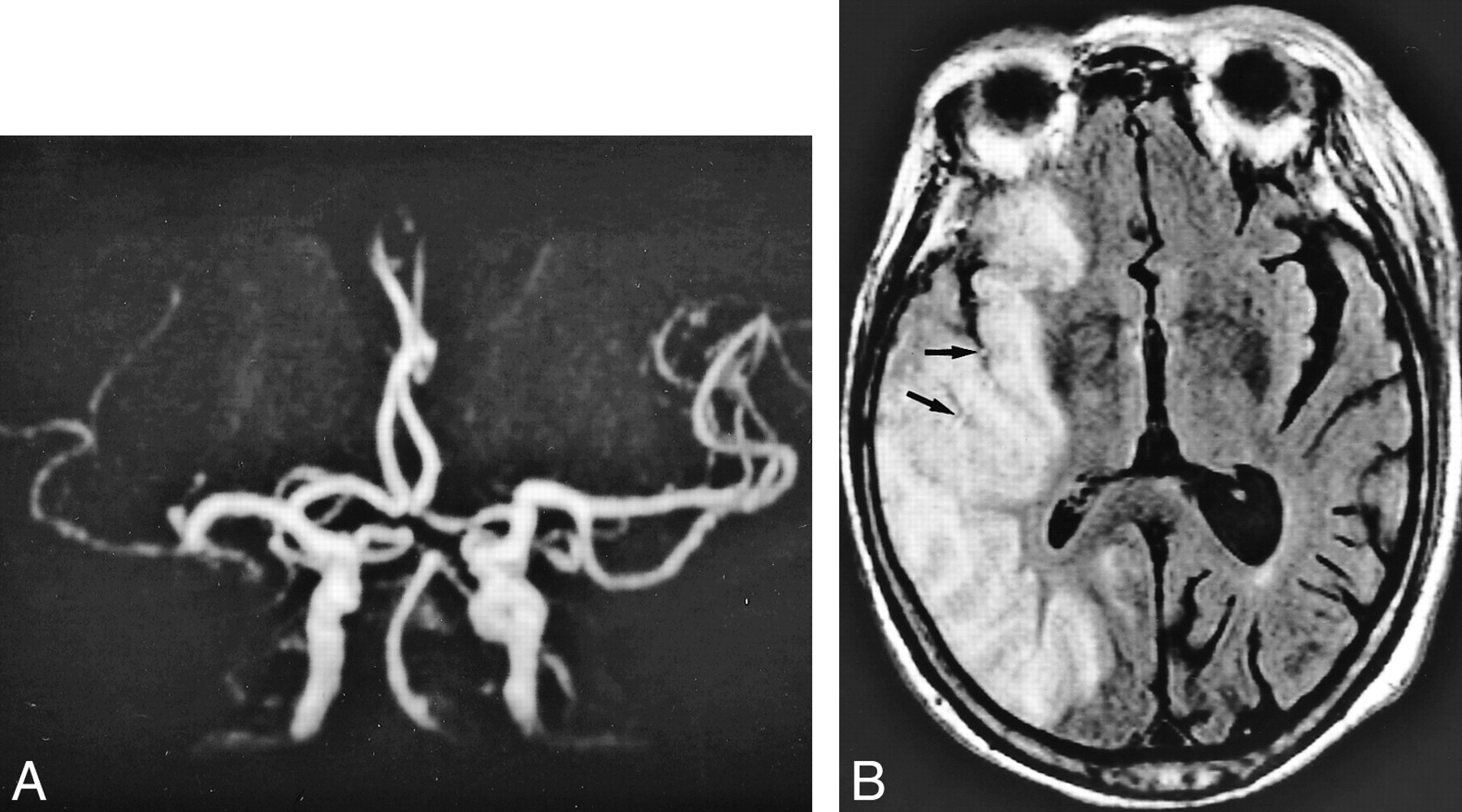
- fig 2.
A 71-year-old woman who underwent imaging 7 hours after onset of right hemiparesis and aphasia.
A and B, FLAIR images (8000/10/1, TI = 2000) show intraarterial signal in M1, M2, and M3 segments of the left middle cerebral artery (arrows).
C, MR angiogram (32/6.8, flip angle = 15 degree) demonstrates lack of TOF in the left internal carotid and middle cerebral arteries.
Perfusion imaging (not shown) showed a hypoperfused area with increased time-to-peak and mean transit time values in the left middle cerebral artery territory as large as the area of intraarterial signal distribution and lack of TOF.
D, Diffusion-weighted image (500/123/1, b = 1200) shows hyperintense lesion in the left basal ganglia and insular cortex.
E, Postcontrast MR angiogram (32/6.8, flip angle = 15 degrees) shows intraluminal enhancement in the left middle cerebral artery except for in the M1 segment. Arrow indicates complete obstruction in the left M1 segment.
F, Final infarction is confirmed in the basal ganglia and insular cortex by CT performed 9 days after stroke symptom onset, corresponding to the initial lesion seen on diffusion-weighted images.
Note lesion in D is smaller than the area of intraarterial signal distribution. Intraarterial signal consists of not only complete obstruction but also slow collateral circulation. The area of final infarct is smaller than that of intraarterial signal distribution. Ischemic penumbra may be present in the mismatch between the intraarterial signal distribution and the diffusion-weighted lesion.
- fig 3.
A 59-year-old man who presented with homonymous hemianopsia. MR imaging was performed 13 hours after stroke symptom onset.
A, MR angiogram (32/6.8, flip angle = 15 degrees) shows lack of TOF in the right posterior communicating artery.
B, On FLAIR images (8000/10/1, TI= 2000), intraarterial signal is visible in the right posterior cerebral artery (arrow); however, signal is discontinuous and isointense to cerebral parenchyma. Final infarction was confirmed in the posterior cerebral territory on T2-weighted image (not shown) obtained 14 days after stroke symptom onset. Note that FLAIR is inferior to MR angiography in detecting occluded artery.
- fig 4.
An 83-year-old woman who underwent imaging 23.5 hours after onset of loss of consciousness.
A, MR angiogram (32/6.8, flip angle = 15 degree) shows occlusion in distal portion of M1 segment of the right middle cerebral artery.
B, FLAIR image (8000/10/1, TI = 2000) already shows hyperintense lesion and cortical swelling in the right middle cerebral artery territory. Intraarterial signal (arrows) is shown in the right middle cerebral artery corresponding to lack of TOF; however, signal is hampered by narrowed sulci owing to vasogenic edema.
- fig 5.
A 62-year-old man who underwent imaging 2.5 hours after onset of loss of consciousness and left hemiparesis.
A and B, FLAIR images (8000/10/1, TI = 2000) show intraarterial signal in the M1 (arrows), M2 (arrowheads), and M3 (thin long arrows) segments of the right middle cerebral artery. Old infarction proceeds in the right insular cortex.
C and D (same level as in A and B), T2-weighted images (4500/96/1) show lack of flow void in M1 (arrows) and M2 (arrowheads) segments; however, evidence of flow void is visible in the M3 segment (thin long arrows).
MR angiogram (not shown) shows lack of TOF in the right middle cerebral artery. CT confirmed final infarction in both perforator and hemispheric-branch territories of the right middle cerebral artery.


Tables

- TABLE 2:
FLAIR to DWI comparison in predicting the area of final infarction

In this issue


Jump to section
Related Articles
Cited By...
- Ivy Sign in Moyamoya Disease: A Comparative Study of the FLAIR Vascular Hyperintensity Sign Against Contrast-Enhanced MRI
- The Association between FLAIR Vascular Hyperintensity and Stroke Outcome Varies with Time from Onset
- Fluid-Attenuated Inversion Recovery Vascular Hyperintensities-Diffusion-Weighted Imaging Mismatch Identifies Acute Stroke Patients Most Likely to Benefit From Recanalization
- Hyperintense Vessels on T2-PROPELLER-FLAIR in Patients with Acute MCA Stroke: Prediction of Arterial Stenosis and Perfusion Abnormality
- Do FLAIR Vascular Hyperintensities beyond the DWI Lesion Represent the Ischemic Penumbra?
- Hyperintense Basilar Artery on FLAIR MR Imaging: Diagnostic Accuracy and Clinical Impact in Patients with Acute Brain Stem Stroke
- Can Diffusion-Weighted Imaging-Fluid-Attenuated Inversion Recovery Mismatch (Positive Diffusion-Weighted Imaging/Negative Fluid-Attenuated Inversion Recovery) at 3 Tesla Identify Patients With Stroke at <4.5 Hours?
- Clinical Significance of Fluid-Attenuated Inversion Recovery Vascular Hyperintensities in Transient Ischemic Attack
- Hyperintense Vessels on Acute Stroke Fluid-Attenuated Inversion Recovery Imaging: Associations With Clinical and Other MRI Findings
- Hyperintense Vessel Sign on Fluid-Attenuated Inversion Recovery MR Imaging Is Reduced by Gadolinium
- Fluid-Attenuated Inversion Recovery Vascular Hyperintensities: An Important Imaging Marker for Cerebrovascular Disease
- Rescue, Combined, and Stand-Alone Thrombectomy in the Management of Large Vessel Occlusion Stroke Using the Solitaire Device: A Prospective 50-Patient Single-Center Study: Timing, Safety, and Efficacy
- Distal hyperintense vessels on FLAIR: An MRI marker for collateral circulation in acute stroke?
- Angiography Reveals That Fluid-Attenuated Inversion Recovery Vascular Hyperintensities Are Due to Slow Flow, Not Thrombus
- Significance of Susceptibility Vessel Sign on T2*-Weighted Gradient Echo Imaging for Identification of Stroke Subtypes
- Evaluation of Hyperintense Vessels on FLAIR MRI for the Diagnosis of Multiple Intracerebral Arterial Stenoses