
Article Figures & Data
Figures
- fig 1.
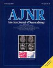
Patient without cerebrovascular disease or relevant hemodynamic alterations.
A, An stdTTP map obtained using DSC-MR imaging with a T2*-weighted echo-planar fast-field–echo sequence (74/30/1 [TR/TE/excitations]) with a 30° flip angle, a 240-mm field of view and a 128 × 128-voxel acquisition matrix of. Central parts of vascular territories are displayed in yellow (time step 1, voxels with short stdTTP) and border zones in dark red (time step 5 and 6, voxels with long stdTTP) color. Each color step codes a time interval of 0.5 s.
B, Corresponding T1-weighted image (500/15/2) after IV administration of contrast medium.
- fig 2.
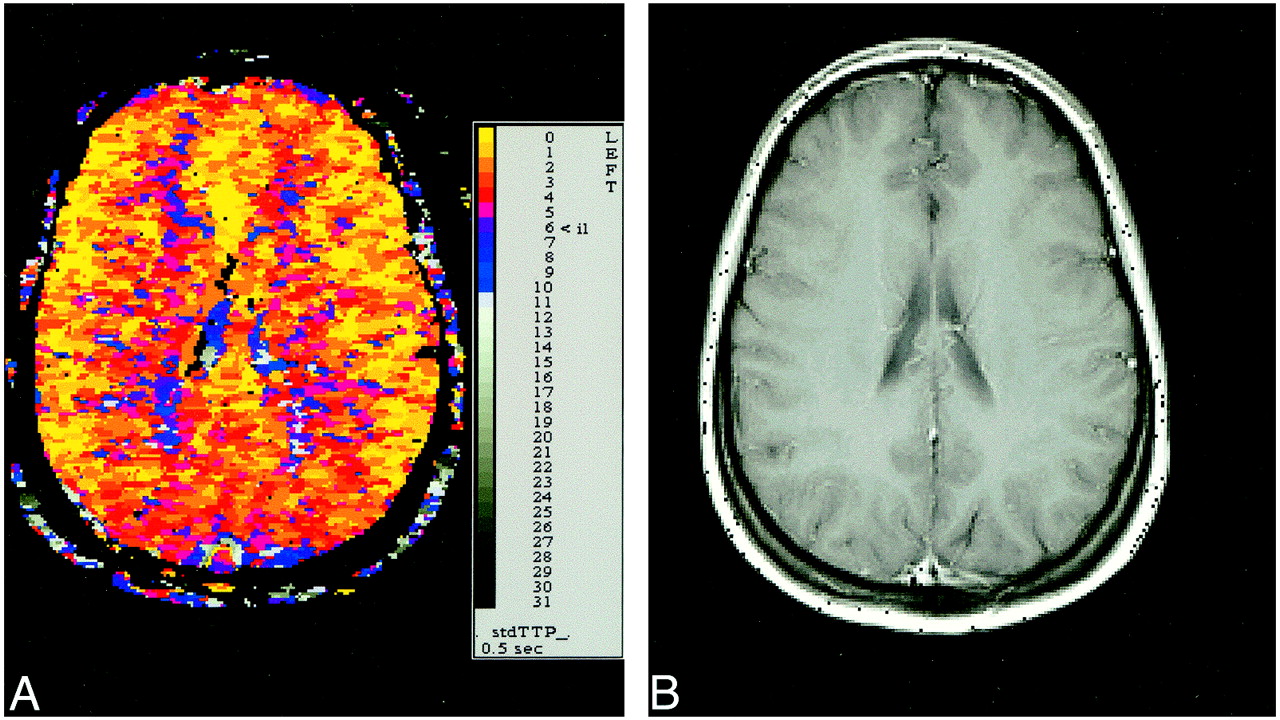
Patient with high-grade stenosis of the left ICA.
A, An stdTTP map obtained using DSC-MR imaging with a T2*-weighted echo-planar fast-field–echo sequence (74/30/1 [TR/TE/excitations]) with a 30° flip angle, a 240-mm field of view and 128 × 128-voxel acquisition matrix. Each color step codes a time interval of 0.5 s. The left hemisphere still shows a small region of normal perfusion within the CVTs (yellow voxels). In the anterior and posterior border zones, stdTTP is significantly prolonged, visible as an increase of voxels shifting color from normally dark red to blue and gray. A slight prolongation of stdTTP in the anterior and posterior border zones in the right hemisphere (color shift to magenta to blue) was not significant.
B, Corresponding T1-weighted image (500/15/2) after IV administration of contrast medium.
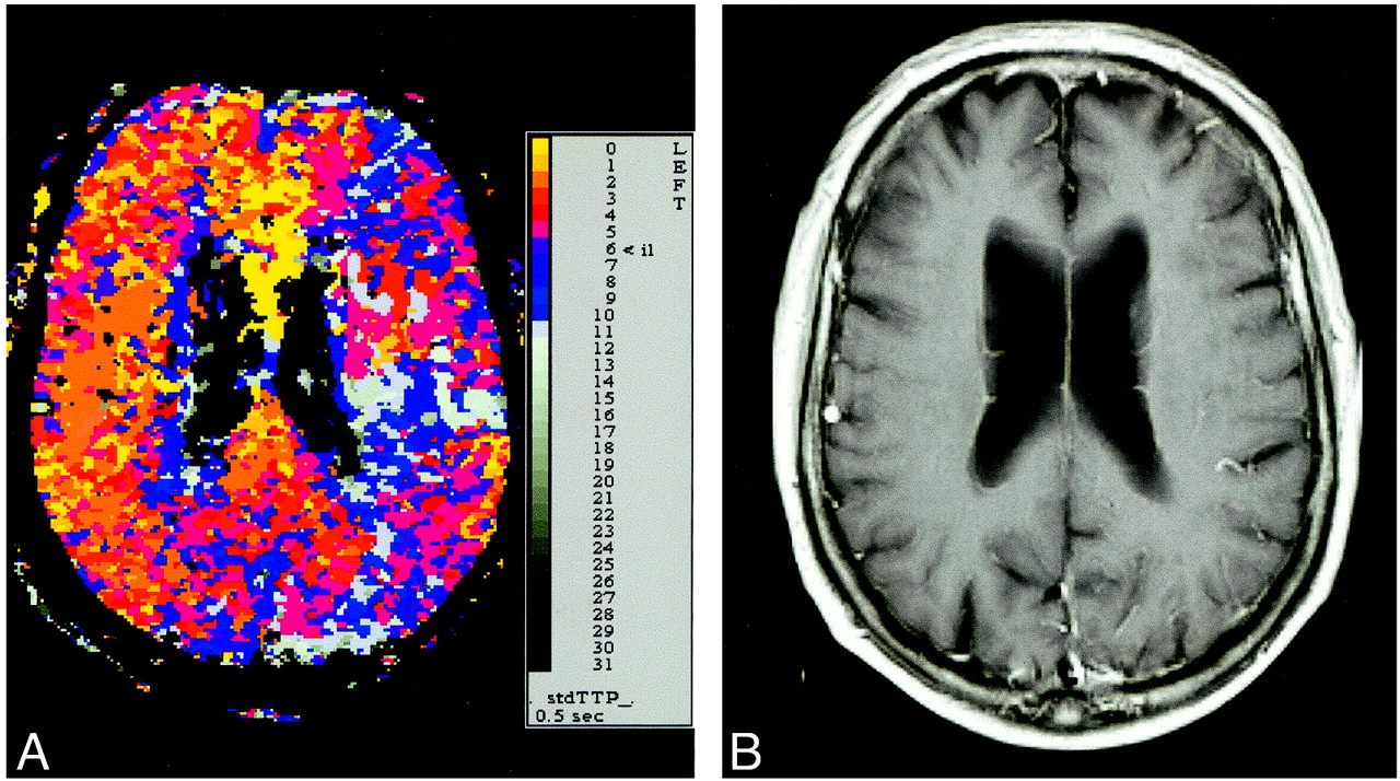
- fig 3.
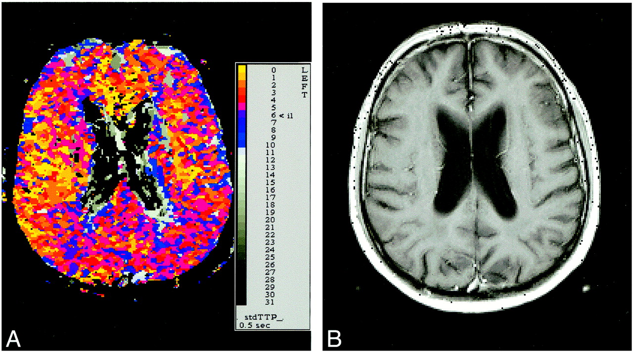
Patient with high-grade stenosis of the right ICA and occlusion of the left ICA.
A, An StdTTP map from DSC-MR imaging using a T2*-weighted echo-planar fast-field–echo sequence (74/30/1 [TR/TE/excitation]) with a 30° flip angle, a 240-mm field of view, and a 128 × 128-voxel acquisition matrix. Each color step codes a time interval of 0.5 s. In the left hemisphere, the stdTTP is markedly prolonged in the anterior and posterior border zones (shift from normal dark red to gray) and in the central territory of the middle cerebral artery (shift from normal yellow to red or magenta). The vascular territory adjacent to the central region shows a shift to blue and gray values comparable with that of border zones. This was interpreted as a result from atherosclerotic small vessel disease, because the morphologic examination showed no related lesion. On the right side, the stdTTP was moderately prolonged, shown as moderate increases in magenta and blue voxels in the border zones.
B, Corresponding T1-weighted image (500/15/2) after IV administration of contrast medium.
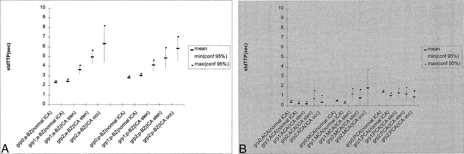
- fig 4.
Mean stdTTTP values with 95% confidence intervals (minimum to maximum).
A, Values measured in border zones. The stdTTP was not significantly prolonged in the anterior (a-BZ) and posterior (p-BZ) border zones in hemispheres supplied by a normal ICA in group 1 (grp 1; a-BZ and p-BZ [normal ICA]) compared with the control (grp 0; a-BZ and p-BZ [normal ICA]). All other groups showed significantly prolonged stdTTP values. In relation to a proposed limit of 3.5 s for the stdTTP value in border zones, this was moderate in hemispheres supplied by a highly stenosed ICA (grp 1; a-BZ and p-BZ [stenotic ICA] and grp 2; a-BZ and p-BZ [stenotic ICA]), but severe in hemispheres isolateral to an occluded ICA (grp 2; a-BZ and p-BZ [occluded ICA]).
B, Values measured in the CVT of the anterior (ACA), middle (MCA), and posterior (PCA) cerebral arteries showed behavior completely different from that of border zones. Only in the CVT of the MCA in group 2 (grp 2; MCA [occluded ICA]), in whom the isolateral ICA was occluded and the contralateral ICA showed a high-grade stenosis, was the stdTTP significantly increased compared with the control group (grp 0).


Tables

StdTTP of patients with cerebrovascular disease
In this issue


Jump to section
Related Articles
Cited By...
- Assessment of Cortical Hemodynamics by Multichannel Near-Infrared Spectroscopy in Steno-Occlusive Disease of the Middle Cerebral Artery
- Internal and Cortical Border-Zone Infarction: Clinical and Diffusion-Weighted Imaging Features
- The Pathophysiology of Watershed Infarction in Internal Carotid Artery Disease: Review of Cerebral Perfusion Studies