Advertisement

Main menu

  • Home
  • Content
    • Current Issue
    • Accepted Manuscripts
    • Article Preview
    • Past Issue Archive
    • Video Articles
    • AJNR Case Collection
    • Case of the Week Archive
    • Case of the Month Archive
    • Classic Case Archive
  • Special Collections
    • AJNR Awards
    • Low-Field MRI
    • Alzheimer Disease
    • ASNR Foundation Special Collection
    • Photon-Counting CT
    • View All
  • Multimedia
    • AJNR Podcasts
    • AJNR SCANtastic
    • Trainee Corner
    • MRI Safety Corner
    • Imaging Protocols
  • For Authors
    • Submit a Manuscript
    • Submit a Video Article
    • Submit an eLetter to the Editor/Response
    • Manuscript Submission Guidelines
    • Statistical Tips
    • Fast Publishing of Accepted Manuscripts
    • Graphical Abstract Preparation
    • Imaging Protocol Submission
    • Author Policies
  • About Us
    • About AJNR
    • Editorial Board
    • Editorial Board Alumni
  • More
    • Become a Reviewer/Academy of Reviewers
    • Subscribers
    • Permissions
    • Alerts
    • Feedback
    • Advertisers
    • ASNR Home

User menu

  • Alerts
  • Log in

Search

  • Advanced search
American Journal of Neuroradiology

American Journal of Neuroradiology

ASHNR American Society of Functional Neuroradiology ASHNR American Society of Pediatric Neuroradiology ASSR
  • Alerts
  • Log in

Advanced Search

  • Home
  • Content
    • Current Issue
    • Accepted Manuscripts
    • Article Preview
    • Past Issue Archive
    • Video Articles
    • AJNR Case Collection
    • Case of the Week Archive
    • Case of the Month Archive
    • Classic Case Archive
  • Special Collections
    • AJNR Awards
    • Low-Field MRI
    • Alzheimer Disease
    • ASNR Foundation Special Collection
    • Photon-Counting CT
    • View All
  • Multimedia
    • AJNR Podcasts
    • AJNR SCANtastic
    • Trainee Corner
    • MRI Safety Corner
    • Imaging Protocols
  • For Authors
    • Submit a Manuscript
    • Submit a Video Article
    • Submit an eLetter to the Editor/Response
    • Manuscript Submission Guidelines
    • Statistical Tips
    • Fast Publishing of Accepted Manuscripts
    • Graphical Abstract Preparation
    • Imaging Protocol Submission
    • Author Policies
  • About Us
    • About AJNR
    • Editorial Board
    • Editorial Board Alumni
  • More
    • Become a Reviewer/Academy of Reviewers
    • Subscribers
    • Permissions
    • Alerts
    • Feedback
    • Advertisers
    • ASNR Home
  • Follow AJNR on Twitter
  • Visit AJNR on Facebook
  • Follow AJNR on Instagram
  • Join AJNR on LinkedIn
  • RSS Feeds

AJNR Awards, New Junior Editors, and more. Read the latest AJNR updates

Research ArticleBrain

Coexistent Neurocysticercosis and Japanese B Encephalitis: MR Imaging Correlation

Paramjeet Singh, Naveen Kalra, Radha Kanta Ratho, Sridhar Shankar, Niranjan Khandelwal and Sudha Suri
American Journal of Neuroradiology June 2001, 22 (6) 1131-1136;
Paramjeet Singh
  • Find this author on Google Scholar
  • Find this author on PubMed
  • Search for this author on this site
Naveen Kalra
  • Find this author on Google Scholar
  • Find this author on PubMed
  • Search for this author on this site
Radha Kanta Ratho
  • Find this author on Google Scholar
  • Find this author on PubMed
  • Search for this author on this site
Sridhar Shankar
  • Find this author on Google Scholar
  • Find this author on PubMed
  • Search for this author on this site
Niranjan Khandelwal
  • Find this author on Google Scholar
  • Find this author on PubMed
  • Search for this author on this site
Sudha Suri
  • Find this author on Google Scholar
  • Find this author on PubMed
  • Search for this author on this site
  • Article
  • Figures & Data
  • Info & Metrics
  • Responses
  • References
  • PDF
Loading

Article Figures & Data

Figures

  • Tables
  • fig 1.
    • Download figure
    • Open in new tab
    • Download powerpoint
    fig 1.

    Images of a 16-year-old man with serologically positive JE and single coexistent NCC.

    A, T2-weighted turbo fluid-attenuated inversion recovery axial section shows bilateral asymmetric thalamic hyperintensity (left more than right) with left globus pallidus involvement.

    B, T2-weighted turbo spin-echo coronal section shows the degenerating cyst in the left frontal region superficially in close association with the leptomeninges with associated edema.
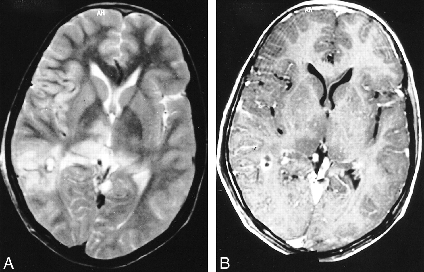
  • fig 2.
    • Download figure
    • Open in new tab
    • Download powerpoint
    fig 2.

    Images of a 13-year-old boy with a solitary cyst and JE.

    A, T2-weighted turbo spin-echo axial section of the brain shows more extensive right thalamic hyperintensity on the same side as the cyst inciting edema. The encephalitic changes in the temporal cortex also lateralize to the same side.

    B, T1-weighted contrast-enhanced axial section shows the enhancement of the wall of the cyst. The thalamic lesions appear hypointense and show no hemorrhage.

  • fig 3.
    • Download figure
    • Open in new tab
    • Download powerpoint
    fig 3.

    Images of a 50-year-old man with JE and positive serology.

    A, T2-weighted turbo spin-echo axial section reveals two cysticercus cysts in the right temporal lobe with thalamic lesion lateralization on the same side. The hyperintensity associated with the cysts (representing edema) involves the right temporal lobe extensively and merges with the thalamic signal changes of JE.

    B, T2-weighted turbo spin-echo axial section reveals the second cysticercus cyst in the right temporal lobe.
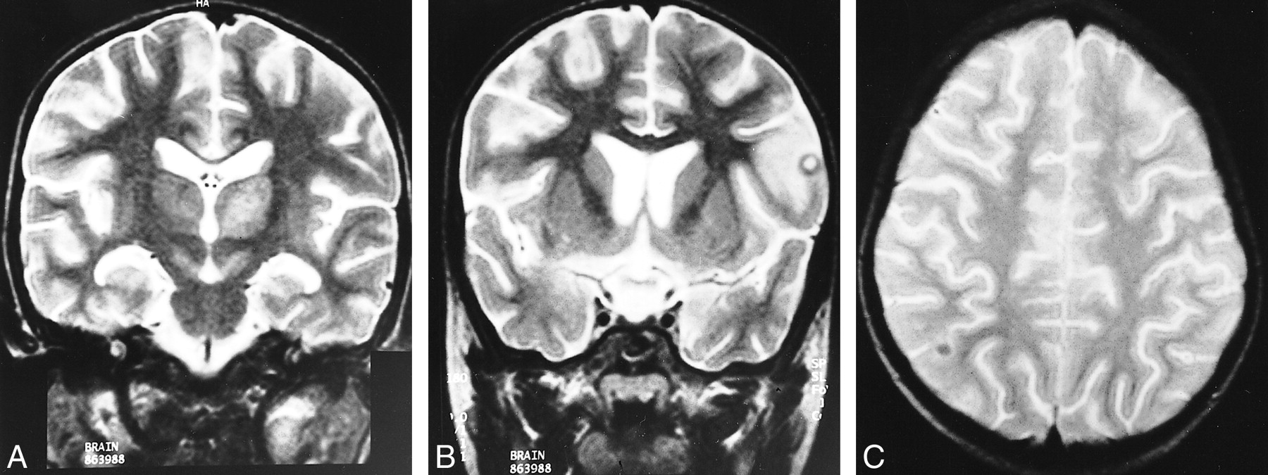
  • fig 4.
    • Download figure
    • Open in new tab
    • Download powerpoint
    fig 4.

    Images of an 8-year-old boy with JE and two cysticercus cysts.

    A, T2-weighted turbo spin-echo coronal section of the brain reveals predominant signal changes in the left thalamus.

    B, T2-weighted turbo spin-echo coronal section shows degenerating cyst with edema in the left frontal cortex, which correlates with the side of dominant JE lesion.

    C, T2-weighted turbo spin-echo axial section shows the other cyst on the contralateral side in granular nodular stage with no inflammation in the surrounding brain.

Tables

  • Figures
  • Table1

    Correlation of distribution and stage of cysts with lateralization of Japanese encephalitis lesions on MR images

PreviousNext
Back to top

In this issue

American Journal of Neuroradiology: 22 (6)
American Journal of Neuroradiology
Vol. 22, Issue 6
1 Jun 2001
  • Table of Contents
  • Index by author
Advertisement
Print
Download PDF
Email Article

Thank you for your interest in spreading the word on American Journal of Neuroradiology.

NOTE: We only request your email address so that the person you are recommending the page to knows that you wanted them to see it, and that it is not junk mail. We do not capture any email address.

Enter multiple addresses on separate lines or separate them with commas.
Coexistent Neurocysticercosis and Japanese B Encephalitis: MR Imaging Correlation
(Your Name) has sent you a message from American Journal of Neuroradiology
(Your Name) thought you would like to see the American Journal of Neuroradiology web site.
CAPTCHA
This question is for testing whether or not you are a human visitor and to prevent automated spam submissions.
Cite this article
Paramjeet Singh, Naveen Kalra, Radha Kanta Ratho, Sridhar Shankar, Niranjan Khandelwal, Sudha Suri
Coexistent Neurocysticercosis and Japanese B Encephalitis: MR Imaging Correlation
American Journal of Neuroradiology Jun 2001, 22 (6) 1131-1136;

Citation Manager Formats

  • BibTeX
  • Bookends
  • EasyBib
  • EndNote (tagged)
  • EndNote 8 (xml)
  • Medlars
  • Mendeley
  • Papers
  • RefWorks Tagged
  • Ref Manager
  • RIS
  • Zotero
0 Responses
Respond to this article
Share
Bookmark this article
Coexistent Neurocysticercosis and Japanese B Encephalitis: MR Imaging Correlation
Paramjeet Singh, Naveen Kalra, Radha Kanta Ratho, Sridhar Shankar, Niranjan Khandelwal, Sudha Suri
American Journal of Neuroradiology Jun 2001, 22 (6) 1131-1136;
del.icio.us logo Twitter logo Facebook logo Mendeley logo
  • Tweet Widget
  • Facebook Like
  • Google Plus One

Jump to section

  • Article
    • Abstract
    • Methods
    • Results
    • Discussion
    • Conclusion
    • Footnotes
    • References
  • Figures & Data
  • Info & Metrics
  • Responses
  • References
  • PDF

Related Articles

  • No related articles found.
  • PubMed
  • Google Scholar

Cited By...

  • Is neurocysticercosis a risk factor in coexistent intracranial disease? An MRI based study
  • Crossref
  • Google Scholar

This article has not yet been cited by articles in journals that are participating in Crossref Cited-by Linking.

More in this TOC Section

  • Progression of Microstructural Damage in Spinocerebellar Ataxia Type 2: A Longitudinal DTI Study
  • SWI or T2*: Which MRI Sequence to Use in the Detection of Cerebral Microbleeds? The Karolinska Imaging Dementia Study
  • Statin Therapy Does Not Affect the Radiographic and Clinical Profile of Patients with TIA and Minor Stroke
Show more Brain

Similar Articles

Advertisement

Indexed Content

  • Current Issue
  • Accepted Manuscripts
  • Article Preview
  • Past Issues
  • Editorials
  • Editor's Choice
  • Fellows' Journal Club
  • Letters to the Editor
  • Video Articles

Cases

  • Case Collection
  • Archive - Case of the Week
  • Archive - Case of the Month
  • Archive - Classic Case

More from AJNR

  • Trainee Corner
  • Imaging Protocols
  • MRI Safety Corner
  • Book Reviews

Multimedia

  • AJNR Podcasts
  • AJNR Scantastics

Resources

  • Turnaround Time
  • Submit a Manuscript
  • Submit a Video Article
  • Submit an eLetter to the Editor/Response
  • Manuscript Submission Guidelines
  • Statistical Tips
  • Fast Publishing of Accepted Manuscripts
  • Graphical Abstract Preparation
  • Imaging Protocol Submission
  • Evidence-Based Medicine Level Guide
  • Publishing Checklists
  • Author Policies
  • Become a Reviewer/Academy of Reviewers
  • News and Updates

About Us

  • About AJNR
  • Editorial Board
  • Editorial Board Alumni
  • Alerts
  • Permissions
  • Not an AJNR Subscriber? Join Now
  • Advertise with Us
  • Librarian Resources
  • Feedback
  • Terms and Conditions
  • AJNR Editorial Board Alumni

American Society of Neuroradiology

  • Not an ASNR Member? Join Now

© 2025 by the American Society of Neuroradiology All rights, including for text and data mining, AI training, and similar technologies, are reserved.
Print ISSN: 0195-6108 Online ISSN: 1936-959X

Powered by HighWire