
Article Figures & Data
Figures
- fig 1.
Transverse T1-weighted image (inversion recovery spin-echo; 1500/20; inversion time, 650 ms; number of acquisitions, one), obtained at the level of the basal ganglia in a patient with liver cirrhosis, shows bilateral increased signal in the globus pallidus. For calculating the globus pallidus index, mean signal intensity was calculated from a region of interest localized in both the putamen (□) and globus pallidus (○).fig 2. Transverse MTR map of a supraventricular level, obtained from two proton density gradient-echo sequences (714/12; flip angle, 20 degrees; number of acquisitions, one), the first with and the second without an off-resonance preparation pulse. MTR values were obtained from four different voxels (□) located in otherwise normal-appearing white matter, as defined on the T2-weighted sequence, within both parietal and frontal lobes.fig 3. Axial T2-weighted image (fast spin-echo; 3550/90; number of acquisitions, two), obtained at a supraventricular level, shows the voxel from which the 1H-MRS image was obtained in the normal appearing white matter in the parietal lobe
- fig 4.
Water-suppressed proton spectra of an 8-mL voxel located in the parietal region, recorded with a stimulated echo-based pulse sequence (1600/20; magnetization transfer, 30; number of acquisitions, 256). The main resonances correspond to N-acetylaspartate (2.0 ppm), glutamic/glutamine (2.1−2.5 ppm), creatine/phosphocreatine (3.02 ppm), choline-containing compounds (3.2 ppm), and myo-inositol (3.55 ppm). Comparison of the spectra shows a decrease in choline and myo-inositol resonances, with an increase in the glutamic/glutamine region in the cirrhotic patient.
A, Healthy control volunteer.
B, Cirrhotic patient.


Tables
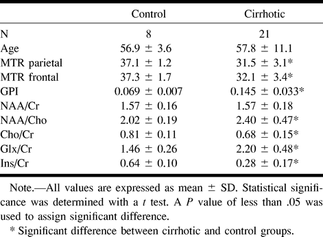
 TABLE 1:
1H-MRS metabolite ratios from the parietal white matter, normalized globus pallidus intensity (GPI) and MTR of different brain regions for the control and cirrhotic groups
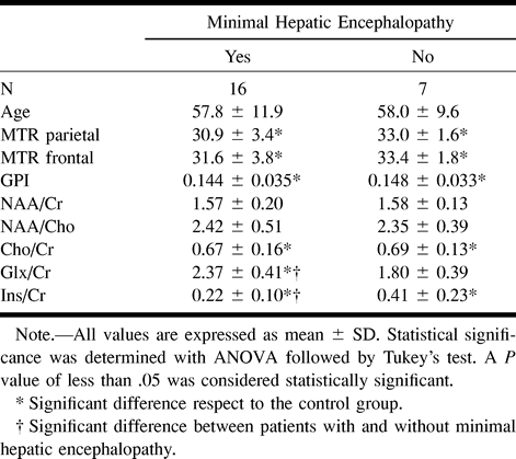
- TABLE 2:
1H-MRS metabolite ratios from the parietal white matter normalized globus pallidus intensity and MTR of different brain regions for cirrhotic patients classified by the existence of a minimal hepatic encephalopathy
In this issue


Jump to section
Related Articles
Cited By...
- Body size interacts with the structure of the central nervous system: A multi-center in vivo neuroimaging study
- Meta-analysis of magnetic resonance spectroscopy in the diagnosis of hepatic encephalopathy
- Growth of White Matter in the Adolescent Brain: Role of Testosterone and Androgen Receptor
- Normalization of T2 signal abnormalities in hemispheric white matter with liver transplant