Advertisement

Main menu

  • Home
  • Content
    • Current Issue
    • Accepted Manuscripts
    • Article Preview
    • Past Issue Archive
    • Video Articles
    • AJNR Case Collection
    • Case of the Week Archive
    • Case of the Month Archive
    • Classic Case Archive
  • Special Collections
    • AJNR Awards
    • Low-Field MRI
    • Alzheimer Disease
    • ASNR Foundation Special Collection
    • Photon-Counting CT
    • View All
  • Multimedia
    • AJNR Podcasts
    • AJNR SCANtastic
    • Trainee Corner
    • MRI Safety Corner
    • Imaging Protocols
  • For Authors
    • Submit a Manuscript
    • Submit a Video Article
    • Submit an eLetter to the Editor/Response
    • Manuscript Submission Guidelines
    • Statistical Tips
    • Fast Publishing of Accepted Manuscripts
    • Graphical Abstract Preparation
    • Imaging Protocol Submission
    • Author Policies
  • About Us
    • About AJNR
    • Editorial Board
    • Editorial Board Alumni
  • More
    • Become a Reviewer/Academy of Reviewers
    • Subscribers
    • Permissions
    • Alerts
    • Feedback
    • Advertisers
    • ASNR Home

User menu

  • Alerts
  • Log in

Search

  • Advanced search
American Journal of Neuroradiology

American Journal of Neuroradiology

ASHNR American Society of Functional Neuroradiology ASHNR American Society of Pediatric Neuroradiology ASSR
  • Alerts
  • Log in

Advanced Search

  • Home
  • Content
    • Current Issue
    • Accepted Manuscripts
    • Article Preview
    • Past Issue Archive
    • Video Articles
    • AJNR Case Collection
    • Case of the Week Archive
    • Case of the Month Archive
    • Classic Case Archive
  • Special Collections
    • AJNR Awards
    • Low-Field MRI
    • Alzheimer Disease
    • ASNR Foundation Special Collection
    • Photon-Counting CT
    • View All
  • Multimedia
    • AJNR Podcasts
    • AJNR SCANtastic
    • Trainee Corner
    • MRI Safety Corner
    • Imaging Protocols
  • For Authors
    • Submit a Manuscript
    • Submit a Video Article
    • Submit an eLetter to the Editor/Response
    • Manuscript Submission Guidelines
    • Statistical Tips
    • Fast Publishing of Accepted Manuscripts
    • Graphical Abstract Preparation
    • Imaging Protocol Submission
    • Author Policies
  • About Us
    • About AJNR
    • Editorial Board
    • Editorial Board Alumni
  • More
    • Become a Reviewer/Academy of Reviewers
    • Subscribers
    • Permissions
    • Alerts
    • Feedback
    • Advertisers
    • ASNR Home
  • Follow AJNR on Twitter
  • Visit AJNR on Facebook
  • Follow AJNR on Instagram
  • Join AJNR on LinkedIn
  • RSS Feeds

AJNR Awards, New Junior Editors, and more. Read the latest AJNR updates

Research ArticleBrain

The Value of Apparent Diffusion Coefficient Maps in Early Cerebral Ischemia

Patricia M. Desmond, Amanda C. Lovell, Andrew A. Rawlinson, Mark W. Parsons, P. Alan Barber, Qing Yang, Ting Li, David G. Darby, Richard P. Gerraty, Steven M. Davis and Brian M. Tress
American Journal of Neuroradiology August 2001, 22 (7) 1260-1267;
Patricia M. Desmond
  • Find this author on Google Scholar
  • Find this author on PubMed
  • Search for this author on this site
Amanda C. Lovell
  • Find this author on Google Scholar
  • Find this author on PubMed
  • Search for this author on this site
Andrew A. Rawlinson
  • Find this author on Google Scholar
  • Find this author on PubMed
  • Search for this author on this site
Mark W. Parsons
  • Find this author on Google Scholar
  • Find this author on PubMed
  • Search for this author on this site
P. Alan Barber
  • Find this author on Google Scholar
  • Find this author on PubMed
  • Search for this author on this site
Qing Yang
  • Find this author on Google Scholar
  • Find this author on PubMed
  • Search for this author on this site
Ting Li
  • Find this author on Google Scholar
  • Find this author on PubMed
  • Search for this author on this site
David G. Darby
  • Find this author on Google Scholar
  • Find this author on PubMed
  • Search for this author on this site
Richard P. Gerraty
  • Find this author on Google Scholar
  • Find this author on PubMed
  • Search for this author on this site
Steven M. Davis
  • Find this author on Google Scholar
  • Find this author on PubMed
  • Search for this author on this site
Brian M. Tress
  • Find this author on Google Scholar
  • Find this author on PubMed
  • Search for this author on this site
  • Article
  • Figures & Data
  • Info & Metrics
  • Responses
  • References
  • PDF
Loading

Article Figures & Data

Figures

  • Tables
  • fig 1.
    • Download figure
    • Open in new tab
    • Download powerpoint
    fig 1.

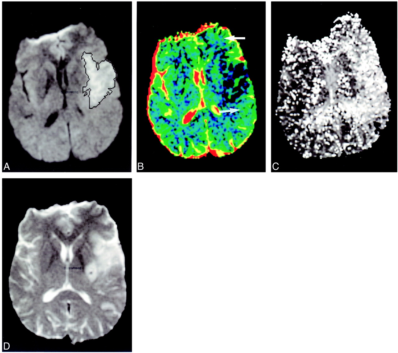
    Regions of abnormality were identified on the TTP (A), T2 outcome (B), and the acute diffusion-weighted scans (C) using region-growing techniques. These were transferred on to the acute ADC map (D), and the region of expansion of the infarct was generated (black outline, right). The region was reflected about the midline axis and regrown to enclose just brain tissue (D, two black regions, left). The region of infarct expansion has reduced ADCs (D, white arrow) compared with the contralateral region and hazy, subtle change on the diffusion-weighted images (C, black arrow)

  • fig 2.
    • Download figure
    • Open in new tab
    • Download powerpoint
    fig 2.

    Graph of mean rADC against region

  • fig 3.
    • Download figure
    • Open in new tab
    • Download powerpoint
    fig 3.

    Infarct expansion. Patient 13 presented 1 hour 30 minutes, after stroke onset. Acute diffusion-weighted images (A) show small region of hyperintensity (black outline). There was a large area of altered perfusion on the TTP map (C). Extensive regions of lowered ADC values are seen on the colored ADC map (B, white arrow) in the regions that progress to infarction (D, subacute scan). (Outcome scan was not available for this patient.) In retrospect, hazy, ill-defined change is seen on the acute diffusion-weighted image (A, black arrowhead)

  • fig 4.
    • Download figure
    • Open in new tab
    • Download powerpoint
    fig 4.

    Minor infarct expansion. Patient 10 presented 3 hours after stroke onset. The initial diffusion-weighted imaging lesion (A) was well defined and only minimally underestimated the final infarct size of the outcome scan (D). A large region of altered perfusion was present on the TTP (C). The larger regions of penumbral preservation show normal ADC values (B, white arrows)

  • fig 5.
    • Download figure
    • Open in new tab
    • Download powerpoint
    fig 5.

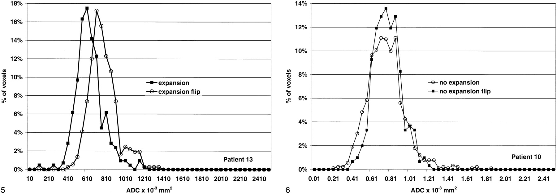
    Histogram analysis of the region of infarct expansion (expansion) and the same region in the normal hemisphere (expansion flip) for patient 13. The curve is shifted to the left with a reduction in the mean ADC value.fig 6. Histogram analysis of the region of penumbral preservation (no expansion) and the comparative region in the normal hemisphere (no expansion flip) for patient 10. The curves are similar

  • fig 7.
    • Download figure
    • Open in new tab
    • Download powerpoint
    fig 7.

    Grouped data.

    A, All regions. Only the region of infarct expansion shows a different distribution of ADC values.

    B, The regions of infarct expansion have lowered ADC values compared with similar regions in the normal hemisphere.

    C, The distribution of the ADC values in regions of penumbral preservation is the same as in the contralateral hemisphere.

Tables

  • Figures
  • Table1

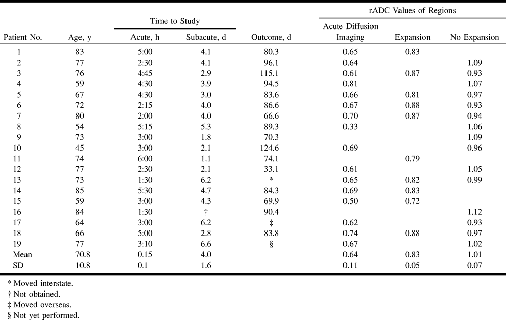
    Summary of patient data, time to MR examination, and values of the ADC ratios on the acute scan in the diffusion abnormality and different regions of the penumbra

PreviousNext
Back to top

In this issue

American Journal of Neuroradiology: 22 (7)
American Journal of Neuroradiology
Vol. 22, Issue 7
1 Aug 2001
  • Table of Contents
  • Index by author
Advertisement
Print
Download PDF
Email Article

Thank you for your interest in spreading the word on American Journal of Neuroradiology.

NOTE: We only request your email address so that the person you are recommending the page to knows that you wanted them to see it, and that it is not junk mail. We do not capture any email address.

Enter multiple addresses on separate lines or separate them with commas.
The Value of Apparent Diffusion Coefficient Maps in Early Cerebral Ischemia
(Your Name) has sent you a message from American Journal of Neuroradiology
(Your Name) thought you would like to see the American Journal of Neuroradiology web site.
CAPTCHA
This question is for testing whether or not you are a human visitor and to prevent automated spam submissions.
Cite this article
Patricia M. Desmond, Amanda C. Lovell, Andrew A. Rawlinson, Mark W. Parsons, P. Alan Barber, Qing Yang, Ting Li, David G. Darby, Richard P. Gerraty, Steven M. Davis, Brian M. Tress
The Value of Apparent Diffusion Coefficient Maps in Early Cerebral Ischemia
American Journal of Neuroradiology Aug 2001, 22 (7) 1260-1267;

Citation Manager Formats

  • BibTeX
  • Bookends
  • EasyBib
  • EndNote (tagged)
  • EndNote 8 (xml)
  • Medlars
  • Mendeley
  • Papers
  • RefWorks Tagged
  • Ref Manager
  • RIS
  • Zotero
0 Responses
Respond to this article
Share
Bookmark this article
The Value of Apparent Diffusion Coefficient Maps in Early Cerebral Ischemia
Patricia M. Desmond, Amanda C. Lovell, Andrew A. Rawlinson, Mark W. Parsons, P. Alan Barber, Qing Yang, Ting Li, David G. Darby, Richard P. Gerraty, Steven M. Davis, Brian M. Tress
American Journal of Neuroradiology Aug 2001, 22 (7) 1260-1267;
del.icio.us logo Twitter logo Facebook logo Mendeley logo
  • Tweet Widget
  • Facebook Like
  • Google Plus One

Jump to section

  • Article
    • Abstract
    • Methods
    • Results
    • Discussion
    • Conclusion
    • Footnotes
    • References
  • Figures & Data
  • Info & Metrics
  • Responses
  • References
  • PDF

Related Articles

  • No related articles found.
  • PubMed
  • Google Scholar

Cited By...

  • Ischemic diffusion lesion reversal is uncommon and rarely alters perfusion-diffusion mismatch
  • Atlas-Based Topographical Scoring for Magnetic Resonance Imaging of Acute Stroke
  • Differential Prognosis of Isolated Cortical Swelling and Hypoattenuation on CT in Acute Stroke
  • Apparent Diffusion Coefficient Thresholds Do Not Predict the Response to Acute Stroke Thrombolysis
  • Refining the Perfusion-Diffusion Mismatch Hypothesis
  • Evolving Paradigms in Neuroimaging of the Ischemic Penumbra
  • A review of structural magnetic resonance neuroimaging
  • Beyond Mismatch: Evolving Paradigms in Imaging the Ischemic Penumbra With Multimodal Magnetic Resonance Imaging
  • Perfusion Thresholds in Acute Stroke Thrombolysis
  • Quantitative Assessment of the Time Course of Infarct Signal Intensity on Diffusion-Weighted Images
  • Crossref
  • Google Scholar

This article has not yet been cited by articles in journals that are participating in Crossref Cited-by Linking.

More in this TOC Section

  • Fast Contrast-Enhanced 4D MRA and 4D Flow MRI Using Constrained Reconstruction (HYPRFlow): Potential Applications for Brain Arteriovenous Malformations
  • Multimodal CT Provides Improved Performance for Lacunar Infarct Detection
  • Optimal MRI Sequence for Identifying Occlusion Location in Acute Stroke: Which Value of Time-Resolved Contrast-Enhanced MRA?
Show more Brain

Similar Articles

Advertisement

Indexed Content

  • Current Issue
  • Accepted Manuscripts
  • Article Preview
  • Past Issues
  • Editorials
  • Editor's Choice
  • Fellows' Journal Club
  • Letters to the Editor
  • Video Articles

Cases

  • Case Collection
  • Archive - Case of the Week
  • Archive - Case of the Month
  • Archive - Classic Case

More from AJNR

  • Trainee Corner
  • Imaging Protocols
  • MRI Safety Corner
  • Book Reviews

Multimedia

  • AJNR Podcasts
  • AJNR Scantastics

Resources

  • Turnaround Time
  • Submit a Manuscript
  • Submit a Video Article
  • Submit an eLetter to the Editor/Response
  • Manuscript Submission Guidelines
  • Statistical Tips
  • Fast Publishing of Accepted Manuscripts
  • Graphical Abstract Preparation
  • Imaging Protocol Submission
  • Evidence-Based Medicine Level Guide
  • Publishing Checklists
  • Author Policies
  • Become a Reviewer/Academy of Reviewers
  • News and Updates

About Us

  • About AJNR
  • Editorial Board
  • Editorial Board Alumni
  • Alerts
  • Permissions
  • Not an AJNR Subscriber? Join Now
  • Advertise with Us
  • Librarian Resources
  • Feedback
  • Terms and Conditions
  • AJNR Editorial Board Alumni

American Society of Neuroradiology

  • Not an ASNR Member? Join Now

© 2025 by the American Society of Neuroradiology All rights, including for text and data mining, AI training, and similar technologies, are reserved.
Print ISSN: 0195-6108 Online ISSN: 1936-959X

Powered by HighWire