
Article Figures & Data
Figures
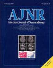
- fig 1.
Mean rCBV of 24 patients with CADASIL, as measured in gray matter and subcortical white matter
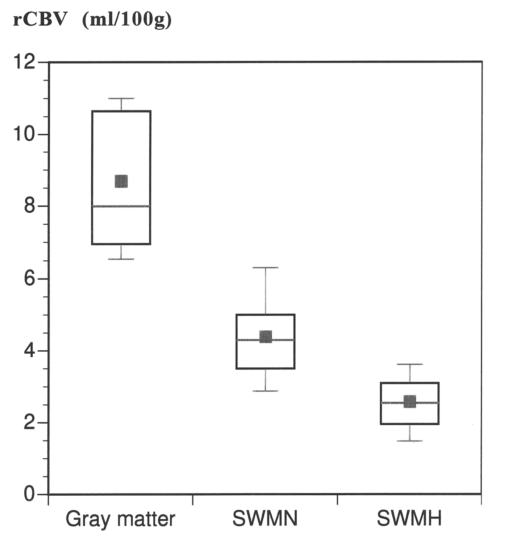
- fig 2.
MR imaging findings of a 53-year-old man with CADASIL (MMSE score, not measurable; Rankin score, 4). A, T2-weighted image (2300/85/1). Abnormal increases in the signal intensity in the subcortical white matter are seen in the frontal and occipital white matter (arrows) and in the basal ganglia bilaterally. B, rCBV map of the same section. Decrease of rCBV in the frontal and occipital lobes is detectable (arrows)
- fig 3.
MR imaging findings of a 50-year-old man with CADASIL (MMSE score, 21; Rankin score, 1). A, T2-weighted image (2300/85/1). Lacunar defects (one arrow) and subcortical white matter abnormalities were detected (two arrows). B, rCBV map of the same section. Bilateral reduction of rCBV in the frontal lobes is visible on the rCBV map (two arrows). Lacunar defect visible on the T2-weighted image was also visible (one arrow). rCBV was calculated to be zero
- fig 4.
rCBV values of 24 patients with CADASIL in normal appearing white matter as a function of age (error bars denote SD; blue dot denotes mean values).fig 5. rCBV determined in the subcortical CADASIL lesions for each age group
- fig 6.
Correlation between disability (Rankin score) of patients with CADASIL and rCBV in white matter that appeared abnormal on T2-weighted images. In our group, no patient had a Rankin score of 3. One patient with a Rankin score of 5 had an rCBV of 2.2 mL/100 g. (* = statistically different from Rankin = 0.)fig 7. Correlation between cognitive performance (MMSE score, 22) and the rCBV in white matter that appeared abnormal on T2-weighted images. (*See text for definition of cognitive impairment.)


In this issue


Jump to section
Related Articles
Cited By...
- Reduced blood flow velocity in lenticulostriate arteries of patients with CADASIL assessed by PC-MRA at 7T
- CADASIL: Experimental Insights From Animal Models
- Neuropathological Correlates of Temporal Pole White Matter Hyperintensities in CADASIL
- Insidious Cognitive Decline in CADASIL
- Positron Emission Tomography Examination of Cerebral Blood Flow and Glucose Metabolism in Young CADASIL Patients