Advertisement

Main menu

  • Home
  • Content
    • Current Issue
    • Accepted Manuscripts
    • Article Preview
    • Past Issue Archive
    • AJNR Case Collection
    • Case of the Week Archive
    • Classic Case Archive
    • Case of the Month Archive
  • Special Collections
    • Spinal CSF Leak Articles (Jan 2020-June 2024)
    • 2024 AJNR Journal Awards
    • Most Impactful AJNR Articles
  • Multimedia
    • AJNR Podcast
    • AJNR Scantastics
    • Video Articles
  • For Authors
    • Submit a Manuscript
    • Author Policies
    • Fast publishing of Accepted Manuscripts
    • Graphical Abstract Preparation
    • Manuscript Submission Guidelines
    • Imaging Protocol Submission
    • Submit a Case for the Case Collection
  • About Us
    • About AJNR
    • Editorial Board
  • More
    • Become a Reviewer/Academy of Reviewers
    • Subscribers
    • Permissions
    • Alerts
    • Feedback
    • Advertisers
    • ASNR Home
  • Other Publications
    • ajnr

User menu

  • Alerts
  • Log in

Search

  • Advanced search
American Journal of Neuroradiology

American Journal of Neuroradiology

ASHNR American Society of Functional Neuroradiology ASHNR American Society of Pediatric Neuroradiology ASSR
  • Alerts
  • Log in

Advanced Search

  • Home
  • Content
    • Current Issue
    • Accepted Manuscripts
    • Article Preview
    • Past Issue Archive
    • AJNR Case Collection
    • Case of the Week Archive
    • Classic Case Archive
    • Case of the Month Archive
  • Special Collections
    • Spinal CSF Leak Articles (Jan 2020-June 2024)
    • 2024 AJNR Journal Awards
    • Most Impactful AJNR Articles
  • Multimedia
    • AJNR Podcast
    • AJNR Scantastics
    • Video Articles
  • For Authors
    • Submit a Manuscript
    • Author Policies
    • Fast publishing of Accepted Manuscripts
    • Graphical Abstract Preparation
    • Manuscript Submission Guidelines
    • Imaging Protocol Submission
    • Submit a Case for the Case Collection
  • About Us
    • About AJNR
    • Editorial Board
  • More
    • Become a Reviewer/Academy of Reviewers
    • Subscribers
    • Permissions
    • Alerts
    • Feedback
    • Advertisers
    • ASNR Home
  • Follow AJNR on Twitter
  • Visit AJNR on Facebook
  • Follow AJNR on Instagram
  • Join AJNR on LinkedIn
  • RSS Feeds

Welcome to the new AJNR, Updated Hall of Fame, and more. Read the full announcements.


AJNR is seeking candidates for the position of Associate Section Editor, AJNR Case Collection. Read the full announcement.


Research ArticleBRAIN

Detection of Intracranial Hemorrhage: Comparison between Gradient-echo Images and b0 Images Obtained from Diffusion-weighted Echo-planar Sequences

Doris D. M. Lin, Christopher G. Filippi, Alex B. Steever and Robert D. Zimmerman
American Journal of Neuroradiology August 2001, 22 (7) 1275-1281;
Doris D. M. Lin
  • Find this author on Google Scholar
  • Find this author on PubMed
  • Search for this author on this site
Christopher G. Filippi
  • Find this author on Google Scholar
  • Find this author on PubMed
  • Search for this author on this site
Alex B. Steever
  • Find this author on Google Scholar
  • Find this author on PubMed
  • Search for this author on this site
Robert D. Zimmerman
  • Find this author on Google Scholar
  • Find this author on PubMed
  • Search for this author on this site
  • Article
  • Figures & Data
  • Info & Metrics
  • Responses
  • References
  • PDF
Loading

Article Figures & Data

Figures

  • Tables
  • fig 1.
    • Download figure
    • Open in new tab
    • Download powerpoint
    fig 1.

    A–C, DWI (10000/102/1, b value of 1000) (A), b0 EPI (DWI without diffusion gradients, 10000/102/1) (B), and GRE (425/15/1, 20° flip angle) (C) MR images of acute hemorrhagic infarction involving the left occipital lobe. The infarction is hyperintense on the DWI scan (A) with central hypointensity reflecting hemorrhage. Hypointensity is well depicted on the b0 (B) and GRE (C) sequences

  • fig 2.
    • Download figure
    • Open in new tab
    • Download powerpoint
    fig 2.

    A–C, DWI scan (A) shows acute (hyperintense) infarction in the left frontal region. On b0 EPI sequence (B) the infarction is relatively hyperintense but somewhat heterogeneous in intensity (scored as negative for hemorrhage on blinded review). The GRE scan (C) clearly shows a hypointense hemorrhagic component within the infarction

  • fig 3.
    • Download figure
    • Open in new tab
    • Download powerpoint
    fig 3.

    A–C, DWI scan (A) shows acute infarction involving the left cerebellar hemisphere, which appears iso- to hyperintense on the b0 EPI scan (B). A focus of prominent hypointensity indicative of hemorrhage is seen in the medial portion of the infarction (vermis) on the GRE image (C)

  • fig 4.
    • Download figure
    • Open in new tab
    • Download powerpoint
    fig 4.

    A–C, DWI san (A) at the level of the atria of the lateral ventricles reveals areas of acute (hyperintense) infarction in the frontoparietal region and an area of relative hypointensity due to encephalomalacia in the left frontal lobe. On the b0 EPI scan (B) the old infarction is hyperintense. Note hypointensity on both the DWI (A) and b0 EPI (B) scans in the right periatrial region, indicative of chronic hematoma. GRE scan (C) reveals multiple punctate foci of hemosiderin deposition that are not apparent on any other pulse sequences. The right periatrial hematoma is hypointense.

    D–F, DWI (D), b0 (E), and GRE (F) images at the level of the foramen of Monro reveal a chronic right thalamic hematoma, which is hypointense on all pulse sequences; however, the numerous foci of hypointensity are seen only on the GRE scan (F).

Tables

  • Figures
  • Table1

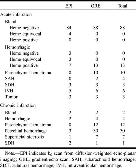
    Table 1. Number and type of lesions identified on EPI and GRE images

  • Table2

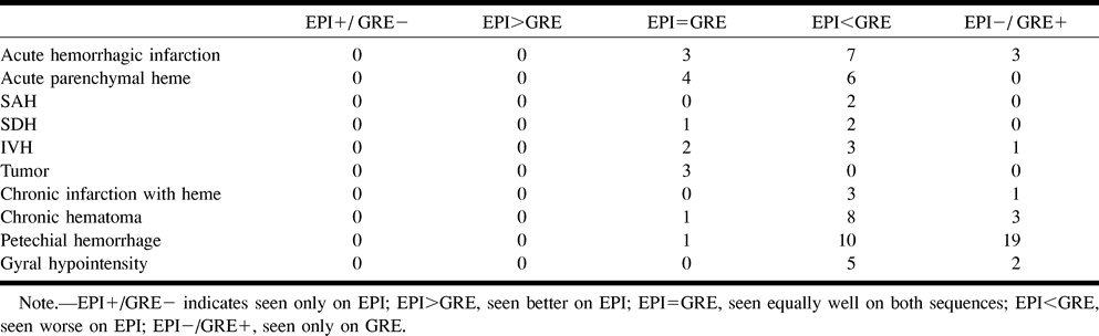
    Table 2. Lesion conspicuity and diagnositc certainty with EPI versus GRE

PreviousNext
Back to top

In this issue

American Journal of Neuroradiology: 22 (7)
American Journal of Neuroradiology
Vol. 22, Issue 7
1 Aug 2001
  • Table of Contents
  • Index by author
Advertisement
Print
Download PDF
Email Article

Thank you for your interest in spreading the word on American Journal of Neuroradiology.

NOTE: We only request your email address so that the person you are recommending the page to knows that you wanted them to see it, and that it is not junk mail. We do not capture any email address.

Enter multiple addresses on separate lines or separate them with commas.
Detection of Intracranial Hemorrhage: Comparison between Gradient-echo Images and b0 Images Obtained from Diffusion-weighted Echo-planar Sequences
(Your Name) has sent you a message from American Journal of Neuroradiology
(Your Name) thought you would like to see the American Journal of Neuroradiology web site.
CAPTCHA
This question is for testing whether or not you are a human visitor and to prevent automated spam submissions.
Cite this article
Doris D. M. Lin, Christopher G. Filippi, Alex B. Steever, Robert D. Zimmerman
Detection of Intracranial Hemorrhage: Comparison between Gradient-echo Images and b0 Images Obtained from Diffusion-weighted Echo-planar Sequences
American Journal of Neuroradiology Aug 2001, 22 (7) 1275-1281;

Citation Manager Formats

  • BibTeX
  • Bookends
  • EasyBib
  • EndNote (tagged)
  • EndNote 8 (xml)
  • Medlars
  • Mendeley
  • Papers
  • RefWorks Tagged
  • Ref Manager
  • RIS
  • Zotero
0 Responses
Respond to this article
Share
Bookmark this article
Detection of Intracranial Hemorrhage: Comparison between Gradient-echo Images and b0 Images Obtained from Diffusion-weighted Echo-planar Sequences
Doris D. M. Lin, Christopher G. Filippi, Alex B. Steever, Robert D. Zimmerman
American Journal of Neuroradiology Aug 2001, 22 (7) 1275-1281;
del.icio.us logo Twitter logo Facebook logo Mendeley logo
  • Tweet Widget
  • Facebook Like
  • Google Plus One

Jump to section

  • Article
    • Abstract
    • Methods
    • Results
    • Discussion
    • Conclusion
    • Acknowledgments
    • Footnotes
    • References
  • Figures & Data
  • Info & Metrics
  • Responses
  • References
  • PDF

Related Articles

  • No related articles found.
  • PubMed
  • Google Scholar

Cited By...

  • An Unusual Late Presentation of Swelling over the Head
  • Distinction between contrast staining and hemorrhage after endovascular stroke treatment: one CT is not enough
  • A review of structural magnetic resonance neuroimaging
  • Contribution of Susceptibility-Weighted Imaging to Acute Stroke Assessment
  • Magnetic Resonance Imaging Improves Detection of Intracerebral Hemorrhage Over Computed Tomography After Intra-Arterial Thrombolysis
  • Stroke Magnetic Resonance Imaging Is Accurate in Hyperacute Intracerebral Hemorrhage: A Multicenter Study on the Validity of Stroke Imaging
  • Intra-Arterial Thrombolysis in 100 Patients With Acute Stroke Due to Middle Cerebral Artery Occlusion
  • Crossref
  • Google Scholar

This article has not yet been cited by articles in journals that are participating in Crossref Cited-by Linking.

More in this TOC Section

  • Progression of Microstructural Damage in Spinocerebellar Ataxia Type 2: A Longitudinal DTI Study
  • Usefulness of Quantitative Susceptibility Mapping for the Diagnosis of Parkinson Disease
  • Evaluating the Effects of White Matter Multiple Sclerosis Lesions on the Volume Estimation of 6 Brain Tissue Segmentation Methods
Show more Brain

Similar Articles

Advertisement

Indexed Content

  • Current Issue
  • Accepted Manuscripts
  • Article Preview
  • Past Issues
  • Editorials
  • Editors Choice
  • Fellow Journal Club
  • Letters to the Editor

Cases

  • Case Collection
  • Archive - Case of the Week
  • Archive - Case of the Month
  • Archive - Classic Case

Special Collections

  • Special Collections

Resources

  • News and Updates
  • Turn around Times
  • Submit a Manuscript
  • Author Policies
  • Manuscript Submission Guidelines
  • Evidence-Based Medicine Level Guide
  • Publishing Checklists
  • Graphical Abstract Preparation
  • Imaging Protocol Submission
  • Submit a Case
  • Become a Reviewer/Academy of Reviewers
  • Get Peer Review Credit from Publons

Multimedia

  • AJNR Podcast
  • AJNR SCANtastic
  • Video Articles

About Us

  • About AJNR
  • Editorial Board
  • Not an AJNR Subscriber? Join Now
  • Alerts
  • Feedback
  • Advertise with us
  • Librarian Resources
  • Permissions
  • Terms and Conditions

American Society of Neuroradiology

  • Not an ASNR Member? Join Now

© 2025 by the American Society of Neuroradiology All rights, including for text and data mining, AI training, and similar technologies, are reserved.
Print ISSN: 0195-6108 Online ISSN: 1936-959X

Powered by HighWire