Advertisement

Main menu

  • Home
  • Content
    • Current Issue
    • Accepted Manuscripts
    • Article Preview
    • Past Issue Archive
    • AJNR Case Collection
    • Case of the Week Archive
    • Classic Case Archive
    • Case of the Month Archive
  • Special Collections
    • Spinal CSF Leak Articles (Jan 2020-June 2024)
    • 2024 AJNR Journal Awards
    • Most Impactful AJNR Articles
  • Multimedia
    • AJNR Podcast
    • AJNR Scantastics
    • Video Articles
  • For Authors
    • Submit a Manuscript
    • Author Policies
    • Fast publishing of Accepted Manuscripts
    • Graphical Abstract Preparation
    • Manuscript Submission Guidelines
    • Imaging Protocol Submission
    • Submit a Case for the Case Collection
  • About Us
    • About AJNR
    • Editorial Board
  • More
    • Become a Reviewer/Academy of Reviewers
    • Subscribers
    • Permissions
    • Alerts
    • Feedback
    • Advertisers
    • ASNR Home
  • Other Publications
    • ajnr

User menu

  • Alerts
  • Log in

Search

  • Advanced search
American Journal of Neuroradiology

American Journal of Neuroradiology

ASHNR American Society of Functional Neuroradiology ASHNR American Society of Pediatric Neuroradiology ASSR
  • Alerts
  • Log in

Advanced Search

  • Home
  • Content
    • Current Issue
    • Accepted Manuscripts
    • Article Preview
    • Past Issue Archive
    • AJNR Case Collection
    • Case of the Week Archive
    • Classic Case Archive
    • Case of the Month Archive
  • Special Collections
    • Spinal CSF Leak Articles (Jan 2020-June 2024)
    • 2024 AJNR Journal Awards
    • Most Impactful AJNR Articles
  • Multimedia
    • AJNR Podcast
    • AJNR Scantastics
    • Video Articles
  • For Authors
    • Submit a Manuscript
    • Author Policies
    • Fast publishing of Accepted Manuscripts
    • Graphical Abstract Preparation
    • Manuscript Submission Guidelines
    • Imaging Protocol Submission
    • Submit a Case for the Case Collection
  • About Us
    • About AJNR
    • Editorial Board
  • More
    • Become a Reviewer/Academy of Reviewers
    • Subscribers
    • Permissions
    • Alerts
    • Feedback
    • Advertisers
    • ASNR Home
  • Follow AJNR on Twitter
  • Visit AJNR on Facebook
  • Follow AJNR on Instagram
  • Join AJNR on LinkedIn
  • RSS Feeds

Welcome to the new AJNR, Updated Hall of Fame, and more. Read the full announcements.


AJNR is seeking candidates for the position of Associate Section Editor, AJNR Case Collection. Read the full announcement.


Research ArticleBRAIN

Findings in a Patient with Herpes Simplex Viral Meningitis Associated with Acute Retinal>> Necrosis Syndrome

Yukiko Tada, Kiyoshi Negoro, Mitsunori Morimatsu, Hajimu Makino and Teruo Nishida
American Journal of Neuroradiology August 2001, 22 (7) 1300-1302;
Yukiko Tada
  • Find this author on Google Scholar
  • Find this author on PubMed
  • Search for this author on this site
Kiyoshi Negoro
  • Find this author on Google Scholar
  • Find this author on PubMed
  • Search for this author on this site
Mitsunori Morimatsu
  • Find this author on Google Scholar
  • Find this author on PubMed
  • Search for this author on this site
Hajimu Makino
  • Find this author on Google Scholar
  • Find this author on PubMed
  • Search for this author on this site
Teruo Nishida
  • Find this author on Google Scholar
  • Find this author on PubMed
  • Search for this author on this site
  • Article
  • Figures & Data
  • Info & Metrics
  • Responses
  • References
  • PDF
Loading

Abstract

Summary: We report the case of a 41-year-old man with clinical findings of viral meningitis associated with acute retinal necrosis syndrome in his right eye. MR images showed right optic nerve enlargement and high-intensity signal abnormalities in the region of the left lateral geniculate body and the left occipital lobe.

Acute retinal necrosis (ARN) syndrome usually affects healthy adults. It involves the acute onset of diffuse uveitis, occlusive retinal vasculitis, vitritis, and rapidly progressive retinal necrosis (1). At end-stage, the syndrome often causes retinal detachment, optic atrophy, and severe visual loss. The origin of the syndrome is thought to be intraocular infection caused by varicella zoster virus (VZV) or herpes simplex virus (HSV) (2). We report the case of a 41-year-old man with viral meningitis associated with ARN syndrome and describe the MR findings.

Case Report

A 41-year-old man was admitted to our hospital on July 12, 1999, because of gradual, progressive loss of visual acuity and narrowing of the visual field of the right eye over the previous 3 days. Headache and periocular pain preceded the symptoms by a few days. He had no relevant family history of disease and no significant events in his personal history.

Ophthalmologic examination on admission showed visual acuity of 20/100 in the right eye and 20/20 in the left eye. The right eye showed a ciliary injection, cells and flare in the anterior chamber, and vitreous opacity. Funduscopic examination revealed characteristic scattered yellowish-white retinal exudates, retinal hemorrhage, retinal vasculitis, and edema of the optic disk. These abnormal retinal findings were more severe on the nasal side than on the aural side. The left fundus was uninvolved.

The patient was alert and well oriented, and neurologic examination revealed neck stiffness and Kernig's sign. Tendon reflexes were normal, and Babinski's sign was absent. Other cranial nerves, the cerebellum, sensation, and the extrapyramidal system were normal. Routine blood tests, urinalysis, electrocardiography, and chest X-ray all were normal. A lumbar puncture performed on July 19 revealed high intracranial pressure (230 mm H2O), 41 leukocytes/mm3 (100% mononuclear cells), a glucose level of 62 mg/dL (3.4 mmol/L), a protein level of 63 mg/dL (0.63 g/L), and negative cultures for bacteria and virus. Antibodies to HSV and VZV were not detectable in the CSF; however, polymerase chain reaction detected HSV DNA in the aqueous humor obtained on July 12.

The patient was diagnosed as having ARN syndrome and began pharmacotherapy of intravenous acyclovir (2250 mg/day), interferon-∝ (3 million IU/day), oral steroids (40 mg/day), and aspirin (80 mg/day). Retinal changes rapidly worsened. Right visual acuity deteriorated to no light perception by July 16, and then gradually improved to hand-motion perception by July 25.

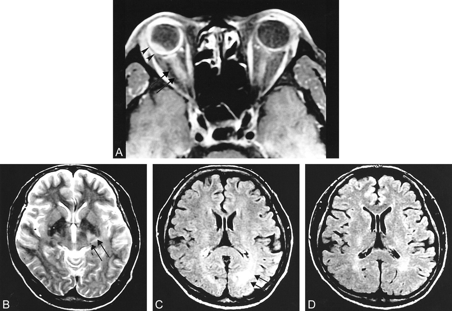
MR studies were performed on July 19 with a 1.5-T system (Magnetom Vision, Siemens-Asahi Medical Technologies, Germany). The MR images of the brain showed right optic-nerve enlargement and a crescent-shaped collection of presumed fluid along the aural side of the right orbit (Fig 1A) on fast short-inversion-time recovery images and high-intensity signal abnormalities in the region of the left lateral geniculate body (Fig 1B) and the left occipital lobe (Fig 1C) on both T2-weighted and fluid-attenuated inversion recovery (FLAIR) images. All signal abnormalities had disappeared by August 2 (Fig 1D).

fig 1.
  • Download figure
  • Open in new tab
  • Download powerpoint
fig 1.

MR images (A–C obtained on July 19; D, on August 2).

A, Axial fast short-inversion-time recovery image with parameters of 520/20 (TR/TE) shows right optic-nerve enlargement (arrow) and a crescent-shaped collection of presumed fluid along the aural side of the right orbit (arrowhead), although funduscopic examination revealed greater retinal abnormalities on the nasal side.

B, Axial T2-weighted image with parameters of 4000/100 shows high-intensity signal in the region of the left lateral geniculate body (arrow).

C, Axial FLAIR image with parameters of 10000/120/2300 (TR/TE/TI) shows high-intensity signal in the region of the left occipital lobe (arrow).

D, Abnormal signal changes observed in the left occipital lobe are no longer visible on FLAIR image taken August 2.

Discussion

Previous case reports have described the occurrence of ARN syndrome after HSV encephalitis (3, 4). On the basis of electron microscopic analysis of the distribution of HSV antigens in the retina, optic nerve, and brain of patients with herpetic retinitis and encephalitis, the virus is generally assumed to spread by retrograde axonal transport through the optic nerve and to invade retinal tissue directly. In contrast, viral meningitis following ARN syndrome is rare.

Three previous reports of CT or MR findings of ARN syndrome have noted optic-nerve enlargement and abnormal enhancement of the optic nerves and chiasm on postcontrast T1-weighted images, and high-intensity signal abnormalities on T2-weighted images in the region of the optic tract and lateral geniculate body (5–7). In our case, the MR images showed signal abnormalities in the region of the lateral geniculate body and occipital lobe that were greater on the left side even though the main lesion was located on the retina of the right eye. These signal changes along the visual pathway might have been construed to show neuronal degeneration, which has been termed Wallerian or transneuronal degeneration (8).

Anatomically, optic nerves and optic tracts contain the axons of the retinal ganglion cells. Visual information is relayed at the lateral geniculate body and projected onto the visual cortex in the occipital lobe through optic radiation. The optic nerves on the nasal side of the retina cross at the chiasm. The laterality of the MR findings in our case was compatible with the results of funduscopic examination, which revealed greater retinal abnormalities on the nasal side of the right eye, although MR images of the eye did show retinal detachment or subretinal fluid collection on the aural side of the right eye (Fig 1A).

Wallerian degeneration results from injury to the cell body or the proximal segment of the axon, with involution of the distal axon and its myelin sheath. Transneuronal degeneration refers to transsynaptic degeneration of the axons and myelin sheath associated with a lesion in either the afferent or efferent connection to the involved neuron. Observations of these types of degeneration in humans have so far been limited to the visual pathway, corticospinal tract, limbic system (mamillary body or fornix atrophy), dentate-rubro-olivary pathway (hypertrophic olivary degeneration), and crossed cerebellar atrophy (8). Regarding the visual pathway, MR images have revealed changes after anterograde degeneration of optic radiations from lesions involving the lateral geniculate body (9). Retrograde degeneration can cause atrophy of the optic nerves or optic tracts from postgeniculate lesions (eg, arteriovenous malformation and periventricular leukomalacia) (10, 11).

According to clinical observations in humans, MR studies cannot detect changes from Wallerian degeneration until about 4 weeks after injury to the fibers (12). From about 4 to 14 weeks after injury, the degenerated tracts appear as low-intensity signal changes on T2-weighted images, which suggests a transient increase in the lipid-protein ratio. After that, the degeneration causes high-intensity signal changes, which are attributed to glial proliferation and increased water content. The MR findings in our case showed high-intensity signal abnormalities on T2-weighted and FLAIR images obtained 7 days after onset, and these signal changes disappeared within about 3 weeks. These findings are incompatible with ordinary changes due to Wallerian degeneration. Therefore, the signal changes of the lateral geniculate body might have been caused mainly by the transient edema of the fibers along the visual pathway after the spread of inflammation or by direct viral invasion from the retina through the optic nerve. The resolution of the edema within the lateral geniculate body and occipital cortex without any evidence of dysfunction or MR findings of encephalomalacia appears impossible, however, and supports Wallerian degeneration as opposed to the edematous hypothesis.

Footnotes

  • 1 Address reprint requests to Yukiko Tada, Department of Neurology, Yamaguchi University School of Medicine, 1-1-1 Minami-Kogushi, Ube, Yamaguchi 755-8505, Japan.

References

  1. ↵
    Urayama A, Yamada N, Sasaki T, et al. Unilateral acute uveitis with retinal periarteritis and detachment. Jpn J Clin Ophthalmol 1971;25:607-619
  2. ↵
    Culbertson WW, Blumenkranz MS, Haines H, Gass DM, Mitchell KB, Norton EW. The acute retinal necrosis syndrome. Part 2: histopathology and etiology. Ophthalmology 1982;89:1317-1325
    PubMed
  3. ↵
    Minckler DS, McLean EB, Shaw CM, Hendrickson A. Herpesvirus hominis encephalitis and retinitis. Arch Ophthalmol 1976;94:89-95
    CrossRefPubMed
  4. Johnson BL, Wisotzkey HM. Neuroretinitis associated with herpes simplex encephalitis in an adult. Am J Ophthalmol 1977;83:481-489
    PubMed
  5. Sergott RC, Belmont JB, Savino PJ, Fisher DH, Bosley TM, Schatz NJ. Optic nerve involvement in the acute retinal necrosis syndrome. Arch Ophthalmol 1985;103:1160-1162
    CrossRefPubMed
  6. Farrell TA, Wolf MD, Folk JC, Pulido JS, Yuh WT. Magnetic resonance imaging in a patient with herpes zoster keratouveitis and contralateral acute retinal necrosis. Am J Ophthalmol 1991;112:735-736
    PubMed
  7. Lewis ML, Culbertson WW, Post JD, Miller D, Kokame GT, Dix RD. Herpes simplex virus type 1: a cause of the acute retinal necrosis syndrome. Ophthalmology 1989;96:875-878
    PubMed
  8. ↵
    Yamada K, Patel U, Shrier DA, Tanaka H, Chang JK, Numaguchi Y. MR imaging of CNS tractopathy: Wallerian and transneuronal degeneration. Am J Roentgenol 1998;171:813-818
    PubMed
  9. ↵
    Savoiardo M, Pareyson D, Grisoli M, Forester M, D'Incerti L, Farina L. The effects of wallerian degeneration of the optic radiations demonstrated by MRI. Neuroradiology 1992;34:323-325
    CrossRefPubMed
  10. ↵
    Kupersmith MJ, Vargas M, Hoyt WF, Berenstein A. Optic tract atrophy with cerebral arteriovenous malformations: direct and transsynaptic degeneration. Neurology 1994;44:80-83
    Abstract/FREE Full Text
  11. Uggetti C, Egitto MG, Fazzi E, et al. Transsynaptic degeneration of lateral geniculate bodies in blind children: in vivo MR demonstration. AJNR Am J Neuroradiol 1997;18:233-238
    Abstract
  12. ↵
    Kuhn MJ, Mikulis DJ, Ayoub DM, Kosofsky BE, Davis K, Taveras JM. Wallerian degeneration after cerebral infarction: evaluation with sequential MR imaging. Radiology 1989;172:179-182
    PubMed
  • Received February 23, 2000.
  • Accepted after revision January 16, 2001.
  • Copyright © American Society of Neuroradiology
View Abstract
PreviousNext
Back to top

In this issue

American Journal of Neuroradiology: 22 (7)
American Journal of Neuroradiology
Vol. 22, Issue 7
1 Aug 2001
  • Table of Contents
  • Index by author
Advertisement
Print
Download PDF
Email Article

Thank you for your interest in spreading the word on American Journal of Neuroradiology.

NOTE: We only request your email address so that the person you are recommending the page to knows that you wanted them to see it, and that it is not junk mail. We do not capture any email address.

Enter multiple addresses on separate lines or separate them with commas.
Findings in a Patient with Herpes Simplex Viral Meningitis Associated with Acute Retinal>> Necrosis Syndrome
(Your Name) has sent you a message from American Journal of Neuroradiology
(Your Name) thought you would like to see the American Journal of Neuroradiology web site.
CAPTCHA
This question is for testing whether or not you are a human visitor and to prevent automated spam submissions.
Cite this article
Yukiko Tada, Kiyoshi Negoro, Mitsunori Morimatsu, Hajimu Makino, Teruo Nishida
Findings in a Patient with Herpes Simplex Viral Meningitis Associated with Acute Retinal>> Necrosis Syndrome
American Journal of Neuroradiology Aug 2001, 22 (7) 1300-1302;

Citation Manager Formats

  • BibTeX
  • Bookends
  • EasyBib
  • EndNote (tagged)
  • EndNote 8 (xml)
  • Medlars
  • Mendeley
  • Papers
  • RefWorks Tagged
  • Ref Manager
  • RIS
  • Zotero
0 Responses
Respond to this article
Share
Bookmark this article
Findings in a Patient with Herpes Simplex Viral Meningitis Associated with Acute Retinal>> Necrosis Syndrome
Yukiko Tada, Kiyoshi Negoro, Mitsunori Morimatsu, Hajimu Makino, Teruo Nishida
American Journal of Neuroradiology Aug 2001, 22 (7) 1300-1302;
del.icio.us logo Twitter logo Facebook logo Mendeley logo
  • Tweet Widget
  • Facebook Like
  • Google Plus One

Jump to section

  • Article
    • Abstract
    • Case Report
    • Discussion
    • Footnotes
    • References
  • Figures & Data
  • Info & Metrics
  • Responses
  • References
  • PDF

Related Articles

  • No related articles found.
  • PubMed
  • Google Scholar

Cited By...

  • Acute retinal necrosis by herpes simplex virus type 1: an unusual presentation of a primary infection
  • Crossref
  • Google Scholar

This article has not yet been cited by articles in journals that are participating in Crossref Cited-by Linking.

More in this TOC Section

  • Progression of Microstructural Damage in Spinocerebellar Ataxia Type 2: A Longitudinal DTI Study
  • Usefulness of Quantitative Susceptibility Mapping for the Diagnosis of Parkinson Disease
  • Evaluating the Effects of White Matter Multiple Sclerosis Lesions on the Volume Estimation of 6 Brain Tissue Segmentation Methods
Show more Brain

Similar Articles

Advertisement

Indexed Content

  • Current Issue
  • Accepted Manuscripts
  • Article Preview
  • Past Issues
  • Editorials
  • Editors Choice
  • Fellow Journal Club
  • Letters to the Editor

Cases

  • Case Collection
  • Archive - Case of the Week
  • Archive - Case of the Month
  • Archive - Classic Case

Special Collections

  • Special Collections

Resources

  • News and Updates
  • Turn around Times
  • Submit a Manuscript
  • Author Policies
  • Manuscript Submission Guidelines
  • Evidence-Based Medicine Level Guide
  • Publishing Checklists
  • Graphical Abstract Preparation
  • Imaging Protocol Submission
  • Submit a Case
  • Become a Reviewer/Academy of Reviewers
  • Get Peer Review Credit from Publons

Multimedia

  • AJNR Podcast
  • AJNR SCANtastic
  • Video Articles

About Us

  • About AJNR
  • Editorial Board
  • Not an AJNR Subscriber? Join Now
  • Alerts
  • Feedback
  • Advertise with us
  • Librarian Resources
  • Permissions
  • Terms and Conditions

American Society of Neuroradiology

  • Not an ASNR Member? Join Now

© 2025 by the American Society of Neuroradiology All rights, including for text and data mining, AI training, and similar technologies, are reserved.
Print ISSN: 0195-6108 Online ISSN: 1936-959X

Powered by HighWire