
Article Figures & Data
Figures
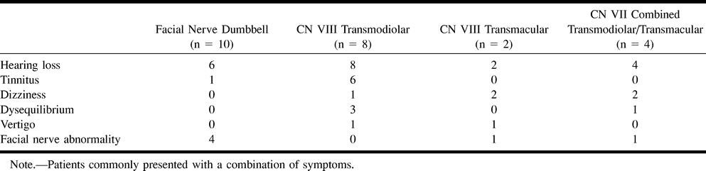
- fig 1.
Dumbbell facial nerve schwannoma. The facial nerve schwannoma (FNS) dumbbells through an enlarged labyrinthine facial canal to involve the geniculate ganglion (GG). Minimal extension of the schwannoma may be present along the greater superficial petrosal nerve (GSPN). The tympanic segment of the facial nerve (TS) is usually not involved. The cisternal portion of the facial nerve (FN) is seen as it courses toward the IAC through the porus acusticus (PA), the entrance to the IAC. Used by permission (37).fig 2. Transmodiolar dumbbell schwannoma. The transmodiolar schwannoma (TMS) extends through the cochlear aperture (CA) and modiolus into the cochlea. The schwannoma involves the cochlear nerve (CN). The normal superior vestibular nerve (SVN) and inferior vestibular nerve (IVN) are present within the IAC. V: Vestibule. Used by permission (37)
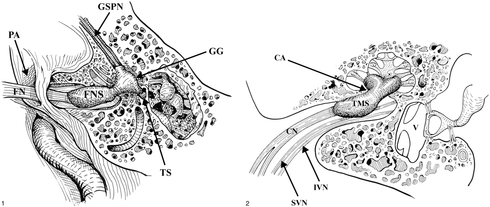
- fig 3.
Dumbbell facial nerve schwannoma.
A, Axial enhanced T1-weighted MR image (800/27/2) at the level of the IAC shows an avidly enhancing mass involving the IAC (open arrow) with an enhancing “tail” involving the labyrinthine segment of the facial nerve (white arrow) as it courses toward the geniculate ganglion.
B, Axial high-resolution FSE T2 MR image (4000/130/1) at the level of the IAC shows a hypointense mass filling the IAC (white arrow) displacing the normal cerebrospinal fluid. The tympanic segment of the facial nerve is barely visible in its bony canal (curved white arrow).
C, Axial high-resolution FSE T2-weighted MR image (4000/130/1) just cephalad to B shows the enlarged labyrinthine segment mass (curved white arrow) as it courses toward the geniculate ganglion. The hypointense intracanalicular portion is again seen (white arrow).
D, Axial CT image at the level of C confirms the enlarged labyrinthine portion of the facial nerve canal (black arrow). Used by permission (37).
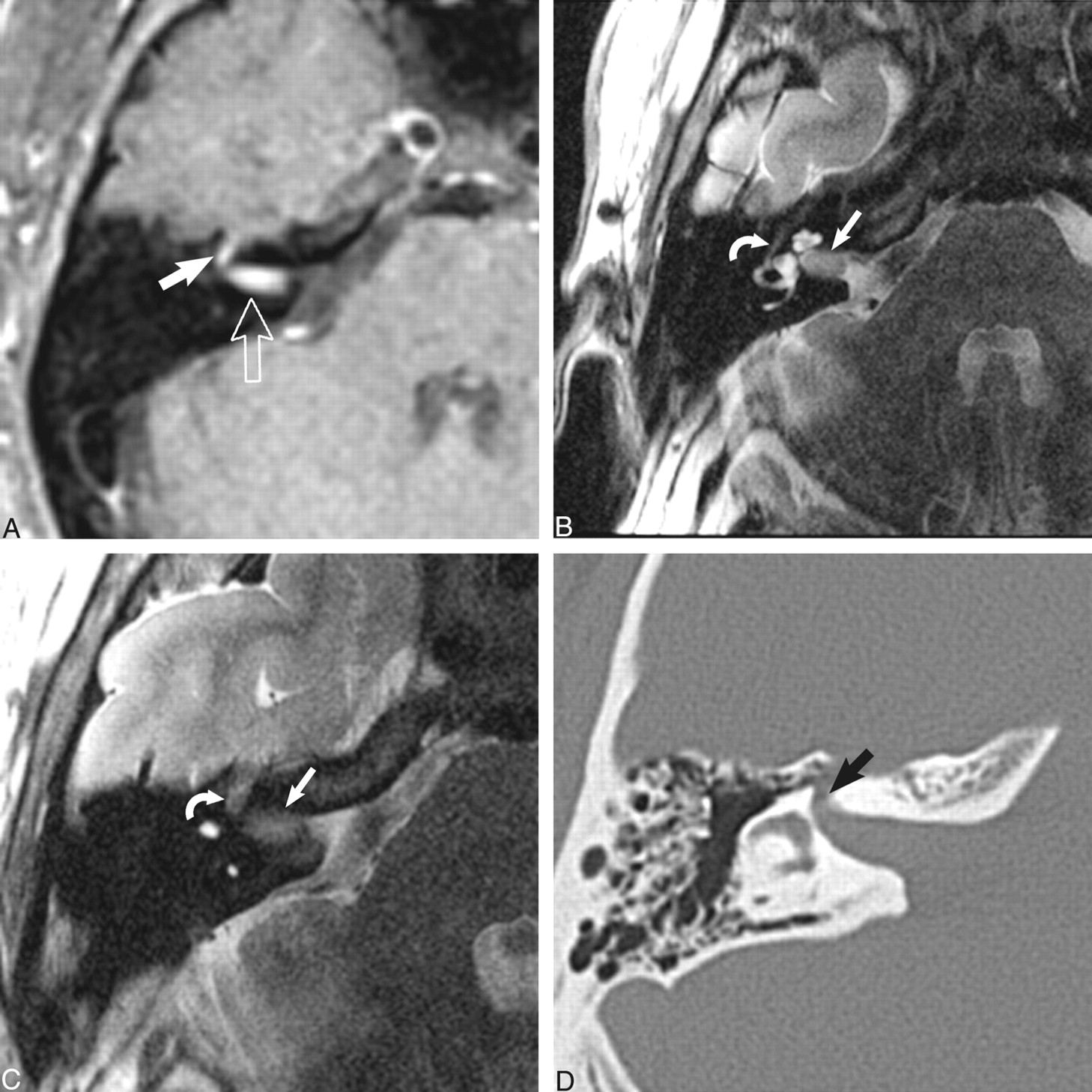
- fig 4.
Transmodiolar dumbbell schwannoma.
A, Axial contrast-enhanced T1-weighted MR image (800/27/2) at the level of the IAC shows an enhancing mass in the fundus of the IAC (open arrow) with extension into the cochlea (large white arrow). The schwannoma extends through the cochlear aperture (small white arrow) and modiolus.
B, Axial high-resolution FSE T2-weighted MR image (4000/102/6) at the level of the IAC shows the transmodiolar schwannoma as a hypointense mass that has replaced the normal fluid of the cochlea (large white arrow). The intracanalicular portion of the mass displaced the normal fluid of the fundus of the IAC (curved white arrow). The vestibule is normal and fluid filled (small white arrow).
C, Axial high-resolution FSE T2-weighted MR image (4000/102/6) shows the normal right membranous labyrinthine structures. The normal cochlea (large open arrow) and vestibule (white arrow) are fluid-filled. The central bony modiolus (curved white arrow) through which the cochlear nerve fibers travel is well seen. The cerebellar flocculus (small open arrow) is a common cerebellopontine angle “pseudomass.” Used by permission (37).
- fig 5.
Transmacular dumbbell schwannoma.
A, Axial contrast-enhanced T1-weighted MR image (800/27/2) at the level of the IAC shows an enhancing mass in the fundus of the IAC (large white arrow) with extension into the vestibule (small white arrow).
B, Axial high-resolution FSE T2-weighted MR image (4000/102/6) at the level of the IAC shows a small nodular mass in the fundus of the IAC (small white arrow). The hypointense mass extends into the vestibule (large white arrow). In addition, the normal left fluid-filled cochlea (large open white arrow) and cochlear aperture (small open white arrow) are seen. Used by permission (37).
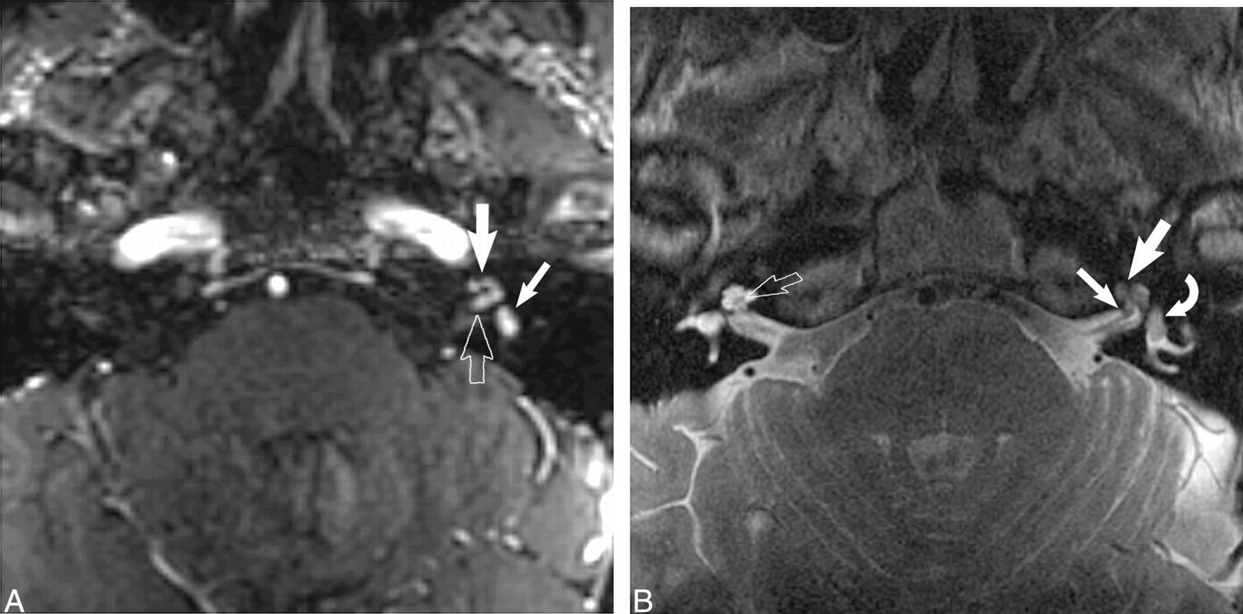
- fig 6.
Combined transmodiolar/transmacular schwannoma.
A, Axial enhanced T1-weighted MR image (39/14/2) at the level of the IAC shows an enhancing mass in the fundus of the IAC (open arrow) with extension into the cochlea (large white arrow) and vestibule (small white arrow).
B, Axial high-resolution FSE T2-weighted MR image (4000/102/6) at the level of the IAC shows the hypointense mass in the cochlea (large white arrow) and vestibule (curved white arrow). A small nodule is also seen within the fundus of the IAC (small white arrow). The normal modiolus is well seen on the right (open right arrow). Used by permission (37).
- fig 7.
Translabyrinthine schwannoma.
A, Axial enhanced T1-weighted MR image (800/27/2) at the level of the IAC shows a large enhancing mass within the cerebellopontine angle-IAC (open arrow) with extension into the cochlea (black arrow) and vestibule (white arrow).
B, Axial CT shows the soft-tissue mass extending into the middle ear (small black arrow) through an enlarged round window (large black arrow). Used by permission (37).


Tables

Presenting symptoms
In this issue


Jump to section
Related Articles
Cited By...
- No citing articles found.