
Article Figures & Data
Figures
- fig 1.
Flare enhancement. MR images obtained preoperatively and on day 1; weeks 5, 9, and 25; and year 4 postoperatively show development of a strong nodular flare very early with massive edema around the precentral resection cavity. Later images show progressive shrinking, leaving scarce residual evidence after 1 year (enhanced T1-weighted coronal and sagittal MR images, patient 1). fig 2. Flare enhancement. MR images obtained on day 1; weeks 2, 4, and 9; and year 2 after surgery. A strong flare develops after complete resection of all enhancing tissue within the frontal lobe. Later, there is shrinking and nearly complete disappearance of the process (enhanced T1-weighted axial MR images, patient 21)
- fig 3.
Flare enhancement and tumor relapse after complete resection. MR images obtained on day 1 and weeks 2, 9, and 41 postoperatively show development of a strong flare after resection of all enhancing tissue within the temporal lobe. There is initial regression of flare, but eventually there is recurrence of local tumor (enhanced T1-weighted axial MR images, patient 18).fig 4. Flare enhancement and tumor relapse after incomplete resection. MR images on day 1 and weeks 2, 4, and 33 after surgery show development of a strong flare after incomplete tumor resection with two remnants within the temporal lobe. The flare enhancement regresses at first, but eventually the tumor recurs from the remnants (enhanced T1-weighted axial MR images, patient 20).fig 5. Benign postsurgical enhancement. MR images from day 1 and weeks 2, 4, and 17 after tumor resection show development of thin, linear, benign enhancement and local tumor recurrence within the temporooccipital region (enhanced T1-weighted axial images, patient 19)
- fig 6.
Volume curves of enhancing tissue during the postoperative course of four patients with flare. There is an early and quick increase in enhancement volume, a high peak at 4 weeks, and slow but continuous decrease during the following months. In patients without tumor recurrence, flare enhancement disappears within 1 year (patients 1 [triangles] and 22 [squares]). In case of tumor recurrence, the regression of flare enhancement is overcome by increasing tumor enhancement (patients 18 [diamonds] and 20 [circles]). The “Screen” point at the bar reflects the preoperative volume of enhancing tumor. For presentation, data for the postoperative images on day 1 or 2 are fixed to week 0, and note the nonlinear time axis.fig 7. Volume curves of benign postsurgical enhancement and residual tumor in two patients with benign postsurgical enhancement and two patients with residual tumor. For benign postsurgical enhancement, there is a delayed and slow increase in enhancement volume, stabilization at a plateau (instead of reaching a peak), and no significant regression. In the later course, the curve is moving upward because of recurrent tumor enhancement (lower two curves; patients 2 [squares] and 19 [diamonds]). Patients with residual tumor without flare or benign postsurgical enhancement show a continuous increase in enhancement volume because of tumor progression (upper two curves; patients 6 [triangles] and 7 [circles]). The “Screen” point at the bar reflects the preoperative enhancing tumor volume. For presentation, data for the postoperative images on day 1 or 2 are fixed to week 0, and note the nonlinear time axis
- fig 8.
Flare enhancement at IMT-SPECT. Scans obtained preoperatively and on day 1, week 9, and year 1 postoperatively show enhancement on MR images (upper line) and amino acid uptake on IMT-SPECT scans (lower line). Although preoperative tumor enhancement on MR images corresponds with an increased amino acid uptake on SPECT scans, the postoperative flare on MR images shows no raised amino acid uptake on SPECT scans (enhanced T1-weighted sagittal MR images and IMT-SPECT scans, patient 1).fig 9. Flare enhancement and residual tumor at IMT-SPECT. Images obtained on day 1 and weeks 2 and 33 postoperatively show enhancement on MR images (upper line) and amino acid uptake on IMT-SPECT scans (lower line). The preoperative MR image shows a large, temporal, enhancing tumor mass. On day 1, two enhancing lesions are visible, which are completely incorporated into a strong flare enhancement at week 2. Tumor enhancement and flare enhancement are indistinguishable on MR images. The SPECT scan at week 2 reveals two “hot” spots of amino acid uptake corresponding to the two tumor remnants visible on MR images at day 1. By week 33, a nodular tumor recurrence has developed from the area of the two tumor remnants (enhanced T1-weighted axial MR images and IMT-SPECT scans, patient 20).fig 10. Flare enhancement and histologic findings. Preoperative MR image shows a large, precentral tumor relapse. At 2 weeks after complete resection, a moderate flare enhancement has developed at the margin of the large resection cavity. At week 4, multiple biopsies from the resection cavity wall (obtained at autopsy) show strong infiltration by inflammatory cells (tissue-resident macrophages and strong perivascular cuffs of lymphocytes) but no tumor (contrast-enhanced T1-weighted sagittal MR images; CD-68 immunostaining, magnetization × 20; patient 3)


Tables
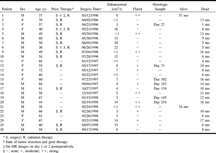
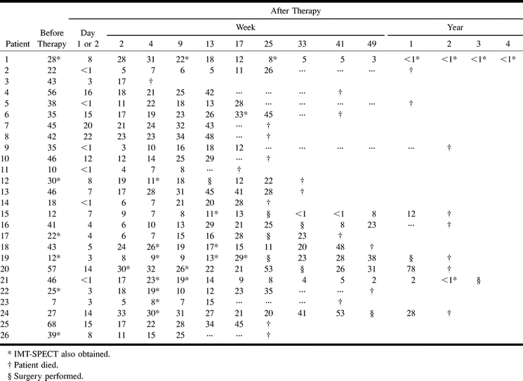
 TABLE 1:
Course and outcome of gene therapy treatments in 26 consecutive patients
In this issue


Jump to section
Related Articles
Cited By...
- Modified RANO, Immunotherapy RANO, and Standard RANO Response to Convection-Enhanced Delivery of IL4R-Targeted Immunotoxin MDNA55 in Recurrent Glioblastoma
- Updated Response Assessment Criteria for High-Grade Gliomas: Response Assessment in Neuro-Oncology Working Group
- End Point Assessment in Gliomas: Novel Treatments Limit Usefulness of Classical Macdonald's Criteria
- Comparison of O-(2-18F-Fluoroethyl)-L-Tyrosine PET and 3-123I-Iodo-{alpha}-Methyl-L-Tyrosine SPECT in Brain Tumors