Advertisement

Main menu

  • Home
  • Content
    • Current Issue
    • Accepted Manuscripts
    • Article Preview
    • Past Issue Archive
    • Video Articles
    • AJNR Case Collection
    • Case of the Week Archive
    • Case of the Month Archive
    • Classic Case Archive
  • Special Collections
    • AJNR Awards
    • Low-Field MRI
    • Alzheimer Disease
    • ASNR Foundation Special Collection
    • Photon-Counting CT
    • View All
  • Multimedia
    • AJNR Podcasts
    • AJNR SCANtastic
    • Trainee Corner
    • MRI Safety Corner
    • Imaging Protocols
  • For Authors
    • Submit a Manuscript
    • Submit a Video Article
    • Submit an eLetter to the Editor/Response
    • Manuscript Submission Guidelines
    • Statistical Tips
    • Fast Publishing of Accepted Manuscripts
    • Graphical Abstract Preparation
    • Imaging Protocol Submission
    • Author Policies
  • About Us
    • About AJNR
    • Editorial Board
    • Editorial Board Alumni
  • More
    • Become a Reviewer/Academy of Reviewers
    • Subscribers
    • Permissions
    • Alerts
    • Feedback
    • Advertisers
    • ASNR Home

User menu

  • Alerts
  • Log in

Search

  • Advanced search
American Journal of Neuroradiology

American Journal of Neuroradiology

ASHNR American Society of Functional Neuroradiology ASHNR American Society of Pediatric Neuroradiology ASSR
  • Alerts
  • Log in

Advanced Search

  • Home
  • Content
    • Current Issue
    • Accepted Manuscripts
    • Article Preview
    • Past Issue Archive
    • Video Articles
    • AJNR Case Collection
    • Case of the Week Archive
    • Case of the Month Archive
    • Classic Case Archive
  • Special Collections
    • AJNR Awards
    • Low-Field MRI
    • Alzheimer Disease
    • ASNR Foundation Special Collection
    • Photon-Counting CT
    • View All
  • Multimedia
    • AJNR Podcasts
    • AJNR SCANtastic
    • Trainee Corner
    • MRI Safety Corner
    • Imaging Protocols
  • For Authors
    • Submit a Manuscript
    • Submit a Video Article
    • Submit an eLetter to the Editor/Response
    • Manuscript Submission Guidelines
    • Statistical Tips
    • Fast Publishing of Accepted Manuscripts
    • Graphical Abstract Preparation
    • Imaging Protocol Submission
    • Author Policies
  • About Us
    • About AJNR
    • Editorial Board
    • Editorial Board Alumni
  • More
    • Become a Reviewer/Academy of Reviewers
    • Subscribers
    • Permissions
    • Alerts
    • Feedback
    • Advertisers
    • ASNR Home
  • Follow AJNR on Twitter
  • Visit AJNR on Facebook
  • Follow AJNR on Instagram
  • Join AJNR on LinkedIn
  • RSS Feeds

AJNR Awards, New Junior Editors, and more. Read the latest AJNR updates

Research ArticleBrain

Tentorial Enhancement on MR Images Is a Sign of Cavernous Sinus Involvement in Patients with Sellar Tumors

Yoko Nakasu, Satoshi Nakasu, Ryuta Ito, Ko-ichi Mitsuya, Osamu Fujimoto and Akira Saito
American Journal of Neuroradiology September 2001, 22 (8) 1528-1533;
Yoko Nakasu
  • Find this author on Google Scholar
  • Find this author on PubMed
  • Search for this author on this site
Satoshi Nakasu
  • Find this author on Google Scholar
  • Find this author on PubMed
  • Search for this author on this site
Ryuta Ito
  • Find this author on Google Scholar
  • Find this author on PubMed
  • Search for this author on this site
Ko-ichi Mitsuya
  • Find this author on Google Scholar
  • Find this author on PubMed
  • Search for this author on this site
Osamu Fujimoto
  • Find this author on Google Scholar
  • Find this author on PubMed
  • Search for this author on this site
Akira Saito
  • Find this author on Google Scholar
  • Find this author on PubMed
  • Search for this author on this site
  • Article
  • Figures & Data
  • Info & Metrics
  • Responses
  • References
  • PDF
Loading

Article Figures & Data

Figures

  • Tables
  • fig 1.
    • Download figure
    • Open in new tab
    • Download powerpoint
    fig 1.

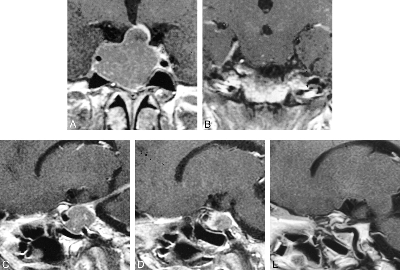
    Case 1: 46-year-old woman with a growth hormone–secreting adenoma.

    A, Coronal T1-weighted MR image after administration of contrast material shows a large sellar tumor compressing the right cavernous sinus. The left cavernous sinus is enhanced and normal in shape.

    B, T1-weighted MR image shows clearly enhanced tentorium on the right side.

    C and D, Sagittal T1-weighted images show linear enhancement of the tentorium from the posterior portion of the cavernous sinus on the right side (C). Note asymmetric tentorial enhancement sign.

    E, Postoperative T1-weighted MR image after administration of contrast material shows enlarged posterior part of the cavernous sinus; however, no tentorial enhancement was seen preoperatively (C).

  • fig 2.
    • Download figure
    • Open in new tab
    • Download powerpoint
    fig 2.

    Case 2: 26-year-old man with a nonfunctioning adenoma.

    A, Coronal T1-weighted MR image shows a sellar tumor invading the left cavernous sinus.

    B and C, Sagittal T1-weighted MR images show tentorial enhancement in connection with the cavernous sinus, more prominent on the left (C) than on the right (B).

  • fig 3.
    • Download figure
    • Open in new tab
    • Download powerpoint
    fig 3.

    Case 3: 64-year-old man with a chordoma.

    A and B, Coronal T1-weighted MR images show sellar tumor infiltrating into both cavernous sinuses and into the right tentorium.

    C and D, Sagittal T1-weighted MR images show bilateral thick tentorial enhancement with dominance on the right side.

Tables

  • Figures
  • TABLE 1:
    • View popup
    • Download powerpoint
    TABLE 1:

    Sellar tumors and their cavernous sinus involvement

  • TABLE 2:
    • View popup
    • Download powerpoint
    TABLE 2:

    Sellar tumors and dural enhancement

  • TABLE 3:
    • View popup
    • Download powerpoint
    TABLE 3:

    Asymmetric tentorial enhancement and cavernous sinus involvement of sellar tumors

PreviousNext
Back to top

In this issue

American Journal of Neuroradiology: 22 (8)
American Journal of Neuroradiology
Vol. 22, Issue 8
1 Sep 2001
  • Table of Contents
  • Index by author
Advertisement
Print
Download PDF
Email Article

Thank you for your interest in spreading the word on American Journal of Neuroradiology.

NOTE: We only request your email address so that the person you are recommending the page to knows that you wanted them to see it, and that it is not junk mail. We do not capture any email address.

Enter multiple addresses on separate lines or separate them with commas.
Tentorial Enhancement on MR Images Is a Sign of Cavernous Sinus Involvement in Patients with Sellar Tumors
(Your Name) has sent you a message from American Journal of Neuroradiology
(Your Name) thought you would like to see the American Journal of Neuroradiology web site.
CAPTCHA
This question is for testing whether or not you are a human visitor and to prevent automated spam submissions.
Cite this article
Yoko Nakasu, Satoshi Nakasu, Ryuta Ito, Ko-ichi Mitsuya, Osamu Fujimoto, Akira Saito
Tentorial Enhancement on MR Images Is a Sign of Cavernous Sinus Involvement in Patients with Sellar Tumors
American Journal of Neuroradiology Sep 2001, 22 (8) 1528-1533;

Citation Manager Formats

  • BibTeX
  • Bookends
  • EasyBib
  • EndNote (tagged)
  • EndNote 8 (xml)
  • Medlars
  • Mendeley
  • Papers
  • RefWorks Tagged
  • Ref Manager
  • RIS
  • Zotero
0 Responses
Respond to this article
Share
Bookmark this article
Tentorial Enhancement on MR Images Is a Sign of Cavernous Sinus Involvement in Patients with Sellar Tumors
Yoko Nakasu, Satoshi Nakasu, Ryuta Ito, Ko-ichi Mitsuya, Osamu Fujimoto, Akira Saito
American Journal of Neuroradiology Sep 2001, 22 (8) 1528-1533;
del.icio.us logo Twitter logo Facebook logo Mendeley logo
  • Tweet Widget
  • Facebook Like
  • Google Plus One

Jump to section

  • Article
    • Abstract
    • Methods
    • Results
    • Discussion
    • Conclusion
    • Acknowledgments
    • Footnotes
    • References
  • Figures & Data
  • Info & Metrics
  • Responses
  • References
  • PDF

Related Articles

  • No related articles found.
  • PubMed
  • Google Scholar

Cited By...

  • A Radiologic Score to Distinguish Autoimmune Hypophysitis from Nonsecreting Pituitary Adenoma Preoperatively
  • Crossref
  • Google Scholar

This article has not yet been cited by articles in journals that are participating in Crossref Cited-by Linking.

More in this TOC Section

  • Usefulness of Quantitative Susceptibility Mapping for the Diagnosis of Parkinson Disease
  • White Matter Alterations in the Brains of Patients with Active, Remitted, and Cured Cushing Syndrome: A DTI Study
  • Qualitative and Quantitative Analysis of MR Imaging Findings in Patients with Middle Cerebral Artery Stroke Implanted with Mesenchymal Stem Cells
Show more Brain

Similar Articles

Advertisement

Indexed Content

  • Current Issue
  • Accepted Manuscripts
  • Article Preview
  • Past Issues
  • Editorials
  • Editor's Choice
  • Fellows' Journal Club
  • Letters to the Editor
  • Video Articles

Cases

  • Case Collection
  • Archive - Case of the Week
  • Archive - Case of the Month
  • Archive - Classic Case

More from AJNR

  • Trainee Corner
  • Imaging Protocols
  • MRI Safety Corner
  • Book Reviews

Multimedia

  • AJNR Podcasts
  • AJNR Scantastics

Resources

  • Turnaround Time
  • Submit a Manuscript
  • Submit a Video Article
  • Submit an eLetter to the Editor/Response
  • Manuscript Submission Guidelines
  • Statistical Tips
  • Fast Publishing of Accepted Manuscripts
  • Graphical Abstract Preparation
  • Imaging Protocol Submission
  • Evidence-Based Medicine Level Guide
  • Publishing Checklists
  • Author Policies
  • Become a Reviewer/Academy of Reviewers
  • News and Updates

About Us

  • About AJNR
  • Editorial Board
  • Editorial Board Alumni
  • Alerts
  • Permissions
  • Not an AJNR Subscriber? Join Now
  • Advertise with Us
  • Librarian Resources
  • Feedback
  • Terms and Conditions
  • AJNR Editorial Board Alumni

American Society of Neuroradiology

  • Not an ASNR Member? Join Now

© 2025 by the American Society of Neuroradiology All rights, including for text and data mining, AI training, and similar technologies, are reserved.
Print ISSN: 0195-6108 Online ISSN: 1936-959X

Powered by HighWire