
Article Figures & Data
Figures
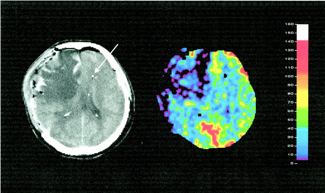
- fig 1.
sXe-enhanced CT blood flow study in a patient with severe head injury. Left, anatomic reference section (arrow indicates tip of TD-rCBF probe); right, corresponding rCBF (mL/100 g per minute) image shows severe right hemispheric hypoperfusion
- fig 2.
Graph shows an original TD-rCBF recording obtained during a 30% sXe/CT blood flow study in a patient with head injury. Baseline (BAS): preinhalation TD-rCBF recording (duration, 42 seconds); wash-in (WI): TD-rCBF during sXe inhalation (duration, 270 seconds)
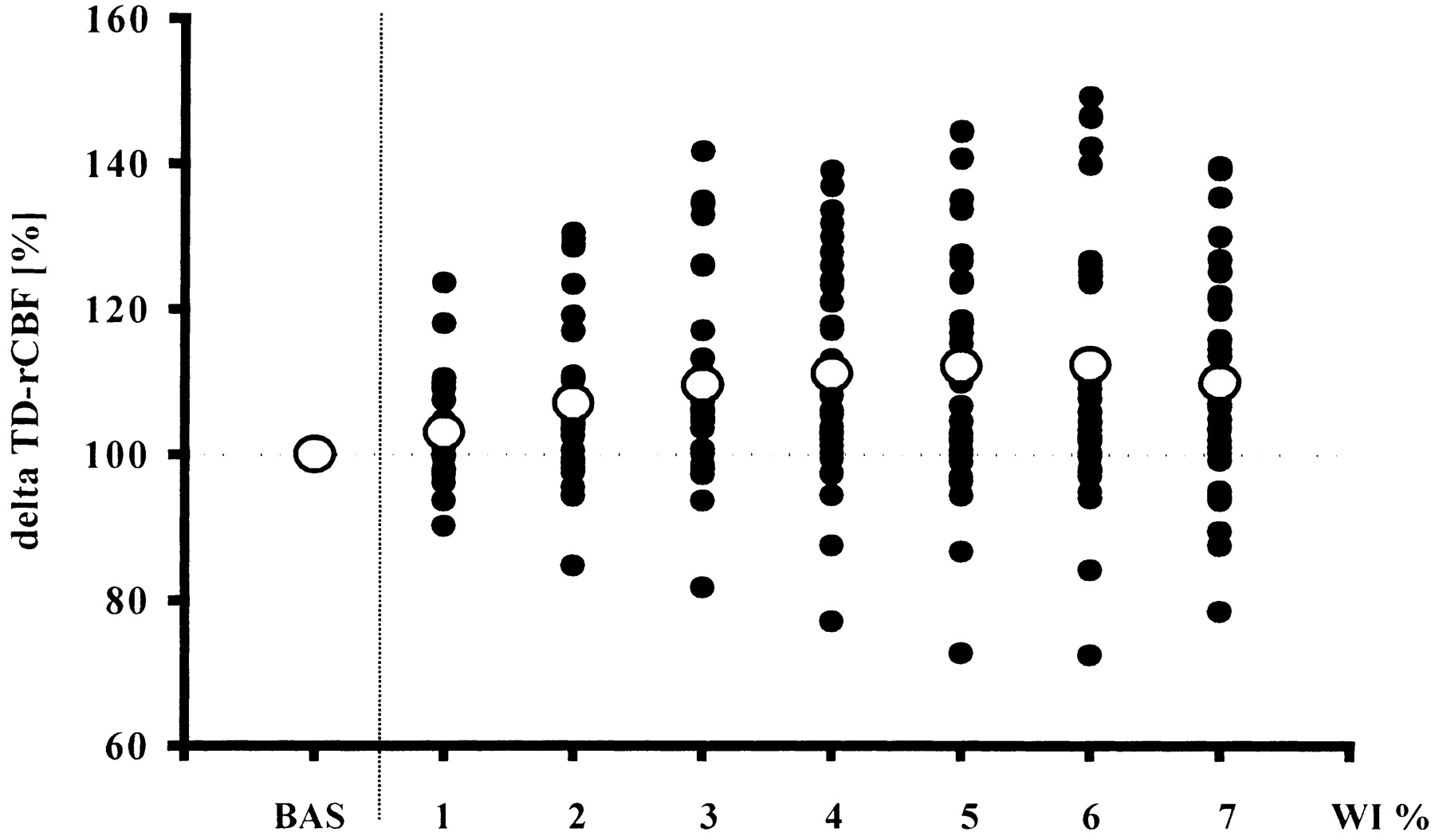
- fig 3.
Relative changes (in %) in baseline values in TD-rCBF due to sXe inhalation as seen in 35 blood flow studies. Solid circles represent individual changes in TD-rCBF at various time periods; open circles depict mean changes in TD-rCBF as compared with baseline. BAS indicates baseline; WI % represents seven periods, each of 38 seconds' duration, of sXe inhalation
- fig 4.
Time course of arterial sXe concentration (open circles) and sXe-induced flow activation (solid circles). The x-axis displays relative values of end-tidal sXe concentration and TD-rCBF, respectively, with each parameter expressed in percentage of maximum response observed


