Advertisement

Main menu

  • Home
  • Content
    • Current Issue
    • Accepted Manuscripts
    • Article Preview
    • Past Issue Archive
    • Video Articles
    • AJNR Case Collection
    • Case of the Week Archive
    • Case of the Month Archive
    • Classic Case Archive
  • Special Collections
    • AJNR Awards
    • Low-Field MRI
    • Alzheimer Disease
    • ASNR Foundation Special Collection
    • Photon-Counting CT
    • View All
  • Multimedia
    • AJNR Podcasts
    • AJNR SCANtastic
    • Trainee Corner
    • MRI Safety Corner
    • Imaging Protocols
  • For Authors
    • Submit a Manuscript
    • Submit a Video Article
    • Submit an eLetter to the Editor/Response
    • Manuscript Submission Guidelines
    • Statistical Tips
    • Fast Publishing of Accepted Manuscripts
    • Graphical Abstract Preparation
    • Imaging Protocol Submission
    • Author Policies
  • About Us
    • About AJNR
    • Editorial Board
    • Editorial Board Alumni
  • More
    • Become a Reviewer/Academy of Reviewers
    • Subscribers
    • Permissions
    • Alerts
    • Feedback
    • Advertisers
    • ASNR Home

User menu

  • Alerts
  • Log in

Search

  • Advanced search
American Journal of Neuroradiology

American Journal of Neuroradiology

ASHNR American Society of Functional Neuroradiology ASHNR American Society of Pediatric Neuroradiology ASSR
  • Alerts
  • Log in

Advanced Search

  • Home
  • Content
    • Current Issue
    • Accepted Manuscripts
    • Article Preview
    • Past Issue Archive
    • Video Articles
    • AJNR Case Collection
    • Case of the Week Archive
    • Case of the Month Archive
    • Classic Case Archive
  • Special Collections
    • AJNR Awards
    • Low-Field MRI
    • Alzheimer Disease
    • ASNR Foundation Special Collection
    • Photon-Counting CT
    • View All
  • Multimedia
    • AJNR Podcasts
    • AJNR SCANtastic
    • Trainee Corner
    • MRI Safety Corner
    • Imaging Protocols
  • For Authors
    • Submit a Manuscript
    • Submit a Video Article
    • Submit an eLetter to the Editor/Response
    • Manuscript Submission Guidelines
    • Statistical Tips
    • Fast Publishing of Accepted Manuscripts
    • Graphical Abstract Preparation
    • Imaging Protocol Submission
    • Author Policies
  • About Us
    • About AJNR
    • Editorial Board
    • Editorial Board Alumni
  • More
    • Become a Reviewer/Academy of Reviewers
    • Subscribers
    • Permissions
    • Alerts
    • Feedback
    • Advertisers
    • ASNR Home
  • Follow AJNR on Twitter
  • Visit AJNR on Facebook
  • Follow AJNR on Instagram
  • Join AJNR on LinkedIn
  • RSS Feeds

AJNR Awards, New Junior Editors, and more. Read the latest AJNR updates

Getting new auth cookie, if you see this message a lot, tell someone!
Research ArticleBrain

Normal Pressure Hydrocephalus: Vascular White Matter Changes on MR Images Must Not Exclude Patients from Shunt Surgery

Mats Tullberg, Christer Jensen, Sven Ekholm and Carsten Wikkelsø
American Journal of Neuroradiology October 2001, 22 (9) 1665-1673;
Mats Tullberg
  • Find this author on Google Scholar
  • Find this author on PubMed
  • Search for this author on this site
Christer Jensen
  • Find this author on Google Scholar
  • Find this author on PubMed
  • Search for this author on this site
Sven Ekholm
  • Find this author on Google Scholar
  • Find this author on PubMed
  • Search for this author on this site
Carsten Wikkelsø
  • Find this author on Google Scholar
  • Find this author on PubMed
  • Search for this author on this site
  • Article
  • Figures & Data
  • Info & Metrics
  • Responses
  • References
  • PDF
Loading

Article Figures & Data

Figures

  • Tables
  • fig 1.
    • Download figure
    • Open in new tab
    • Download powerpoint
    fig 1.

    MR imaging of representative patients with NPH, before and after shunting.

    Periventricular hyperintensity (PVH): P1 = normal; P2S = thin (5−10 mm) continuous PVH, smooth lateral borders; P2I = thin (5−10 mm) continuous PVH, irregular lateral borders; P3S = thick (>10 mm) continuous PVH, smooth lateral borders; P3I = thick (>10 mm) continuous PVH, irregular lateral borders. Deep white matter hyperintensity (DWMH): D1 = No DWMH or few (≤3) small discontinuous areas; D2 = several (>3) small (<10 mm) discontinuous areas of DWMH; D3 = several (>3) small and large (≥10 mm) areas of DWMH, beginning confluence; D4 = confluent DWMH.

    Patient 1. Top row. Preoperative MR image (left) shows smooth PVH (P2S) at the anterior third and irregular PVH (P2I and P3I) at the posterior third of the lateral ventricles (TR/TE/excitations, 1960/60/6). Postoperative MR image (right) shows normalization (P1) of smooth PVH anteriorly and irregular PVH posteriorly. The patient improved (MoD, 0.34) after surgery (TR/TE/excitations, 2000/48/6).

    Patient 2. Second row. Preoperative MR image (left) shows irregular PVH (P3I) at the anterior third of the lateral ventricles. There is no DWMH (D1) to the right and several small areas of DWMH (D2) to the left (TR/TE/excitations, 2035/60/4). Postoperatively (right), there is complete normalization of anterior PVH (P1), whereas DWMH remains unchanged. The patient improved (MoD, 0.73) after surgery (TR/TE/excitations, 1960/60/6).

    Patient 3. Third row. Preoperative MR image (left) shows marked irregular PVH (P3I) at the anterior and central third of lateral ventricles and confluent DWMH (D4) bilaterally (TR/TE/excitations, 2168/30/1). Postoperative MR image (right) is unchanged. The patient deteriorated (MoD,−0.70) after surgery. Note the flow void sign indicating a patent shunt (TR/TE/excitations, 2034/60/4).

    Patient 4. Fourth row. Preoperative MR image (left) shows marked irregular PVH (P3I) at the anterior and posterior third of the lateral ventricles (TR/TE/excitations, 1959/60/4). Postoperative MR image (right) shows smooth PVH at the anterior third (P2S) and normalization of PVH at posterior third (P1). The patient improved slightly (MoD, 0.52) after surgery (TR/TE/excitations, 1959/60/6).
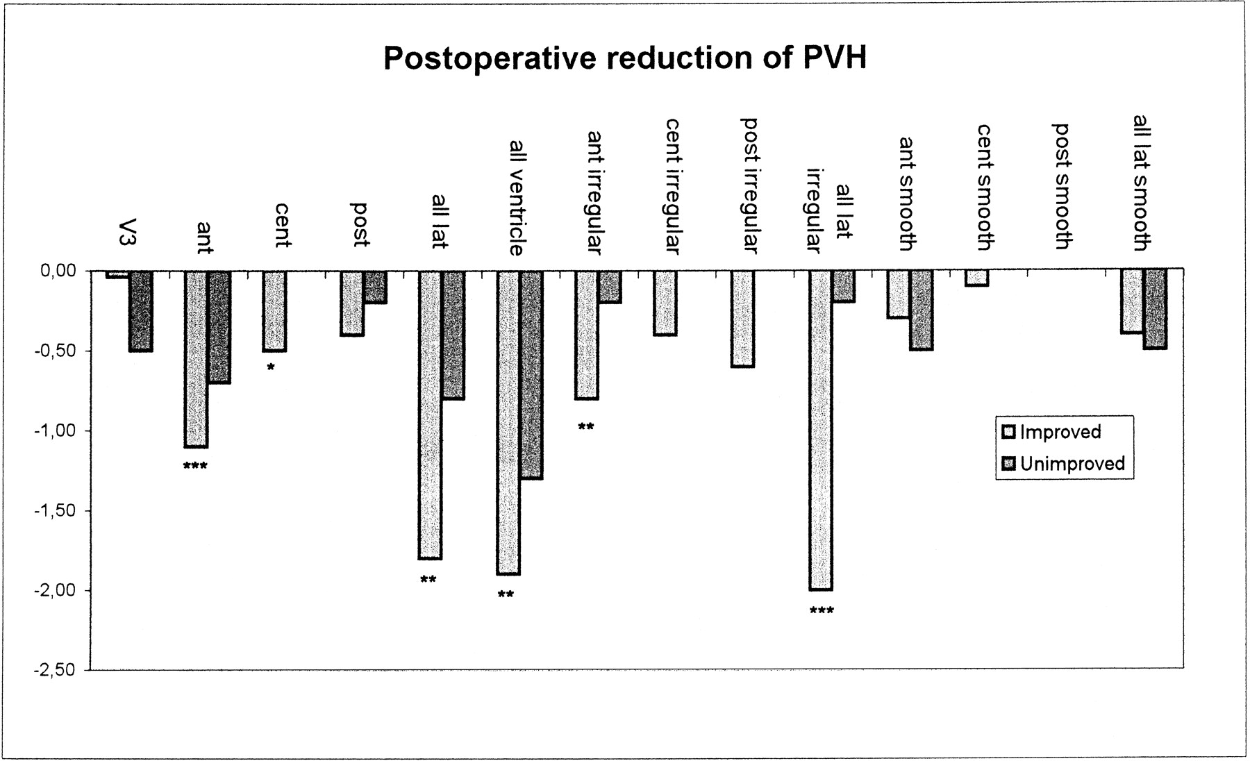
  • fig 2.
    • Download figure
    • Open in new tab
    • Download powerpoint
    fig 2.

    Postoperative reduction in PVH. Note the reduction in irregular but not smooth PVH around the frontal horns in improved patients

  • fig 3.
    • Download figure
    • Open in new tab
    • Download powerpoint
    fig 3.

    Postoperative reduction in ventricular size is numerically equal in both improved and unimproved patients, although significant only in the larger group of improved patients

Tables

  • Figures
  • TABLE 1:
    • View popup
    • Download powerpoint
    TABLE 1:

    Baseline characteristics and results of shunt surgery, overall and by clinical outcome of surgery

  • TABLE 2:
    • View popup
    • Download powerpoint
    TABLE 2:

    MR imaging results

PreviousNext
Back to top

In this issue

American Journal of Neuroradiology: 22 (9)
American Journal of Neuroradiology
Vol. 22, Issue 9
1 Oct 2001
  • Table of Contents
  • Index by author
Advertisement
Print
Download PDF
Email Article

Thank you for your interest in spreading the word on American Journal of Neuroradiology.

NOTE: We only request your email address so that the person you are recommending the page to knows that you wanted them to see it, and that it is not junk mail. We do not capture any email address.

Enter multiple addresses on separate lines or separate them with commas.
Normal Pressure Hydrocephalus: Vascular White Matter Changes on MR Images Must Not Exclude Patients from Shunt Surgery
(Your Name) has sent you a message from American Journal of Neuroradiology
(Your Name) thought you would like to see the American Journal of Neuroradiology web site.
CAPTCHA
This question is for testing whether or not you are a human visitor and to prevent automated spam submissions.
Cite this article
Mats Tullberg, Christer Jensen, Sven Ekholm, Carsten Wikkelsø
Normal Pressure Hydrocephalus: Vascular White Matter Changes on MR Images Must Not Exclude Patients from Shunt Surgery
American Journal of Neuroradiology Oct 2001, 22 (9) 1665-1673;

Citation Manager Formats

  • BibTeX
  • Bookends
  • EasyBib
  • EndNote (tagged)
  • EndNote 8 (xml)
  • Medlars
  • Mendeley
  • Papers
  • RefWorks Tagged
  • Ref Manager
  • RIS
  • Zotero
0 Responses
Respond to this article
Share
Bookmark this article
Normal Pressure Hydrocephalus: Vascular White Matter Changes on MR Images Must Not Exclude Patients from Shunt Surgery
Mats Tullberg, Christer Jensen, Sven Ekholm, Carsten Wikkelsø
American Journal of Neuroradiology Oct 2001, 22 (9) 1665-1673;
del.icio.us logo Twitter logo Facebook logo Mendeley logo
  • Tweet Widget
  • Facebook Like
  • Google Plus One

Jump to section

  • Article
    • Abstract
    • Methods
    • Results
    • Discussion
    • Conclusion
    • Footnotes
    • References
  • Figures & Data
  • Info & Metrics
  • Responses
  • References
  • PDF

Related Articles

  • No related articles found.
  • PubMed
  • Google Scholar

Cited By...

  • Clinical Improvement after Shunt Surgery in Patients with Idiopathic Normal Pressure Hydrocephalus Can Be Quantified by Diffusion Tensor Imaging
  • CSF biomarkers distinguish idiopathic normal pressure hydrocephalus from its mimics
  • Increased Water Content in Periventricular Caps in Patients without Acute Hydrocephalus
  • Absence of Disproportionately Enlarged Subarachnoid Space Hydrocephalus, a Sharp Callosal Angle, or Other Morphologic MRI Markers Should Not Be Used to Exclude Patients with Idiopathic Normal Pressure Hydrocephalus from Shunt Surgery
  • High-throughput analysis of sulfatides in cerebrospinal fluid using automated extraction and UPLC-MS/MS
  • Vascular factors in suspected normal pressure hydrocephalus: A population-based study
  • Preoperative Prognostic Value of MRI Findings in 108 Patients with Idiopathic Normal Pressure Hydrocephalus
  • Low-dose acetazolamide reverses periventricular white matter hyperintensities in iNPH
  • Proton MR spectroscopy and white matter hyperintensities in idiopathic normal pressure hydrocephalus and other dementias
  • Idiopathic normal pressure hydrocephalus
  • Brain energy metabolism and intracranial pressure in idiopathic adult hydrocephalus syndrome
  • Intracerebral microdialysis and CSF hydrodynamics in idiopathic adult hydrocephalus syndrome
  • Crossref
  • Google Scholar

This article has not yet been cited by articles in journals that are participating in Crossref Cited-by Linking.

More in this TOC Section

  • Enhanced Axonal Metabolism during Early Natalizumab Treatment in Relapsing-Remitting Multiple Sclerosis
  • Progression of Microstructural Damage in Spinocerebellar Ataxia Type 2: A Longitudinal DTI Study
  • SWI or T2*: Which MRI Sequence to Use in the Detection of Cerebral Microbleeds? The Karolinska Imaging Dementia Study
Show more Brain

Similar Articles

Advertisement

Indexed Content

  • Current Issue
  • Accepted Manuscripts
  • Article Preview
  • Past Issues
  • Editorials
  • Editor's Choice
  • Fellows' Journal Club
  • Letters to the Editor
  • Video Articles

Cases

  • Case Collection
  • Archive - Case of the Week
  • Archive - Case of the Month
  • Archive - Classic Case

More from AJNR

  • Trainee Corner
  • Imaging Protocols
  • MRI Safety Corner
  • Book Reviews

Multimedia

  • AJNR Podcasts
  • AJNR Scantastics

Resources

  • Turnaround Time
  • Submit a Manuscript
  • Submit a Video Article
  • Submit an eLetter to the Editor/Response
  • Manuscript Submission Guidelines
  • Statistical Tips
  • Fast Publishing of Accepted Manuscripts
  • Graphical Abstract Preparation
  • Imaging Protocol Submission
  • Evidence-Based Medicine Level Guide
  • Publishing Checklists
  • Author Policies
  • Become a Reviewer/Academy of Reviewers
  • News and Updates

About Us

  • About AJNR
  • Editorial Board
  • Editorial Board Alumni
  • Alerts
  • Permissions
  • Not an AJNR Subscriber? Join Now
  • Advertise with Us
  • Librarian Resources
  • Feedback
  • Terms and Conditions
  • AJNR Editorial Board Alumni

American Society of Neuroradiology

  • Not an ASNR Member? Join Now

© 2025 by the American Society of Neuroradiology All rights, including for text and data mining, AI training, and similar technologies, are reserved.
Print ISSN: 0195-6108 Online ISSN: 1936-959X

Powered by HighWire