
Article Figures & Data
Figures
- fig 1.
Graph shows that the increased frequency of ischemia and/or infarction changes with increasing time after ictus. No patients had ischemic changes at days 0 or 1, but the frequency increased to 86% (six of seven cases) in patients presenting 8 d or more after SAH
- fig 2.
MR images obtained 1 d after SAH in patient 3 show acute SAH with no change in cerebral blood flow. Nonenhanced CT scan (not shown) showed a typical anterior circulation hemorrhage.
A, Fast spin-echo fluid-attenuated inversion recovery (6000/96/1800 [TR/TE/TI]) MR image shows SAH in the basal cisterns as areas of high signal intensity.
B, Field-echo T2*-weighted (588/30 [TR/TE]) MR image shows SAH in the basal cisterns as areas of low signal intensity.
C, Diffusion-weighted image shows no restriction of diffusion.
D, Perfusion image shows two selected regions of interest.
E, Map of the time to peak shows normal, symmetric patterns.
F, Map of the cerebral blood flow shows normal, symmetric patterns.
G, Maximum intensity projection of the time-of-flight MR angiogram data shows an anterior communicating aneurysm (arrow).
H, Plot of time versus signal change shows a symmetric curve.
- fig 3.
Images in patient 22, 6 d after SAH.
A, Hemorrhage is visible on this CT scan of the left sylvian fissure and perimesencephalic cisterns.
B, Spin-echo T1-weighted (501/16 [TR/TE[) MR image shows a high-signal-intensity change, due to T1 shortening, in the hemorrhage.
C, Diffusion-weighted image shows no abnormality.
D, Cerebral perfusion is reduced, as shown by increased time to peak in the left hemisphere on this map.
- fig 4.
Images in patient 26, who presented 8 d after SAH.
A, CT scan shows a subtle finding of isoattenuating material (arrow) in the suprasellar cistern.
B, Axial fluid-attenuated inversion recovery (6000/96/1800) MR image shows extensive SAH as areas of high signal intensity in the suprasellar cistern and left sylvian fissure.
C, T2-weighted (3096/80 [TR/TE]) MR image shows areas of abnormal high signal intensity around the left insular cortex.
D, Diffusion-weighted MR image shows areas of high signal intensity due to infarction.
E, ADC map also shows that this finding is due to infarction.
F, Time-to-peak map from the perfusion-weighted MR sequence shows reduced perfusion in the entire left hemisphere.


Tables
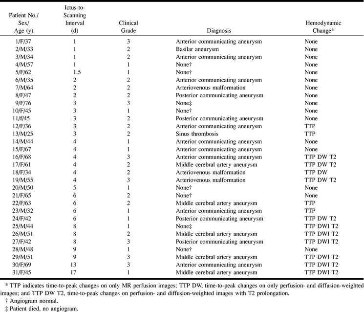
 TABLE 1:
Case summaries of 31 patients with SAH confirmed at CT or lumbar puncture


