
Article Figures & Data
Figures
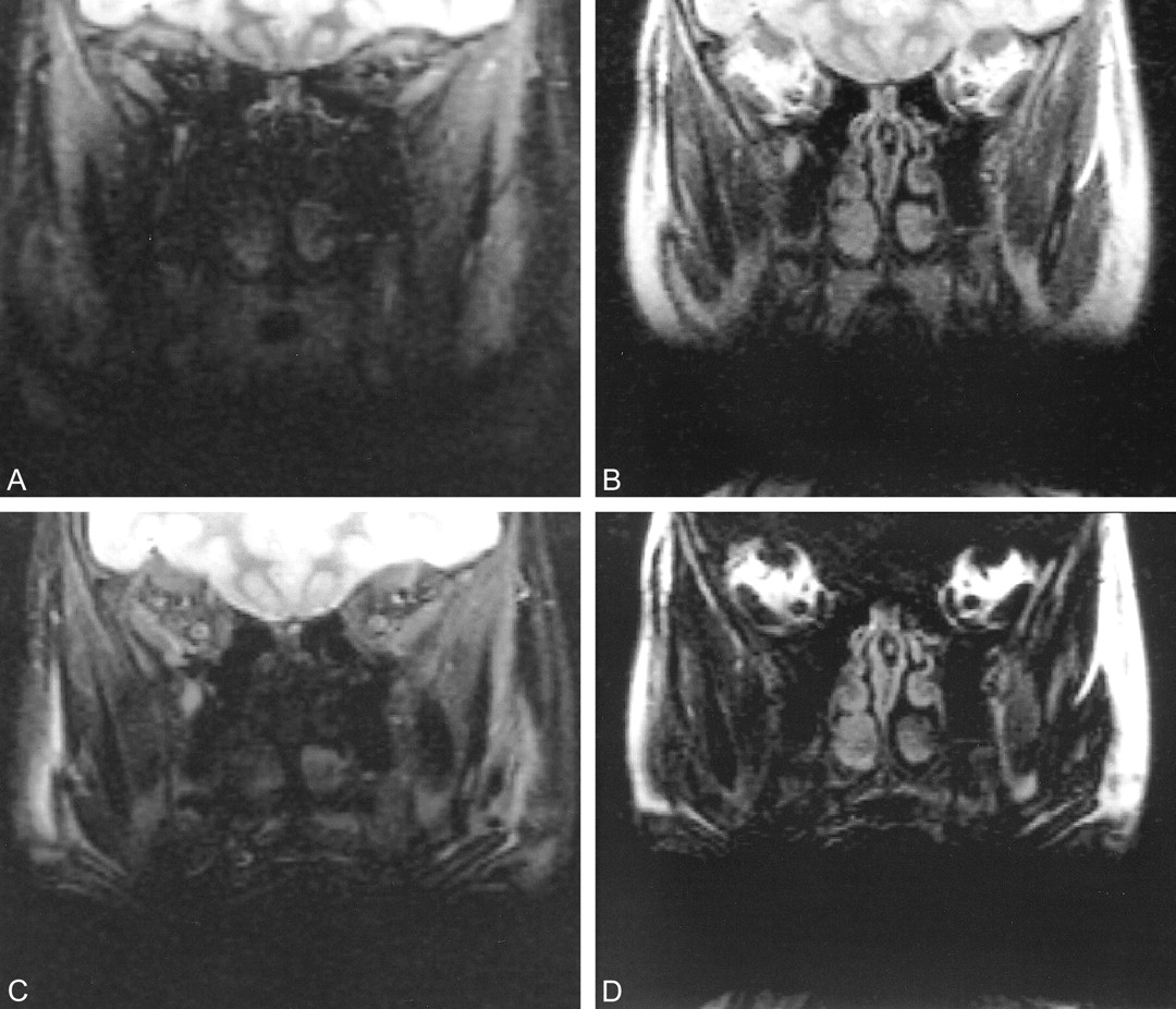
- fig 1.
Coronal MR images of the retrobulbar space in a 3-year-old boy.
A, FSEIR image (2500/30/8/2) shows that nulling of fat from the inversion pulse effectively isolates the signal from the optic nerves. Structure is evident within the right optic nerve with these sequence settings.
B, 3PD water-plus-fat image (3000/85/8/1) shows both optic nerves as hypointense areas within a bright fat background. The internal structure is well depicted. Note the substantial signal from the sinuses, which is greatly nulled on the FSEIR image.
C, 3PD pure water image (3000/85/8/1) shows the optic nerves isolated from the surrounding fat. Substantial signal is present from water-containing tissue in the sinuses. Because of its relatively short T1, this signal is lost with the delay time in the FSEIR image.
D, 3PD pure fat image (3000/85/8/1) shows dramatic contrast and sharply defined margins between the water-containing structures (optic nerves and extraocular muscles) and retrobulbar fat.
- fig 2.
Coronal MR images of the retrobulbar space in a 12-year-old girl with dental braces.
A, FSEIR image shows that the evaluation of orbits is limited by artifact from the dental braces. Note the improved contrast between gray and white matter because of the additional T1 weighting caused by the inversion pulse.
B, 3PD water-plus-fat image (3000/85/8/1) offers dramatically improved signal; the artifact from the dental braces has a sharper margin.
C, 3PD pure water image (3000/85/8/1) shows superior anatomic detail compared with FSEIR.
D, 3PD pure fat image (3000/85/8/1) shows substantial signal from fat-containing structures outside the retrobulbar space.


Tables

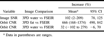
Comparison of image SNR and orbit CNR for individual 3PD images and FSEIR
In this issue


Jump to section
Related Articles
Cited By...
- Optimal Fat Suppression in Head and Neck MRI: Comparison of Multipoint Dixon with 2 Different Fat-Suppression Techniques, Spectral Presaturation and Inversion Recovery, and STIR
- Improving Fat-Suppressed T2-Weighted Imaging of the Head and Neck with 2 Fast Spin-Echo Dixon Techniques: Initial Experiences