
Article Figures & Data
Figures
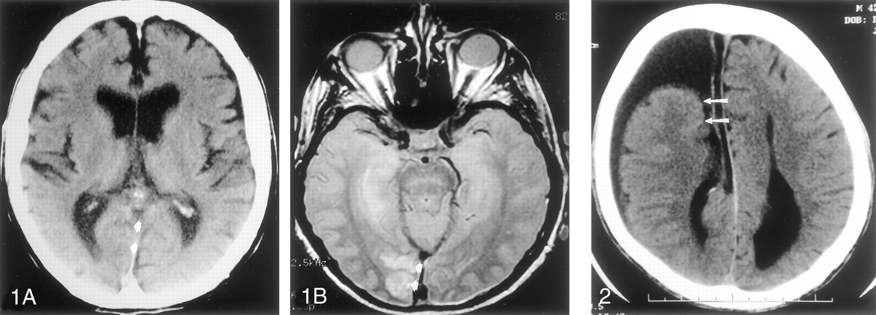
- Fig 1.
Axial CT scan (A) and MR image (B) show false-negative finding involving ischemic disease.
A, Acute right occipital infarct is visible as both hypoattenuating gray matter and hypoattenuating white matter, with associated sulcal effacement (arrows).
B, Proton density–weighted (2400/30 [TR/TE]) MR image confirms the findings (arrows).
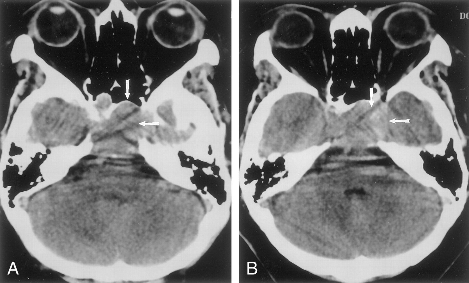
- Fig 2.
Axial CT image obtained in a patient with schizencephaly and callosal dysgenesis shows an error in synthesis that was considered significant. Note the communication of the right lateral ventricle with the subarachnoid space (arrows) and the characteristic configuration of the occipital horns.
- Fig 3.
Contiguous 5-mm non–contrast-enhanced routine axial CT scans demonstrate a large mass (arrows) in the sella in a case of pituitary macroadenoma.
- Fig 4.
Axial images show metastatic disease interpreted as infarction.
A, CT image shows a right frontal lobe mass (arrows).
B, On the CT section adjacent to A, vasogenic edema (arrows) is evident.
C, Contrast-enhanced MR image more clearly shows the mass (arrows).


Tables
Category Rate of Agreement by Resident Year (%) 1st 2nd 3rd 4th Agree 90 92 94 99 Disagree-insignificant 8 7 6 1 Disagree-significant 2 1 0 0 Note.—The level of training had a significant effect on the rate of agreement (P = .032).
- TABLE 2:
Sources of errors by residents in the interpretation of scans in disagree-significant and disagree-insignificant categories
Type of error Number of Error Percentage (%) Of All Scans Of All Errors Fracture 18 1.4 16 Chronic ischemic change 12 0.9 11 Atrophy 8 0.6 7 Related to neoplasm 3 0.2 3 Note.—The total scans was 1324. The total number of errors by residents was 113.
- TABLE 3:
Accuracy of initial interpretations of 1324 emergency cranial CT scans by residents
Diagnosis Findings True-Positive False-Positive False-Negative True-Negative Acute cerebral ischemia 7 2 1 1314 Hemorrhage 27 10 2 1285 Note.—For acute cerebral ischemia, specificity and sensitivity were 87.5% and 99.8%, respectively; for hemorrhage, 93.1% and 99.2%, respectively.
In this issue


Jump to section
Related Articles
Cited By...
- Review of deep learning algorithms for the automatic detection of intracranial hemorrhages on computed tomography head imaging
- Risk Factors for Perceptual-versus-Interpretative Errors in Diagnostic Neuroradiology
- Emergency department interpretation of CT of the brain: a systematic review
- Comparing the accuracy of initial head CT reporting by radiologists, radiology trainees, neuroradiographers and emergency doctors