
Article Figures & Data
Figures
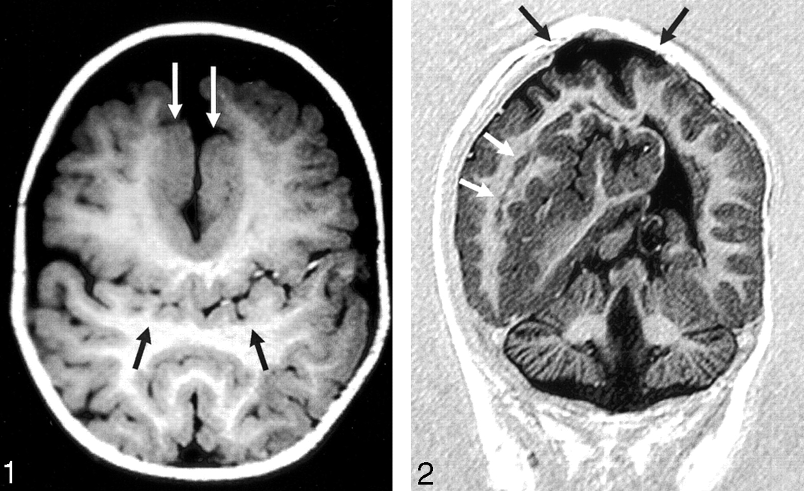
- Fig 1.
Axial T1-weighted image (550/14/2 [TR/TE/excitations]) in the posterior frontal and parietal regions shows the abnormal sylvian fissures communicating across the midline over the vertex (black arrows). Flow-related enhancement is seen within branches of the middle cerebral arteries, confirming that this represents the sylvian fissure. Thickened dysplastic cortex is present along the anterior IHF (white arrows).
- Fig 2.
Coronal echo-planar inversion recovery image (2000/30/400 [TR/TE/TI]) depicts the large area of subcortical dysplasia and associated gray matter heterotopia (white arrows). The site of the repaired cephalocele can be seen at the vertex (black arrows). The inferior vermis is absent.
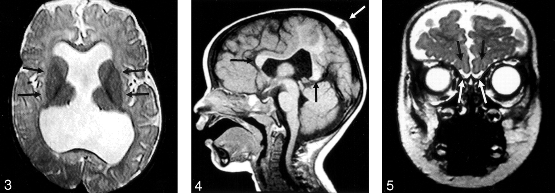
- Fig 3.
Axial spin-echo T2-weighted image (2530/80/2 [TR/TE/excitations]) shows the widely separated deep gray nuclei (arrows), unlike those seen in classic HPE.
- Fig 4.
Sagittal T1-weighted image (400/15/1) through the midline reveals the genu and splenium of the corpus callosum (black arrows). The body of the callosum is absent in the region of non-cleaved parenchyma. An atretic cephalocele is also present (white arrow). Note also that the anterior recess of the third ventricle and basal forebrain are normal, unlike that seen in classic HPE.
- Fig 5.
Coronal fast spin-echo T2-weighted image (3200/117/1) through the inferior frontal lobes depicts the normal appearance of the anterior IHF, olfactory sulci (black arrows), and olfactory bulbs (white arrows).
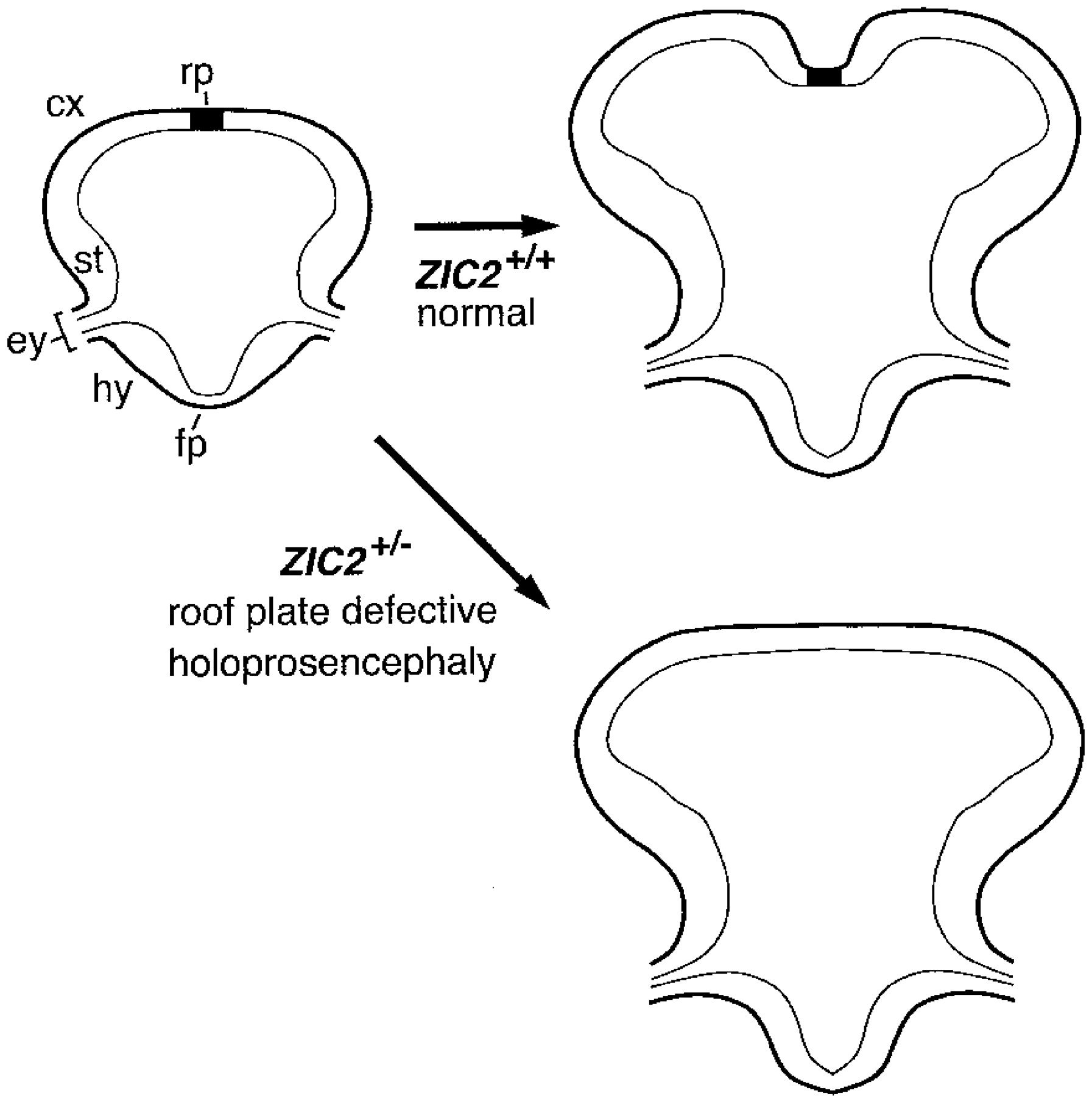
- Fig 6.
Illustration of roof plate differentiation, which depends on ZIC2 function.
After neural tube closure (left), ZIC2 promotes differentiation of the embryonic roof plate (rp) in the dorsal midline of the neural tube. Normally, a low rate of mitotic activity and a high rate of apoptosis in the roof plate of the forebrain contribute to interhemispheric cleavage (upper right). If one ZIC2 allele is mutant (ZIC2+/-), differentiation of the roof plate is compromised, resulting in HPE (lower right).
cx indicates cortex; ey, eye; fp, floor plate; hy, hypothalamus; st, striatum.


In this issue


Jump to section
Related Articles
Cited By...
- Monozygotic twins with de novo ZIC2 gene mutations discordant for the type of holoprosencephaly
- Asymptomatic Interhypothalamic Adhesions in Children
- In Utero MR Imaging of Fetal Holoprosencephaly: A Structured Approach to Diagnosis and Classification
- Retinoic acid signaling regulates development of the dorsal forebrain midline and the choroid plexus in the chick
- New findings for phenotype-genotype correlations in a large European series of holoprosencephaly cases
- Septopreoptic Holoprosencephaly: A Mild Subtype Associated with Midline Craniofacial Anomalies
- The Cavum Septi Pellucidi: Why Is It Important?
- Mutations in the BMP pathway in mice support the existence of two molecular classes of holoprosencephaly
- Central Roles of the Roof Plate in Telencephalic Development and Holoprosencephaly
- Middle interhemispheric variant of holoprosencephaly: A very mild clinical case
- Facial hemangioma and cerebral corticovascular dysplasia: A syndrome associated with epilepsy
- Middle interhemispheric variant of holoprosencephaly: A distinct cliniconeuroradiologic subtype
- MRI shows abnormal white matter maturation in classical holoprosencephaly
- Neuroanatomy of holoprosencephaly as predictor of function: Beyond the face predicting the brain