Advertisement

Main menu

  • Home
  • Content
    • Current Issue
    • Accepted Manuscripts
    • Article Preview
    • Past Issue Archive
    • AJNR Case Collection
    • Case of the Week Archive
    • Classic Case Archive
    • Case of the Month Archive
  • Special Collections
    • Spinal CSF Leak Articles (Jan 2020-June 2024)
    • 2024 AJNR Journal Awards
    • Most Impactful AJNR Articles
  • Multimedia
    • AJNR Podcast
    • AJNR Scantastics
    • Video Articles
  • For Authors
    • Submit a Manuscript
    • Author Policies
    • Fast publishing of Accepted Manuscripts
    • Graphical Abstract Preparation
    • Manuscript Submission Guidelines
    • Imaging Protocol Submission
    • Submit a Case for the Case Collection
  • About Us
    • About AJNR
    • Editorial Board
  • More
    • Become a Reviewer/Academy of Reviewers
    • Subscribers
    • Permissions
    • Alerts
    • Feedback
    • Advertisers
    • ASNR Home
  • Other Publications
    • ajnr

User menu

  • Alerts
  • Log in

Search

  • Advanced search
American Journal of Neuroradiology

American Journal of Neuroradiology

ASHNR American Society of Functional Neuroradiology ASHNR American Society of Pediatric Neuroradiology ASSR
  • Alerts
  • Log in

Advanced Search

  • Home
  • Content
    • Current Issue
    • Accepted Manuscripts
    • Article Preview
    • Past Issue Archive
    • AJNR Case Collection
    • Case of the Week Archive
    • Classic Case Archive
    • Case of the Month Archive
  • Special Collections
    • Spinal CSF Leak Articles (Jan 2020-June 2024)
    • 2024 AJNR Journal Awards
    • Most Impactful AJNR Articles
  • Multimedia
    • AJNR Podcast
    • AJNR Scantastics
    • Video Articles
  • For Authors
    • Submit a Manuscript
    • Author Policies
    • Fast publishing of Accepted Manuscripts
    • Graphical Abstract Preparation
    • Manuscript Submission Guidelines
    • Imaging Protocol Submission
    • Submit a Case for the Case Collection
  • About Us
    • About AJNR
    • Editorial Board
  • More
    • Become a Reviewer/Academy of Reviewers
    • Subscribers
    • Permissions
    • Alerts
    • Feedback
    • Advertisers
    • ASNR Home
  • Follow AJNR on Twitter
  • Visit AJNR on Facebook
  • Follow AJNR on Instagram
  • Join AJNR on LinkedIn
  • RSS Feeds

Welcome to the new AJNR, Updated Hall of Fame, and more. Read the full announcements.


AJNR is seeking candidates for the position of Associate Section Editor, AJNR Case Collection. Read the full announcement.


Research ArticleBRAIN

Comparison of Independent Component Analysis and Conventional Hypothesis-Driven Analysis for Clinical Functional MR Image Processing

Michelle A. Quigley, Victor M. Haughton, John Carew, Dietmar Cordes, Chad H. Moritz and M. Elizabeth Meyerand
American Journal of Neuroradiology January 2002, 23 (1) 49-58;
Michelle A. Quigley
  • Find this author on Google Scholar
  • Find this author on PubMed
  • Search for this author on this site
Victor M. Haughton
  • Find this author on Google Scholar
  • Find this author on PubMed
  • Search for this author on this site
John Carew
  • Find this author on Google Scholar
  • Find this author on PubMed
  • Search for this author on this site
Dietmar Cordes
  • Find this author on Google Scholar
  • Find this author on PubMed
  • Search for this author on this site
Chad H. Moritz
  • Find this author on Google Scholar
  • Find this author on PubMed
  • Search for this author on this site
M. Elizabeth Meyerand
  • Find this author on Google Scholar
  • Find this author on PubMed
  • Search for this author on this site
  • Article
  • Figures & Data
  • Info & Metrics
  • Responses
  • References
  • PDF
Loading

Article Figures & Data

Figures

  • Tables
  • Fig 1.
    • Download figure
    • Open in new tab
    • Download powerpoint
    Fig 1.

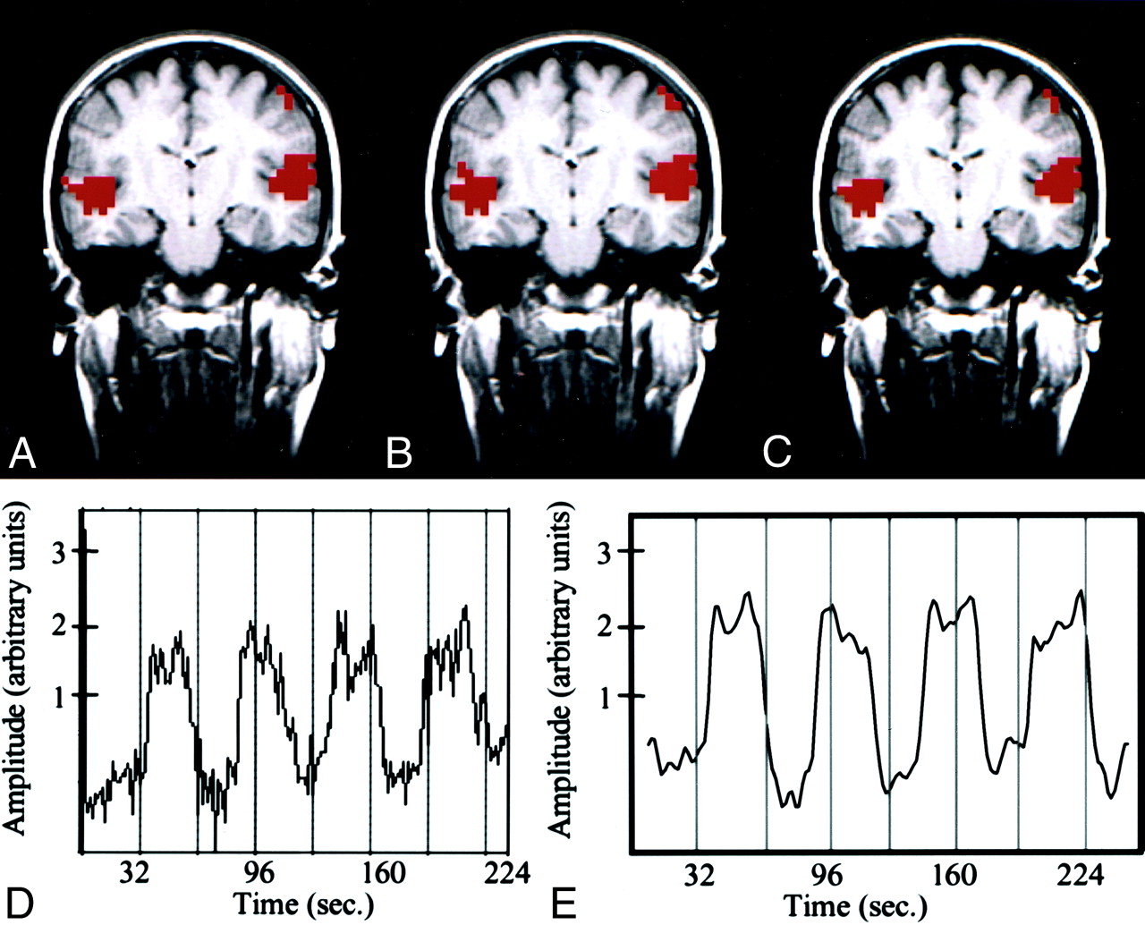
    Text-listening task in a patient with cortical dysplasia involving the left occipital lobe.

    A–C, fMR images. The z score map processed with conventional analysis (A) and that processed with ICA (B) are similar. Both show bilateral activation in the auditory cortices. The intersect map of pixels identified in A and B is shown in C; it demonstrates a 91% CR between the maps in A and B.

    D, Time course plot from an activated voxel in the conventional analysis shows that the changes in signal are temporally correlated with task performance.

    E, Temporal pattern of a selected independent component in the ICA with the highest z score closely resembles the time-course plot of the activated voxel in D. A .70 correlation between the time course of the ICA component and the boxcar reference function was found.

  • Fig 2.
    • Download figure
    • Open in new tab
    • Download powerpoint
    Fig 2.

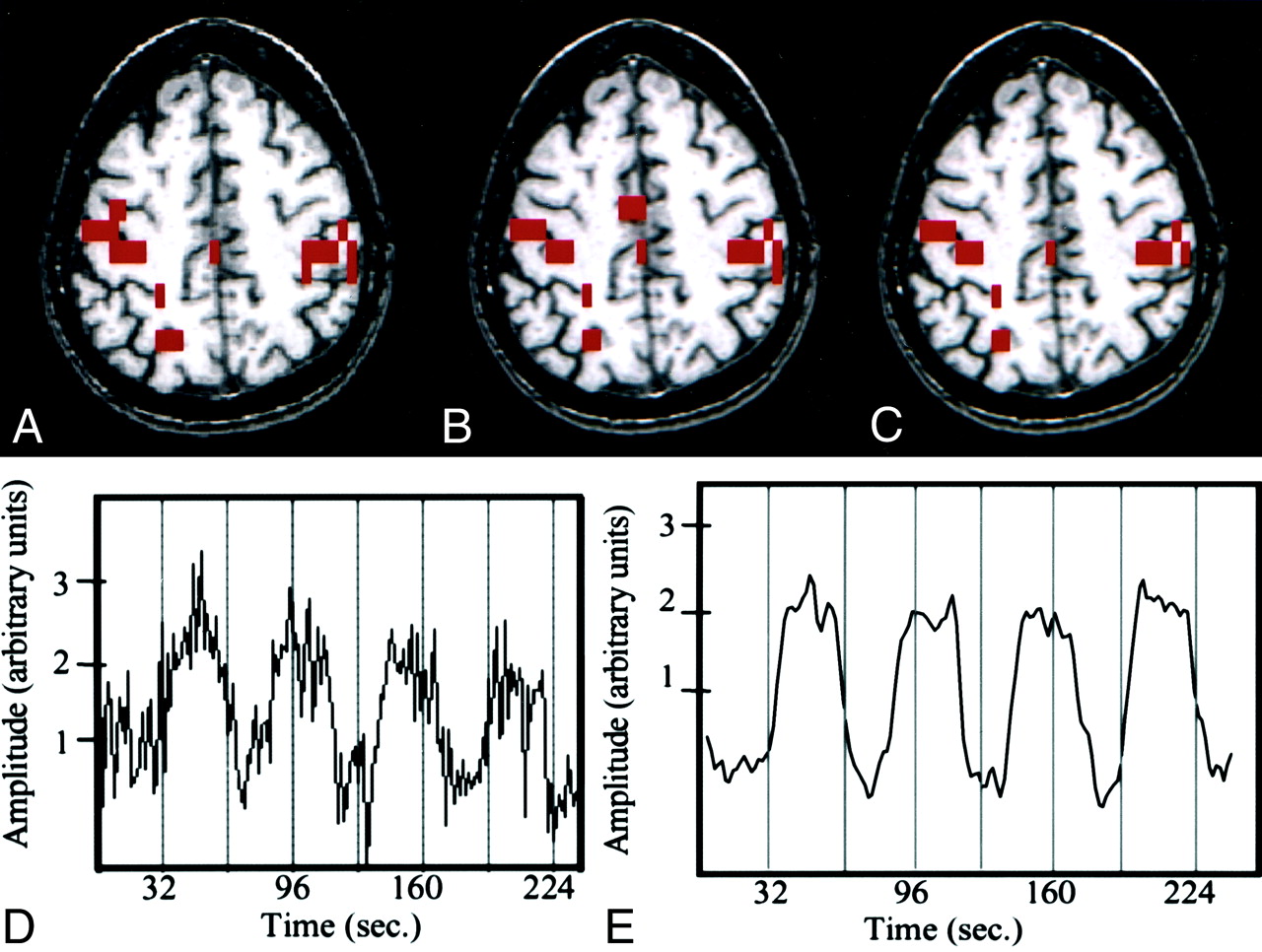
    Finger-tapping task in a patient with a left temporal glioma.

    A–C, fMR images. The image processed with reference function analysis (A) shows activation in the sensorimotor cortices and sensorimotor area. The image processed with ICA (B) shows a similar pattern of activation. The intersect map of pixels identified on both the image processed conventionally and that processed with ICA (C) has a high CR (83%). Note the similarity of the three maps.

    D, Time course plot of the activated voxel shows that the changes in signal are temporally correlated with task performance.

    E, Temporal pattern of a selected ICA component (representing the highest z score) has a .64 correlation between the time course of the component and the boxcar reference function.

  • Fig 3.
    • Download figure
    • Open in new tab
    • Download powerpoint
    Fig 3.

    Word-generation task in a patient with a left arteriovenous malformation.

    A–C, fMR images. The image processed by using the reference function (A) and the image processed with ICA (B) are similar. The intersect map of pixels (C) shows that 80% of the pixels identified with the methods were the same (80% CR between the maps).

    D, Time course plot selected from an activated voxel shows that the fluctuation in signal is temporally related to task performance.

    E, Temporal pattern of a selected ICA component has a pattern similar to that of the time course in the activated pixel.

  • Fig 4.
    • Download figure
    • Open in new tab
    • Download powerpoint
    Fig 4.

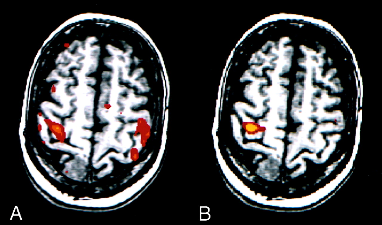
    fMR images in a patient with a right meningioma. For this task, predominant activation typically is seen in the sensorimotor cortex of one hemisphere.

    A, Image processed with reference function analysis. The patient moved the left hand according to the on-off commands; however, the patient moved the right hand when instructed to move right hand and when instructed to move the left hand. Therefore, this image shows anomalous activation.

    B, However, the image processed with the ICA component specific for activation in the right hemisphere shows the expected unilateral activation pattern.

  • Fig 5.
    • Download figure
    • Open in new tab
    • Download powerpoint
    Fig 5.

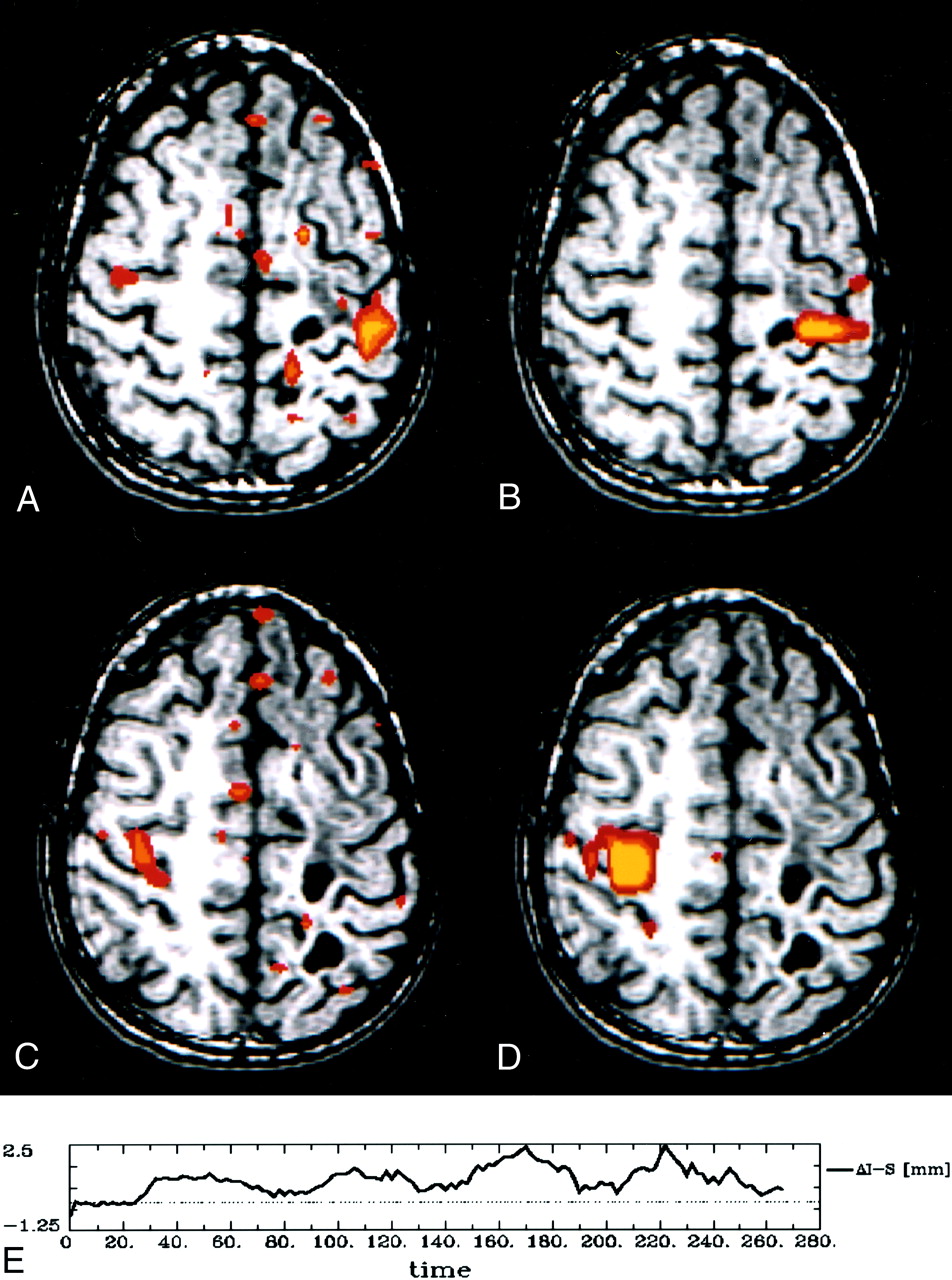
    Findings in a patient with focal seizures and a history of resection of a primitive neuroectodermal tumor and presumptive postoperative complications. When performing the finger-tapping task with either hand, the patient appeared to move.

    A–D, fMR images. The z score maps processed with reference to the standard boxcar (A and C) show activation in the sensorimotor cortex and motion-related artifact. The z score maps based on an independent component identified with ICA (B and D) show activation with less motion artifact.

    E, The patient’s motion in the inferior-superior direction while performing the finger-tapping task is documented on this graph of inferior-superior motion versus time.

  • Fig 6.
    • Download figure
    • Open in new tab
    • Download powerpoint
    Fig 6.

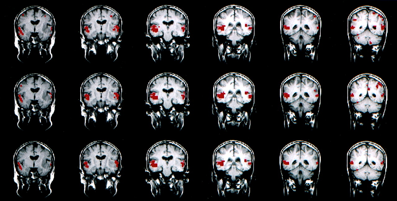
    fMR images obtained with the text-listening task in a patient with a cortical dysplasia involving the left occipital lobe. (Images in the leftmost column are those in Figure 1, with the addition of the next three consecutive sections.)

    Top row, The z score maps obtained with conventional hypothesis-driven analysis.

    Middle row, The z score maps obtained with ICA show good agreement with those in the top row.

    Bottom row, The intersect map reflects an 87% CR for these four consecutive sections.

  • Fig 7.
    • Download figure
    • Open in new tab
    • Download powerpoint
    Fig 7.

    fMR images obtained with a text-listening task in the patient with a left temporal glioma.

    Top row, Consecutive z score map obtained with conventional analysis.

    Middle row, Consecutive z score map obtained with ICA.

    Bottom row, Concurrence in 81% with for these six sections, as reflected in the intersect map.

  • Fig 8.
    • Download figure
    • Open in new tab
    • Download powerpoint
    Fig 8.

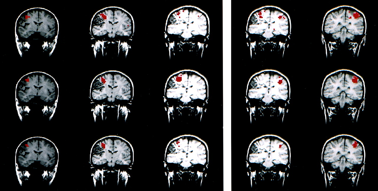
    fMR images in a right-handed patient with a right parasylvian arteriovenous malformation (depicted in the figure) performing the finger-tapping task. In this case, ICA revealed two independent components for the right and left sensorimotor patterns. Therefore, two comparisons were made. The set of images on the left shows the right sensorimotor component, while the set on the right shows the left component. The conventional z score map (top row), spatial ICA map (middle row), and intersect map (bottom row) for the right sensorimotor cortex show a concurrence of 59% for the three sections shown. The left motor sensoricortex reflects a 49% concurrence for the two sections shown.

  • Fig 9.
    • Download figure
    • Open in new tab
    • Download powerpoint
    Fig 9.

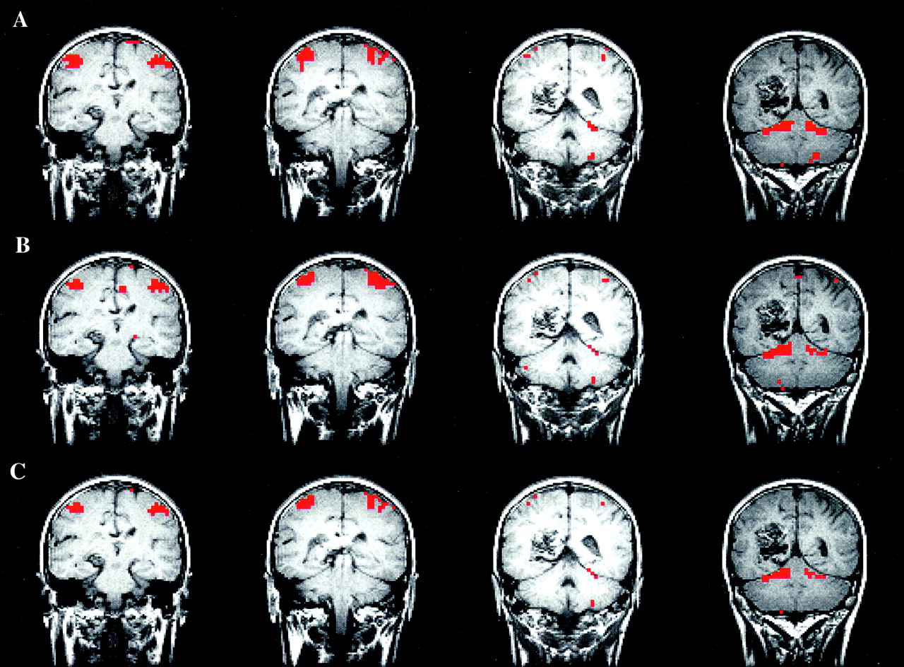
    fMR images in a left-handed patient with a left posterior parietal arteriovenous malformation (depicted in the Figure) performing the finger-tapping task. Note that the four consecutive sections of the conventional z score map (top row), spatial ICA map (middle row), and intersect map (bottom row) are similar. In this case, just one independent component showed the left and right sensorimotor cortices in the same component. This component even included activation in the cerebellum that was found in both analyses. The CR in this four-section example was 73%.

Tables

  • Figures
    • View popup
    TABLE 1:

    CRs in the comparison of conventional z maps and ICA maps determined with single sections

    Patient No.Auditory Cortex CR (%)Motor Cortex CR (%)Left Language Cortex CR (%)Average CR (%)
    LeftRightLeftRight
    1797967797876
    2989881816885
    3929283832575
    4848476766477
    5868667828080
    691913552None67
    776768471None77
    874745757None65
    9909067674071
    10616166663858
    11626252376455
    1255556363None59
    Average797967685770
    • Note.—CRs are the highest CR determined from a section selected from those that showed activation in the relevant cortex. Combined averages in auditory, motor, and left language cortex were 79%, 68%, and 57%, respectively.

    • View popup
    TABLE 2:

    CRs in the comparison of conventional z maps and ICA maps determined with all sections

    Patient No.Auditory Cortex CR (%)Motor Cortex CR (%)Left Language Cortex CR (%)Average CR (%)
    LeftRightLeftRight
    175 (7)75 (7)49 (2)59 (3)78 (1)67
    259 (8)59 (8)73 (4)73 (4)68 (1)66
    381 (6)81 (6)71 (5)71 (5)25 (1)66
    463 (6)63 (6)75 (2)75 (2)50 (2)65
    568 (10)68 (10)47 (3)51 (3)80 (1)63
    687 (4)87 (4)35 (1)48 (2)None64
    758 (5)58 (5)63 (2)67 (2)None62
    864 (5)64 (5)49 (2)49 (2)None57
    961 (8)61 (8)55 (4)55 (4)38 (2)54
    1046 (7)46 (7)57 (4)57 (4)38 (1)49
    1138 (5)38 (5)52 (1)31 (2)64 (1)45
    1234 (6)34 (6)30 (4)30 (4)None32
    Average616155565558
    • Note.—CRs are the averages of all sections that showed activation in the relevant cortex. Data in parentheses are the number of sections used in the calculation. Combined averages in auditory, motor, and left language cortex were 61%, 56%, and 55%, respectively.

PreviousNext
Back to top

In this issue

American Journal of Neuroradiology: 23 (1)
American Journal of Neuroradiology
Vol. 23, Issue 1
1 Jan 2002
  • Table of Contents
  • Index by author
Advertisement
Print
Download PDF
Email Article

Thank you for your interest in spreading the word on American Journal of Neuroradiology.

NOTE: We only request your email address so that the person you are recommending the page to knows that you wanted them to see it, and that it is not junk mail. We do not capture any email address.

Enter multiple addresses on separate lines or separate them with commas.
Comparison of Independent Component Analysis and Conventional Hypothesis-Driven Analysis for Clinical Functional MR Image Processing
(Your Name) has sent you a message from American Journal of Neuroradiology
(Your Name) thought you would like to see the American Journal of Neuroradiology web site.
CAPTCHA
This question is for testing whether or not you are a human visitor and to prevent automated spam submissions.
Cite this article
Michelle A. Quigley, Victor M. Haughton, John Carew, Dietmar Cordes, Chad H. Moritz, M. Elizabeth Meyerand
Comparison of Independent Component Analysis and Conventional Hypothesis-Driven Analysis for Clinical Functional MR Image Processing
American Journal of Neuroradiology Jan 2002, 23 (1) 49-58;

Citation Manager Formats

  • BibTeX
  • Bookends
  • EasyBib
  • EndNote (tagged)
  • EndNote 8 (xml)
  • Medlars
  • Mendeley
  • Papers
  • RefWorks Tagged
  • Ref Manager
  • RIS
  • Zotero
0 Responses
Respond to this article
Share
Bookmark this article
Comparison of Independent Component Analysis and Conventional Hypothesis-Driven Analysis for Clinical Functional MR Image Processing
Michelle A. Quigley, Victor M. Haughton, John Carew, Dietmar Cordes, Chad H. Moritz, M. Elizabeth Meyerand
American Journal of Neuroradiology Jan 2002, 23 (1) 49-58;
del.icio.us logo Twitter logo Facebook logo Mendeley logo
  • Tweet Widget
  • Facebook Like
  • Google Plus One

Jump to section

  • Article
    • Abstract
    • Methods
    • Results
    • Discussion
    • Conclusion
    • References
  • Figures & Data
  • Info & Metrics
  • Responses
  • References
  • PDF

Related Articles

  • No related articles found.
  • PubMed
  • Google Scholar

Cited By...

  • Reduction of Motion Artifacts and Noise Using Independent Component Analysis in Task-Based Functional MRI for Preoperative Planning in Patients with Brain Tumor
  • Comparison of Hypothesis- and a Novel Hybrid Data/Hypothesis-Driven Method of Functional MR Imaging Analysis in Patients with Brain Gliomas
  • Functional Connectivity MR Imaging of the Language Network in Patients with Drug-Resistant Epilepsy
  • Dissociable Intrinsic Connectivity Networks for Salience Processing and Executive Control
  • Default-mode network activity distinguishes Alzheimer's disease from healthy aging: Evidence from functional MRI
  • Crossref
  • Google Scholar

This article has not yet been cited by articles in journals that are participating in Crossref Cited-by Linking.

More in this TOC Section

  • Progression of Microstructural Damage in Spinocerebellar Ataxia Type 2: A Longitudinal DTI Study
  • Usefulness of Quantitative Susceptibility Mapping for the Diagnosis of Parkinson Disease
  • Evaluating the Effects of White Matter Multiple Sclerosis Lesions on the Volume Estimation of 6 Brain Tissue Segmentation Methods
Show more Brain

Similar Articles

Advertisement

Indexed Content

  • Current Issue
  • Accepted Manuscripts
  • Article Preview
  • Past Issues
  • Editorials
  • Editors Choice
  • Fellow Journal Club
  • Letters to the Editor

Cases

  • Case Collection
  • Archive - Case of the Week
  • Archive - Case of the Month
  • Archive - Classic Case

Special Collections

  • Special Collections

Resources

  • News and Updates
  • Turn around Times
  • Submit a Manuscript
  • Author Policies
  • Manuscript Submission Guidelines
  • Evidence-Based Medicine Level Guide
  • Publishing Checklists
  • Graphical Abstract Preparation
  • Imaging Protocol Submission
  • Submit a Case
  • Become a Reviewer/Academy of Reviewers
  • Get Peer Review Credit from Publons

Multimedia

  • AJNR Podcast
  • AJNR SCANtastic
  • Video Articles

About Us

  • About AJNR
  • Editorial Board
  • Not an AJNR Subscriber? Join Now
  • Alerts
  • Feedback
  • Advertise with us
  • Librarian Resources
  • Permissions
  • Terms and Conditions

American Society of Neuroradiology

  • Not an ASNR Member? Join Now

© 2025 by the American Society of Neuroradiology All rights, including for text and data mining, AI training, and similar technologies, are reserved.
Print ISSN: 0195-6108 Online ISSN: 1936-959X

Powered by HighWire