Advertisement

Main menu

  • Home
  • Content
    • Current Issue
    • Accepted Manuscripts
    • Article Preview
    • Past Issue Archive
    • AJNR Case Collection
    • Case of the Week Archive
    • Classic Case Archive
    • Case of the Month Archive
  • Special Collections
    • Spinal CSF Leak Articles (Jan 2020-June 2024)
    • 2024 AJNR Journal Awards
    • Most Impactful AJNR Articles
  • Multimedia
    • AJNR Podcast
    • AJNR Scantastics
    • Video Articles
  • For Authors
    • Submit a Manuscript
    • Author Policies
    • Fast publishing of Accepted Manuscripts
    • Graphical Abstract Preparation
    • Manuscript Submission Guidelines
    • Imaging Protocol Submission
    • Submit a Case for the Case Collection
  • About Us
    • About AJNR
    • Editorial Board
  • More
    • Become a Reviewer/Academy of Reviewers
    • Subscribers
    • Permissions
    • Alerts
    • Feedback
    • Advertisers
    • ASNR Home
  • Other Publications
    • ajnr

User menu

  • Alerts
  • Log in

Search

  • Advanced search
American Journal of Neuroradiology

American Journal of Neuroradiology

ASHNR American Society of Functional Neuroradiology ASHNR American Society of Pediatric Neuroradiology ASSR
  • Alerts
  • Log in

Advanced Search

  • Home
  • Content
    • Current Issue
    • Accepted Manuscripts
    • Article Preview
    • Past Issue Archive
    • AJNR Case Collection
    • Case of the Week Archive
    • Classic Case Archive
    • Case of the Month Archive
  • Special Collections
    • Spinal CSF Leak Articles (Jan 2020-June 2024)
    • 2024 AJNR Journal Awards
    • Most Impactful AJNR Articles
  • Multimedia
    • AJNR Podcast
    • AJNR Scantastics
    • Video Articles
  • For Authors
    • Submit a Manuscript
    • Author Policies
    • Fast publishing of Accepted Manuscripts
    • Graphical Abstract Preparation
    • Manuscript Submission Guidelines
    • Imaging Protocol Submission
    • Submit a Case for the Case Collection
  • About Us
    • About AJNR
    • Editorial Board
  • More
    • Become a Reviewer/Academy of Reviewers
    • Subscribers
    • Permissions
    • Alerts
    • Feedback
    • Advertisers
    • ASNR Home
  • Follow AJNR on Twitter
  • Visit AJNR on Facebook
  • Follow AJNR on Instagram
  • Join AJNR on LinkedIn
  • RSS Feeds

Welcome to the new AJNR, Updated Hall of Fame, and more. Read the full announcements.


AJNR is seeking candidates for the position of Associate Section Editor, AJNR Case Collection. Read the full announcement.


Research ArticleBRAIN

High-Resolution Line Scan Diffusion Tensor MR Imaging of White Matter Fiber Tract Anatomy

Hatsuho Mamata, Yoshiaki Mamata, Carl-Fredrik Westin, Martha E. Shenton, Ron Kikinis, Ferenc A. Jolesz and Stephan E. Maier
American Journal of Neuroradiology January 2002, 23 (1) 67-75;
Hatsuho Mamata
  • Find this author on Google Scholar
  • Find this author on PubMed
  • Search for this author on this site
Yoshiaki Mamata
  • Find this author on Google Scholar
  • Find this author on PubMed
  • Search for this author on this site
Carl-Fredrik Westin
  • Find this author on Google Scholar
  • Find this author on PubMed
  • Search for this author on this site
Martha E. Shenton
  • Find this author on Google Scholar
  • Find this author on PubMed
  • Search for this author on this site
Ron Kikinis
  • Find this author on Google Scholar
  • Find this author on PubMed
  • Search for this author on this site
Ferenc A. Jolesz
  • Find this author on Google Scholar
  • Find this author on PubMed
  • Search for this author on this site
Stephan E. Maier
  • Find this author on Google Scholar
  • Find this author on PubMed
  • Search for this author on this site
  • Article
  • Figures & Data
  • Info & Metrics
  • Responses
  • References
  • PDF
Loading

Article Figures & Data

Figures

  • Fig 1.
    • Download figure
    • Open in new tab
    • Download powerpoint
    Fig 1.

    When viewed in multiple sections, the appearance of the association fibers on the principal eigenvector maps is consistent with known anatomy.

    A, Sagittal 110 × 110-mm subimage close to the midline, passing through the cingulum (pink). Note that the fibers of the corpus callosum run at a slight angle through the plane (blue dots with short lines).

    B, More lateral sagittal section obtained through the arcuate fasciculus (green), inferior longitudinal fasciculus (blue), and uncinate fasciculus (orange).

    C, Coronal section obtained at the level of the posterior limb of the internal capsule. Fibers of the cingulum (pink), arcuate fasciculus (green), and uncinate fasciculus (orange) pass through the section (dots). Fibers of the posterior limb of the internal capsule (yellow lines) are running in-plane.

    D, High-resolution 55 × 55-mm coronal subimage of the thalamic region. The strionigral and pallidonigral fibers are shown in light blue.

    E, High-resolution axial surface coil image of the calcarine area at a 120 × 60-mm field of view. U-fibers connecting visual cortices are well seen.
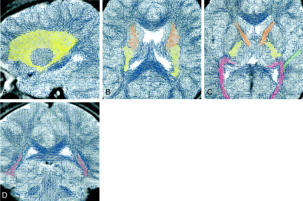
  • Fig 2.
    • Download figure
    • Open in new tab
    • Download powerpoint
    Fig 2.

    Subimages (110 × 110-mm field of view) of the projection fibers.

    A, Lateral sagittal section passing through the corona radiata (yellow lines).

    B, Axial section obtained through the genu and splenium of the corpus callosum. Corona radiata fibers within the posterior limb (yellow) of the internal capsule can be distinguished by their strong through-plane component (dots), whereas fibers within the anterior limb (orange) of the corpus callosum run in-plane (lines).

    C, Axial section obtained at the level of the basal ganglia. Fibers in the anterior limb of the internal capsule (orange) are running in-plane (lines), whereas in the posterior limb of the internal capsule (yellow), the fibers run through-plane (dots). Parts of the optic (pink) and auditory (green) radiation can also be seen.

    D, Coronal section obtained through the trigone of the lateral ventricles. Pink dots indicate through-plane fibers that are part of the optic radiation.

  • Fig 3.
    • Download figure
    • Open in new tab
    • Download powerpoint
    Fig 3.

    Commissural fibers of the corpus callosum are observed in sections obtained near the lateral ventricle.

    A, Coronal section obtained through the insula (110 × 110-mm subimage). In this section, the cingulum (pink) and fibers (blue) interconnecting the frontoparietal cortices of each hemisphere with the corpus callosum can be seen. The fibers of the anterior commissure (green) radiate into the temporal lobes.

    B, High resolution coronal section (55 × 55-mm subimage) obtained at the level of the anterior commissure (green).
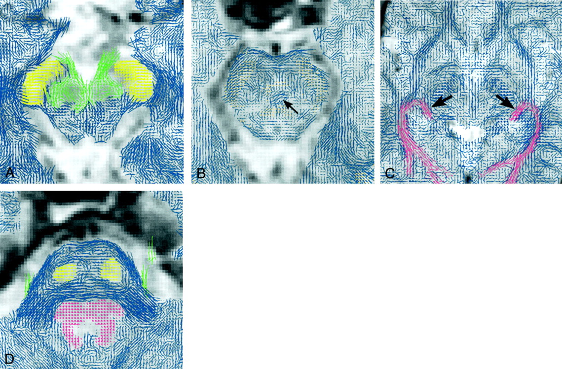
  • Fig 4.
    • Download figure
    • Open in new tab
    • Download powerpoint
    Fig 4.

    Subimages (55 × 55-mm field of view) of the brain stem.

    A, Axial section obtained at the level of the upper midbrain. Very fine fibers that originate from the oculomotor nucleus run through and around the red nucleus and converge to the roots of the oculomotor nerves (green). Note that fibers of the cerebral peduncles are running at a slight angle through the plane (yellow dots with lines).

    B, Axial section obtained at the level of the lower midbrain. The arrow indicates the decussation of the superior cerebellar peduncle.

    C, Axial section (110 × 110-mm field of view) obtained at the level of the midbrain-cerebrum junction. The fibers of the optic tract (arrows) connect to the lateral geniculate bodies. Fibers of the optic radiation (pink) pass lateral to the optic tract.

    D, Axial section obtained at the level of the pons, through the roots of the trigeminal nerve (green). Fibers of the craniocaudal tracts (superior cerebellar peduncle, medial longitudinal fasciculus, tectospinal tract, dorsal and ventral trigeminothalamic tracts, central tegmental tract, spinothalamic tract, and rubrospinal tract) can be seen in the posterior part of the pons (pink). The yellow dots indicate fibers of the pyramidal and the frontopontine tracts.

  • Fig 5.
    • Download figure
    • Open in new tab
    • Download powerpoint
    Fig 5.

    Subimages of the brain stem.

    A, Coronal section (110 × 55-mm subimage) of the brain stem obtained at the level of the motor decussation. Fibers of the motor tract cross to the contralateral side at the level of the lower medulla.

    B, Axial subimage (27.5 × 27.5-mm field of view) of the medulla obtained at the level of the motor decussation. Fibers of the motor tract cross to the contralateral side. Short lines with blue dots indicate low anisotropy.

    C, Midsagittal subimage (55 × 55-mm field of view) of the brain stem. The pyramidal tract is shown in yellow and the medial longitudinal fasciculus in pink.

    D, Sagittal section (55 × 55-mm field of view subimage) of the brain stem at a lateral position. Fibers of the pyramidal tract (yellow) enter the pons. The arrowheads indicate fine fibers of the sensory decussation (internal arcuate fibers), running in an anteroposterior direction.
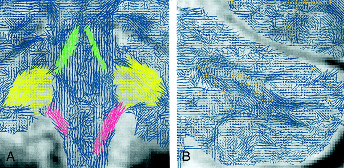
  • Fig 6.
    • Download figure
    • Open in new tab
    • Download powerpoint
    Fig 6.

    Subimages (55 × 55-mm field of view) of the cerebellum.

    A, Coronal section obtained at the level of the fourth ventricle floor. Fibers of the superior (green) and inferior (pink) cerebellar peduncles pass in a craniocaudal direction toward the cerebellar hemisphere, and fibers of the middle cerebellar peduncle (yellow) pass at an oblique angle through the section (dots with lines).

    B, More posterior coronal section of the cerebellum. Fibers from the cerebellar peduncles branch out toward the cerebellar cortices.

PreviousNext
Back to top

In this issue

American Journal of Neuroradiology: 23 (1)
American Journal of Neuroradiology
Vol. 23, Issue 1
1 Jan 2002
  • Table of Contents
  • Index by author
Advertisement
Print
Download PDF
Email Article

Thank you for your interest in spreading the word on American Journal of Neuroradiology.

NOTE: We only request your email address so that the person you are recommending the page to knows that you wanted them to see it, and that it is not junk mail. We do not capture any email address.

Enter multiple addresses on separate lines or separate them with commas.
High-Resolution Line Scan Diffusion Tensor MR Imaging of White Matter Fiber Tract Anatomy
(Your Name) has sent you a message from American Journal of Neuroradiology
(Your Name) thought you would like to see the American Journal of Neuroradiology web site.
CAPTCHA
This question is for testing whether or not you are a human visitor and to prevent automated spam submissions.
Cite this article
Hatsuho Mamata, Yoshiaki Mamata, Carl-Fredrik Westin, Martha E. Shenton, Ron Kikinis, Ferenc A. Jolesz, Stephan E. Maier
High-Resolution Line Scan Diffusion Tensor MR Imaging of White Matter Fiber Tract Anatomy
American Journal of Neuroradiology Jan 2002, 23 (1) 67-75;

Citation Manager Formats

  • BibTeX
  • Bookends
  • EasyBib
  • EndNote (tagged)
  • EndNote 8 (xml)
  • Medlars
  • Mendeley
  • Papers
  • RefWorks Tagged
  • Ref Manager
  • RIS
  • Zotero
0 Responses
Respond to this article
Share
Bookmark this article
High-Resolution Line Scan Diffusion Tensor MR Imaging of White Matter Fiber Tract Anatomy
Hatsuho Mamata, Yoshiaki Mamata, Carl-Fredrik Westin, Martha E. Shenton, Ron Kikinis, Ferenc A. Jolesz, Stephan E. Maier
American Journal of Neuroradiology Jan 2002, 23 (1) 67-75;
del.icio.us logo Twitter logo Facebook logo Mendeley logo
  • Tweet Widget
  • Facebook Like
  • Google Plus One

Jump to section

  • Article
    • Abstract
    • Methods
    • Results
    • Discussion
    • Conclusion
    • Footnotes
    • References
  • Figures & Data
  • Info & Metrics
  • Responses
  • References
  • PDF

Related Articles

  • No related articles found.
  • PubMed
  • Google Scholar

Cited By...

  • Diffusion Tensor Imaging and Tractography Utilized in the Resection of a Midbrain Cavernous Malformation
  • Local structural connectivity directs seizure spread in focal epilepsy
  • Perceptual learning treatment in patients with anisometropic amblyopia: a neuroimaging study
  • Is depression a disconnection syndrome? Meta-analysis of diffusion tensor imaging studies in patients with MDD
  • Acute Damage to the Posterior Limb of the Internal Capsule on Diffusion Tensor Tractography as an Early Imaging Predictor of Motor Outcome after Stroke
  • Tract-Based Analysis of Callosal, Projection, and Association Pathways in Pediatric Patients with Multiple Sclerosis: A Preliminary Study
  • Pearls & Oy-sters: The medial longitudinal fasciculus in ocular motor physiology
  • Ipsilateral motor dysfunction from unilateral stroke: implications for the functional neuroanatomy of hemiparesis
  • Hierarchical Processing in Spoken Language Comprehension
  • Crossref
  • Google Scholar

This article has not yet been cited by articles in journals that are participating in Crossref Cited-by Linking.

More in this TOC Section

  • Progression of Microstructural Damage in Spinocerebellar Ataxia Type 2: A Longitudinal DTI Study
  • Usefulness of Quantitative Susceptibility Mapping for the Diagnosis of Parkinson Disease
  • Evaluating the Effects of White Matter Multiple Sclerosis Lesions on the Volume Estimation of 6 Brain Tissue Segmentation Methods
Show more Brain

Similar Articles

Advertisement

Indexed Content

  • Current Issue
  • Accepted Manuscripts
  • Article Preview
  • Past Issues
  • Editorials
  • Editors Choice
  • Fellow Journal Club
  • Letters to the Editor

Cases

  • Case Collection
  • Archive - Case of the Week
  • Archive - Case of the Month
  • Archive - Classic Case

Special Collections

  • Special Collections

Resources

  • News and Updates
  • Turn around Times
  • Submit a Manuscript
  • Author Policies
  • Manuscript Submission Guidelines
  • Evidence-Based Medicine Level Guide
  • Publishing Checklists
  • Graphical Abstract Preparation
  • Imaging Protocol Submission
  • Submit a Case
  • Become a Reviewer/Academy of Reviewers
  • Get Peer Review Credit from Publons

Multimedia

  • AJNR Podcast
  • AJNR SCANtastic
  • Video Articles

About Us

  • About AJNR
  • Editorial Board
  • Not an AJNR Subscriber? Join Now
  • Alerts
  • Feedback
  • Advertise with us
  • Librarian Resources
  • Permissions
  • Terms and Conditions

American Society of Neuroradiology

  • Not an ASNR Member? Join Now

© 2025 by the American Society of Neuroradiology All rights, including for text and data mining, AI training, and similar technologies, are reserved.
Print ISSN: 0195-6108 Online ISSN: 1936-959X

Powered by HighWire