Advertisement

Main menu

  • Home
  • Content
    • Current Issue
    • Accepted Manuscripts
    • Article Preview
    • Past Issue Archive
    • Video Articles
    • AJNR Case Collection
    • Case of the Week Archive
    • Case of the Month Archive
    • Classic Case Archive
  • Special Collections
    • AJNR Awards
    • Low-Field MRI
    • Alzheimer Disease
    • ASNR Foundation Special Collection
    • Photon-Counting CT
    • View All
  • Multimedia
    • AJNR Podcasts
    • AJNR SCANtastic
    • Trainee Corner
    • MRI Safety Corner
    • Imaging Protocols
  • For Authors
    • Submit a Manuscript
    • Submit a Video Article
    • Submit an eLetter to the Editor/Response
    • Manuscript Submission Guidelines
    • Statistical Tips
    • Fast Publishing of Accepted Manuscripts
    • Graphical Abstract Preparation
    • Imaging Protocol Submission
    • Author Policies
  • About Us
    • About AJNR
    • Editorial Board
    • Editorial Board Alumni
  • More
    • Become a Reviewer/Academy of Reviewers
    • Subscribers
    • Permissions
    • Alerts
    • Feedback
    • Advertisers
    • ASNR Home

User menu

  • Alerts
  • Log in

Search

  • Advanced search
American Journal of Neuroradiology

American Journal of Neuroradiology

ASHNR American Society of Functional Neuroradiology ASHNR American Society of Pediatric Neuroradiology ASSR
  • Alerts
  • Log in

Advanced Search

  • Home
  • Content
    • Current Issue
    • Accepted Manuscripts
    • Article Preview
    • Past Issue Archive
    • Video Articles
    • AJNR Case Collection
    • Case of the Week Archive
    • Case of the Month Archive
    • Classic Case Archive
  • Special Collections
    • AJNR Awards
    • Low-Field MRI
    • Alzheimer Disease
    • ASNR Foundation Special Collection
    • Photon-Counting CT
    • View All
  • Multimedia
    • AJNR Podcasts
    • AJNR SCANtastic
    • Trainee Corner
    • MRI Safety Corner
    • Imaging Protocols
  • For Authors
    • Submit a Manuscript
    • Submit a Video Article
    • Submit an eLetter to the Editor/Response
    • Manuscript Submission Guidelines
    • Statistical Tips
    • Fast Publishing of Accepted Manuscripts
    • Graphical Abstract Preparation
    • Imaging Protocol Submission
    • Author Policies
  • About Us
    • About AJNR
    • Editorial Board
    • Editorial Board Alumni
  • More
    • Become a Reviewer/Academy of Reviewers
    • Subscribers
    • Permissions
    • Alerts
    • Feedback
    • Advertisers
    • ASNR Home
  • Follow AJNR on Twitter
  • Visit AJNR on Facebook
  • Follow AJNR on Instagram
  • Join AJNR on LinkedIn
  • RSS Feeds

AJNR Awards, New Junior Editors, and more. Read the latest AJNR updates

OtherSpine Imaging and Spine Image-Guided Interventions

Lumbar Disk Pseudotumor: An Unusual Presentation of Lumbar Spinal Fracture and Stenosis

Robert A. Koenigsberg, Perry Black, Scott H. Faro and Jeffrey Rykken
American Journal of Neuroradiology November 2002, 23 (10) 1662-1664;
Robert A. Koenigsberg
  • Find this author on Google Scholar
  • Find this author on PubMed
  • Search for this author on this site
Perry Black
  • Find this author on Google Scholar
  • Find this author on PubMed
  • Search for this author on this site
Scott H. Faro
  • Find this author on Google Scholar
  • Find this author on PubMed
  • Search for this author on this site
Jeffrey Rykken
  • Find this author on Google Scholar
  • Find this author on PubMed
  • Search for this author on this site
  • Article
  • Figures & Data
  • Info & Metrics
  • Responses
  • References
  • PDF
Loading

Abstract

Summary: We present an unusual case of a primary lumbar disk-space mass that presumably developed secondary to a chronic hyperextension spinal fracture associated with spinal stenosis. This injury resulted in the appearance of a lumbar intervertebral disk-space mass or pseudotumor. The pseudotumor most likely resulted from a prior spinal fracture, leading to a fused hyperextension deformity in a patient with underlying chronic degenerative spinal disease.

The lumbar-spine intervertebral disk space is rarely involved with primary tumor (1). When seen, involvement is usually a result of contiguous neoplastic invasion, either from vertebral body neoplasia with direct invasion of the disk space or from paraspinal tumor with resultant disk-space involvement. Infectious diseases, such as tuberculosis or those caused by Staphylococcus aureus, frequently involve lumbar intervertebral spaces, but they do not typically result in intervertebral disk-space expansion. We present an unusual case in which a primary lumbar disk-space mass arose secondary to a chronic hyperextension spinal fracture associated with spinal stenosis. This injury resulted in the appearance of a lumbar intervertebral disk-space mass or pseudotumor.

Case Report

An 84-year-old woman with chronic cardiac problems presented with a history of low back pain of several years’ duration. In the 2 months before her hospital admission, she had increasing low back pain to the extent that she could no longer walk. She had minimal pain in the lower extremities. Her bladder and bowel function were maintained, and the patient had no history of weight loss. On examination, power in the lower extremities was 4 on a scale of 5. Knee and ankle reflexes were absent; plantar reflexes were flexor. The patient’s sensation was intact. She required intravenous narcotic medication by means of a patient-controlled anesthesia pump.

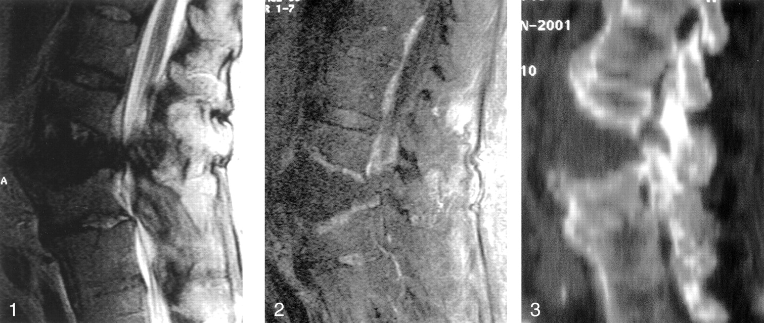
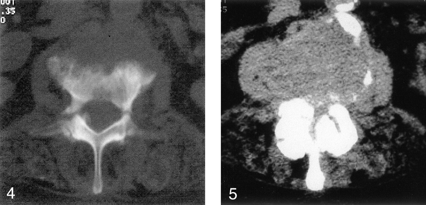
MR imaging demonstrated an expansile mass at the L2-L3 level (Fig 1). The mass demonstrated low signal intensity on T1-weighted images, and remained low in signal intensity on T2-weighted images (Fig 2). Interestingly, the signal intensity in the intervertebral disk was preserved along the lower L2 endplate on T2-weighted images, yet it remained indistinguishable on T1-weighted images. The mass did not enhance with the injection of contrast material, although the vertebral body endplates did. The CSF signal intensity in the central canal was obliterated because of associated central canal stenosis. CT showed that the mass was soft-tissue material that was isoattenuated relative to skeletal muscle (Fig 3). The study further showed bilateral L2 pedicle fractures, with deformed L2 and L3 vertebral-body endplates associated with sclerosis. Central canal stenosis due to a combination of the disk-space mass and posterior-element changes was present. Air attenuation or vacuum phenomenon was not seen within the disk space (Fig 4). Both the L2-L3 and L3-L4 disk levels showed severe degenerative spinal stenosis, as demonstrated on both MR images and CT scans (Fig 5). Further, extensive laminar calcification was present along the margins of the vertebral body; this finding was suggestive of longstanding arthropathy, that is, diffuse idiopathic skeletal hypertrophy.

Fig 1.
  • Download figure
  • Open in new tab
  • Download powerpoint
Fig 1.

MR sagittal T2-weighted image shows an expansile mass at the L2-L3 disk level that represents the pseudotumor. The disk attached to the inferior L2 endplate has increased signal intensity. Spinal stenosis is present at the L2-L3 and L3-L4 levels.

Fig 2.
  • Download figure
  • Open in new tab
  • Download powerpoint
Fig 2.

Gadolinium-enhanced T1-weighted sagittal MR image shows endplate enhancement at the L2-L3 level.

Fig 3.
  • Download figure
  • Open in new tab
  • Download powerpoint
Fig 3.

Sagittal CT reconstruction of the lumbar spine shows a disk-space mass with an extension deformity. Note the laminar calcification along the anterior vertebral margins disrupted by the disk-space mass and the facet malalignment.

Fig 4.
  • Download figure
  • Open in new tab
  • Download powerpoint
Fig 4.

Axial CT scan (bone window) at the L2 level shows bilateral pedicle fractures. Note the moth-eaten appearance of the L2 endplate simulating an underlying destructive mass.

Fig 5.
  • Download figure
  • Open in new tab
  • Download powerpoint
Fig 5.

Axial soft tissue–window CT scan (soft tissue window) at the level of the L2-L3 disk space demonstrates severe central-canal stenosis. This finding was also apparent at the L3-L4 level (not shown).

Clinically, in view of the expansile lesion at the L2-L3 level, a neoplasm was suspected. Results of a metastatic workup were negative. The patient’s pain was thought to be largely related to the severe lumbar central-canal and foraminal stenosis. A decompressive laminectomy was performed from L2 to L5, with the finding of severe central canal stenosis, particularly at L2 and L3. The L2-L3 disk space consisted of a large, empty cavity. No tumor was seen, but some fragments of blood clot and whitish-yellow fragments of tissue were noted. At pathologic analysis, these tissue fragments were reported as fibrous exudate with reactive granulation (Figs 6 and 7). Cultures of the tissue in the depths of the L2-L3 disk proved to be sterile. The patient tolerated the procedure well, with considerable relief of her pain at rest, but she continued to have moderate pain during trunk movement. She died 2 months later in a rehabilitation facility. Autopsy was not performed.

Fig 6.
  • Download figure
  • Open in new tab
  • Download powerpoint
Fig 6.

Low-power photomicrograph shows reparative granulation tissue (solid arrows) and a proliferation of capillaries. Bone (open arrow on the left) and cartilage (open arrows on the right) are adjacent to the granulation tissue.

Fig 7.
  • Download figure
  • Open in new tab
  • Download powerpoint
Fig 7.

Medium-power photomicrograph shows extensive fibrin stroma (pink) with scattered fibroblasts (elongated cells) and inflammatory cells (lymphocytes and polymorphonuclear cells).

Discussion

Vertebral pseudotumors may be classified into those of vertebral origin and those of disk-space origin. Those of vertebral origin, as cited in the literature, include those related to spinal tuberculosis; cystic masses such as a hydatid cyst; synovitis, acne, pustulosis, hyperostosis, and osteitis (SAPHO) syndrome; eosinophilic granuloma; Langerhans cell histiocytosis; fibrous dysplasia; and osteoporotic vertebral collapse (2). Tuberculous pseudotumors can present as destructive vertebral masses that may not radiographically appear to be of infectious origin. Cystic bone lesions, such as hydatid cysts, can occupy a significant bulk of any given vertebral element to mimic a more aggressive lesion (3).

The SAPHO syndrome may mimic vertebral tumors (eg, sclerotic bone tumors) because of the erosive changes with adjacent sclerosis and the hyperostosis that can appear similar to blastic vertebral metastasis (2). In cases of SAPHO, additional lesions can be identified with conventional radiography, bone scintigraphy, CT, or MR imaging. Again, the findings may be suggestive of metastatic disease when this disease process is actually benign. Eosinophilic granuloma, which is a localized form of Langerhans cell histiocytosis, may appear as a pseudotumoral vertebral lesion and is most commonly found in children younger than 10 years (2). These lesions frequently appear with vertebral collapse or vertebra plana (4). Such cases can also be seen in young adults, predominantly in male patients (2, 5). Space-occupying masses are seen in fibrous dysplasia. In such cases, T1-weighted MR images may show decreased signal intensity, and T2-weighted images may show variable signal intensity, depending on the degree of lesion ossification. Chordoma, a mass that arises from a remnant of the notochord, can also appear as a disk-space mass. These tumors occur chiefly in the occipital-cervical and sacrococcygeal regions, and they less frequently appear in the lumbar, cervical, and thoracic regions (7).

The detection of gas within a vertebral disk, especially in the lumbar region, is often consistent with the vacuum phenomenon (8–11). This finding represents an accumulation of gas between the vertebral bodies that is typically more than 90% nitrogen (12). In the presence of a vacuum phenomenon, one can almost exclude the possibility of neoplastic growth because the presence of gas in the disk space may represent a gas-producing infection (13, 14). Importantly, the presence of gas is only rarely associated with infectious spondylitis, but it has been reported in cases of infection with Clostridium species, Brucella organisms, Mycobacteria tuberculosis, Peptococcus species, Streptococcus viridans, Escherichia coli, and Staphylococcus epidermidis (13).

This case involved an expansile soft-tissue mass within the disk space that suggested the possibility of a tumor. Our general experience has been that the height of the intervertebral disk space concomitantly and minimally increases in proportion to the adjacent vertebral compression fractures. However, in this case, the extension fracture deformity widely expanded the space, tearing the inferior attachment of the intervertebral disk. We postulate that the widened space then became occupied with chronic fibrotic tissue, resulting in soft-tissue attenuation at CT. The MR signal intensity mimicked changes seen in processes such as epidermoid or chronic mucocele (that displaying isointensity on T1-weighted images and hypointensity on T2-weighted images).

Conclusion

We believe that this pseudotumor arose secondary to an extension spinal fracture deformity in a patient with degenerative spinal rigidity with spinal stenosis. Interestingly, an air or vacuum phenomenon was not present within the pathologic disk space on any imaging study despite the presence of degenerative disk disease.

Acknowledgments

The authors gratefully acknowledge the histopathologic contribution by Dr Lorenzo Galindo, Department of Pathology, Drexel University College of Medicine.

References

  1. ↵
    Tsuguo Y, Yamauchi Y, Kouichi A, et al. Histopathologic study on tumor infiltration into the intervertebral disc. Spine 1989;14:1245–1248
    PubMed
  2. ↵
    Laredo JD, Quessar AE, Bossard P, et al. Vertebral tumors and pseudotumors. Radiologic Clinics of North America 2001;39:137–163
    CrossRefPubMed
  3. ↵
    Duran H, Fernandez L, Gomez-Castrasena F, et al. Osseous hydatidosis. J Bone Joint Surg Am 1978;60:685–690
    PubMed
  4. ↵
    Canadell J, Villas C, Martinez-Denegri J, et al. Vertebral eosinophilic granuloma. Spine 1986;11:767–769
    PubMed
  5. ↵
    Casson IR, Blair D, Gerard G, et al. Eosinophilic granuloma of the cervical spine in an adult. NY State J Med 1981;81:1102–1104
    PubMed
  6. Stäbler A, Reiser MF. Imaging of spinal infection. Radiol Clin N Am 2001;39:115–135
    CrossRefPubMed
  7. ↵
    Bjornsson J, Wold LE, Ebersold MJ, et al. Chordoma of the mobile spine: a clinicopathologic analysis of 40 patients. Cancer 1993;71:735–740
    CrossRefPubMed
  8. ↵
    Golimbu C, Firooznia H, Rafii M. The intravertebral vacuum sign. Spine 1986;11:1040–1043
    CrossRefPubMed
  9. Maldague BE, Noel HM, Malghem JJ. The intravertebral vacuum cleft: a sign of ischemic vertebral collapse. Radiology 1978;129:23–29
    CrossRefPubMed
  10. Resnick D, Niwayama G, Guerra, J Jr., et al. Spinal vacuum phenomena: anatomical study and review. Radiology 1981;139:341–348
    PubMed
  11. ↵
    Sartoris DJ. What is the significance of the “vertebral vacuum phenomenon”? AJR Am J Roentgenol 1994;163:218
    PubMed
  12. ↵
    Ford LT, Gilula LA, Murphy WA, Gado M. Analysis of gas in vacuum lumbar disc. Am J Roentgenol 1977;128:1056–1057
    PubMed
  13. ↵
    Bielicki DK, Sartoris D, Resnick D, et al. Intraosseous and intradiscal gas in association with spinal infection: report of three cases. AJR Am J Roentgenol 1986;147:83–86
    CrossRefPubMed
  14. ↵
    Wagner SC, Schweitzer ME, Morrison WB, et al. Can imaging findings help differentiate spinal neuropathic arthropathy from disk space infection? Initial experience. Radiology 2000;214:693–699
    PubMed
  15. Daffner RH, Lupetin AR, Dash N, et al. MRI in the detection of malignant infiltration of bone marrow. AJR Am J Roentgenol 1986;146:353–358
    CrossRefPubMed
  16. Lundon K, Bolton K. Structure and function of the lumbar intervertebral disk in health, aging, and pathologic conditions. J Orthop Sports Physical Ther 2001;31:291–306
  17. Tehranzadeh J, Andrews C, and Wong E. Lumbar spine imaging: normal variants, imaging pitfalls, and artifacts. Radiol Clin North Am 2001;38:1207–1253
  • Received February 20, 2002.
  • Accepted after revision July 9, 2002.
  • Copyright © American Society of Neuroradiology
View Abstract
PreviousNext
Back to top

In this issue

American Journal of Neuroradiology: 23 (10)
American Journal of Neuroradiology
Vol. 23, Issue 10
1 Nov 2002
  • Table of Contents
  • Index by author
Advertisement
Print
Download PDF
Email Article

Thank you for your interest in spreading the word on American Journal of Neuroradiology.

NOTE: We only request your email address so that the person you are recommending the page to knows that you wanted them to see it, and that it is not junk mail. We do not capture any email address.

Enter multiple addresses on separate lines or separate them with commas.
Lumbar Disk Pseudotumor: An Unusual Presentation of Lumbar Spinal Fracture and Stenosis
(Your Name) has sent you a message from American Journal of Neuroradiology
(Your Name) thought you would like to see the American Journal of Neuroradiology web site.
CAPTCHA
This question is for testing whether or not you are a human visitor and to prevent automated spam submissions.
Cite this article
Robert A. Koenigsberg, Perry Black, Scott H. Faro, Jeffrey Rykken
Lumbar Disk Pseudotumor: An Unusual Presentation of Lumbar Spinal Fracture and Stenosis
American Journal of Neuroradiology Nov 2002, 23 (10) 1662-1664;

Citation Manager Formats

  • BibTeX
  • Bookends
  • EasyBib
  • EndNote (tagged)
  • EndNote 8 (xml)
  • Medlars
  • Mendeley
  • Papers
  • RefWorks Tagged
  • Ref Manager
  • RIS
  • Zotero
0 Responses
Respond to this article
Share
Bookmark this article
Lumbar Disk Pseudotumor: An Unusual Presentation of Lumbar Spinal Fracture and Stenosis
Robert A. Koenigsberg, Perry Black, Scott H. Faro, Jeffrey Rykken
American Journal of Neuroradiology Nov 2002, 23 (10) 1662-1664;
del.icio.us logo Twitter logo Facebook logo Mendeley logo
  • Tweet Widget
  • Facebook Like
  • Google Plus One

Jump to section

  • Article
    • Abstract
    • Case Report
    • Discussion
    • Conclusion
    • Acknowledgments
    • References
  • Figures & Data
  • Info & Metrics
  • Responses
  • References
  • PDF

Related Articles

  • No related articles found.
  • PubMed
  • Google Scholar

Cited By...

  • No citing articles found.
  • Crossref
  • Google Scholar

This article has not yet been cited by articles in journals that are participating in Crossref Cited-by Linking.

More in this TOC Section

  • Management Outcomes For VO Spine Biopsy
  • Characteristics of SIH Type I Culprit Lesions
  • Advanced Imaging of Type 2 Spinal CSF Leaks
Show more Spine Imaging and Spine Image-Guided Interventions

Similar Articles

Advertisement

Indexed Content

  • Current Issue
  • Accepted Manuscripts
  • Article Preview
  • Past Issues
  • Editorials
  • Editor's Choice
  • Fellows' Journal Club
  • Letters to the Editor
  • Video Articles

Cases

  • Case Collection
  • Archive - Case of the Week
  • Archive - Case of the Month
  • Archive - Classic Case

More from AJNR

  • Trainee Corner
  • Imaging Protocols
  • MRI Safety Corner
  • Book Reviews

Multimedia

  • AJNR Podcasts
  • AJNR Scantastics

Resources

  • Turnaround Time
  • Submit a Manuscript
  • Submit a Video Article
  • Submit an eLetter to the Editor/Response
  • Manuscript Submission Guidelines
  • Statistical Tips
  • Fast Publishing of Accepted Manuscripts
  • Graphical Abstract Preparation
  • Imaging Protocol Submission
  • Evidence-Based Medicine Level Guide
  • Publishing Checklists
  • Author Policies
  • Become a Reviewer/Academy of Reviewers
  • News and Updates

About Us

  • About AJNR
  • Editorial Board
  • Editorial Board Alumni
  • Alerts
  • Permissions
  • Not an AJNR Subscriber? Join Now
  • Advertise with Us
  • Librarian Resources
  • Feedback
  • Terms and Conditions
  • AJNR Editorial Board Alumni

American Society of Neuroradiology

  • Not an ASNR Member? Join Now

© 2025 by the American Society of Neuroradiology All rights, including for text and data mining, AI training, and similar technologies, are reserved.
Print ISSN: 0195-6108 Online ISSN: 1936-959X

Powered by HighWire