Advertisement

Main menu

  • Home
  • Content
    • Current Issue
    • Accepted Manuscripts
    • Article Preview
    • Past Issue Archive
    • AJNR Case Collection
    • Case of the Week Archive
    • Classic Case Archive
    • Case of the Month Archive
  • Special Collections
    • Spinal CSF Leak Articles (Jan 2020-June 2024)
    • 2024 AJNR Journal Awards
    • Most Impactful AJNR Articles
  • Multimedia
    • AJNR Podcast
    • AJNR Scantastics
    • Video Articles
  • For Authors
    • Submit a Manuscript
    • Author Policies
    • Fast publishing of Accepted Manuscripts
    • Graphical Abstract Preparation
    • Manuscript Submission Guidelines
    • Imaging Protocol Submission
    • Submit a Case for the Case Collection
  • About Us
    • About AJNR
    • Editorial Board
  • More
    • Become a Reviewer/Academy of Reviewers
    • Subscribers
    • Permissions
    • Alerts
    • Feedback
    • Advertisers
    • ASNR Home
  • Other Publications
    • ajnr

User menu

  • Alerts
  • Log in

Search

  • Advanced search
American Journal of Neuroradiology

American Journal of Neuroradiology

ASHNR American Society of Functional Neuroradiology ASHNR American Society of Pediatric Neuroradiology ASSR
  • Alerts
  • Log in

Advanced Search

  • Home
  • Content
    • Current Issue
    • Accepted Manuscripts
    • Article Preview
    • Past Issue Archive
    • AJNR Case Collection
    • Case of the Week Archive
    • Classic Case Archive
    • Case of the Month Archive
  • Special Collections
    • Spinal CSF Leak Articles (Jan 2020-June 2024)
    • 2024 AJNR Journal Awards
    • Most Impactful AJNR Articles
  • Multimedia
    • AJNR Podcast
    • AJNR Scantastics
    • Video Articles
  • For Authors
    • Submit a Manuscript
    • Author Policies
    • Fast publishing of Accepted Manuscripts
    • Graphical Abstract Preparation
    • Manuscript Submission Guidelines
    • Imaging Protocol Submission
    • Submit a Case for the Case Collection
  • About Us
    • About AJNR
    • Editorial Board
  • More
    • Become a Reviewer/Academy of Reviewers
    • Subscribers
    • Permissions
    • Alerts
    • Feedback
    • Advertisers
    • ASNR Home
  • Follow AJNR on Twitter
  • Visit AJNR on Facebook
  • Follow AJNR on Instagram
  • Join AJNR on LinkedIn
  • RSS Feeds

Welcome to the new AJNR, Updated Hall of Fame, and more. Read the full announcements.


AJNR is seeking candidates for the position of Associate Section Editor, AJNR Case Collection. Read the full announcement.


Research ArticleBRAIN

Preoperative Assessment of Intracranial Tumors with Perfusion MR and a Volumetric Interpolated Examination: A Comparative Study with DSA

Stephan G. Wetzel, Soonmee Cha, Meng Law, Glyn Johnson, John Golfinos, Peter Lee and Peter Kim Nelson
American Journal of Neuroradiology November 2002, 23 (10) 1767-1774;
Stephan G. Wetzel
  • Find this author on Google Scholar
  • Find this author on PubMed
  • Search for this author on this site
Soonmee Cha
  • Find this author on Google Scholar
  • Find this author on PubMed
  • Search for this author on this site
Meng Law
  • Find this author on Google Scholar
  • Find this author on PubMed
  • Search for this author on this site
Glyn Johnson
  • Find this author on Google Scholar
  • Find this author on PubMed
  • Search for this author on this site
John Golfinos
  • Find this author on Google Scholar
  • Find this author on PubMed
  • Search for this author on this site
Peter Lee
  • Find this author on Google Scholar
  • Find this author on PubMed
  • Search for this author on this site
Peter Kim Nelson
  • Find this author on Google Scholar
  • Find this author on PubMed
  • Search for this author on this site
  • Article
  • Figures & Data
  • Info & Metrics
  • Responses
  • References
  • PDF
Loading

Article Figures & Data

Figures

  • Tables
  • Fig 1.
    • Download figure
    • Open in new tab
    • Download powerpoint
    Fig 1.

    Relationship between maximum rCBV and angiographic vascularity. Scores are as follows: 0 indicates occult; 1, mild blush; 2, moderate blush; and 3, exuberant. The correlation between the two parameters is shown by the dotted line (r = 0.75; P < .05). The average rCBV and the standard deviation are displayed for each group of angiographic vascularity scores.

  • Fig 2.
    • Download figure
    • Open in new tab
    • Download powerpoint
    Fig 2.

    Images in a 57-year-old patient with left parietal glioblastoma multiforme.

    A, Color overlay map reconstructed from pMRI (1000/54) shows marked hyperperfusion of the lesion compared with normal white matter.

    B, Para-axial source image of the 3D VIBE acquisition (8.8/4.4; flip angle, 18°) shows a contrast-enhancing tumor with a necrotic center. Note the prominent cerebral vein posterior to the tumor (arrow).

    C, Real-time MIP image with 30-mm thickness was reconstructed in position of the image in A and shows the relation of the tumor to the cortical veins and the superior sagittal sinus. The more distal part of the overlying cortical vein (arrow) is not included in the volume. As a result of the MIP algorithm, the tumor appears solid.

    D, Coronal source image shows an overlying cortical vein (arrow) in relation to the tumor. The line in parasagittal plane shows the orientation of the image in E.

    E, Real-time parasagittal MIP image with 10-mm thickness shows the distal part of the vein in its course (arrows).

    F, Venous-phase DSA image obtained with an injection in the left internal carotid artery shows persistent tumor blush (arrow). Compare the display of the overlying cortical vein (arrowheads) to the image in E.

  • Fig 3.
    • Download figure
    • Open in new tab
    • Download powerpoint
    Fig 3.

    Images in a 46-year-old patient with left parasagittal meningioma.

    A, Real-time para-axial MIP image with 30-mm thickness shows occlusion (open arrow) of the superior sagittal sinus by the tumor, which has lower signal intensity compared with that of the sinus. Note the extensive collateral cortical veins (solid arrows) draining the frontal part of the sinus.

    B, Venous-phase DSA obtained with an injection in the left common carotid artery shows occlusion of the sinus (solid arrow) and prominent frontal collateral cortical veins (open arrow).
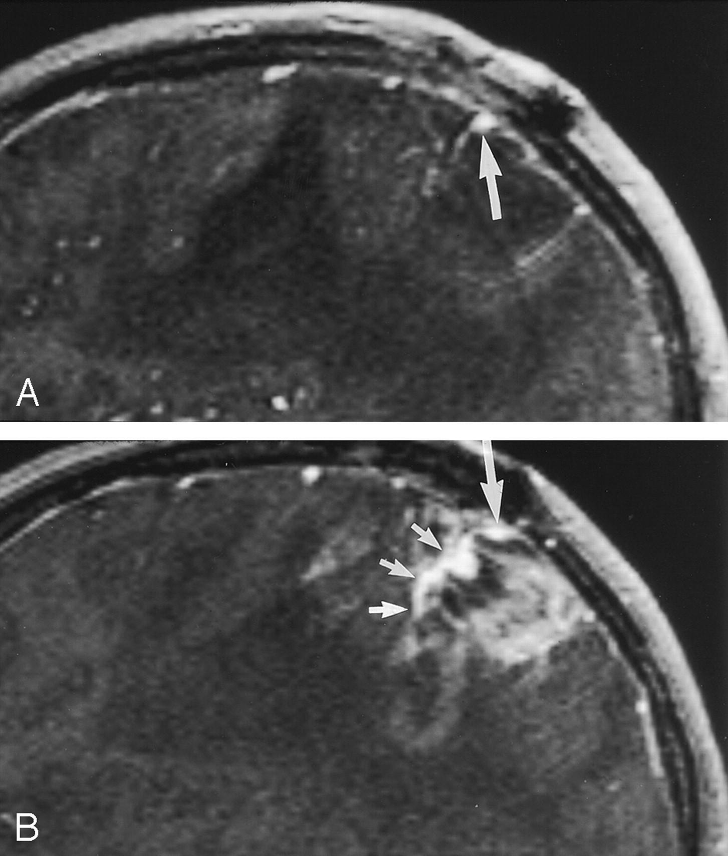
  • Fig 4.
    • Download figure
    • Open in new tab
    • Download powerpoint
    Fig 4.

    Sagittal (A) and parasagittal (B) images in a 44-year-old patient with recurrent left parietal glioblastoma multiforme. Sagittal source images lateral to the contrast-enhancing part of the tumor show a cortical vein (large arrow), which blends with the tumor in the medial part (small arrows in B) and can not be distinguished from the tumor. On conventional angiograms (not shown), the vein was patent.
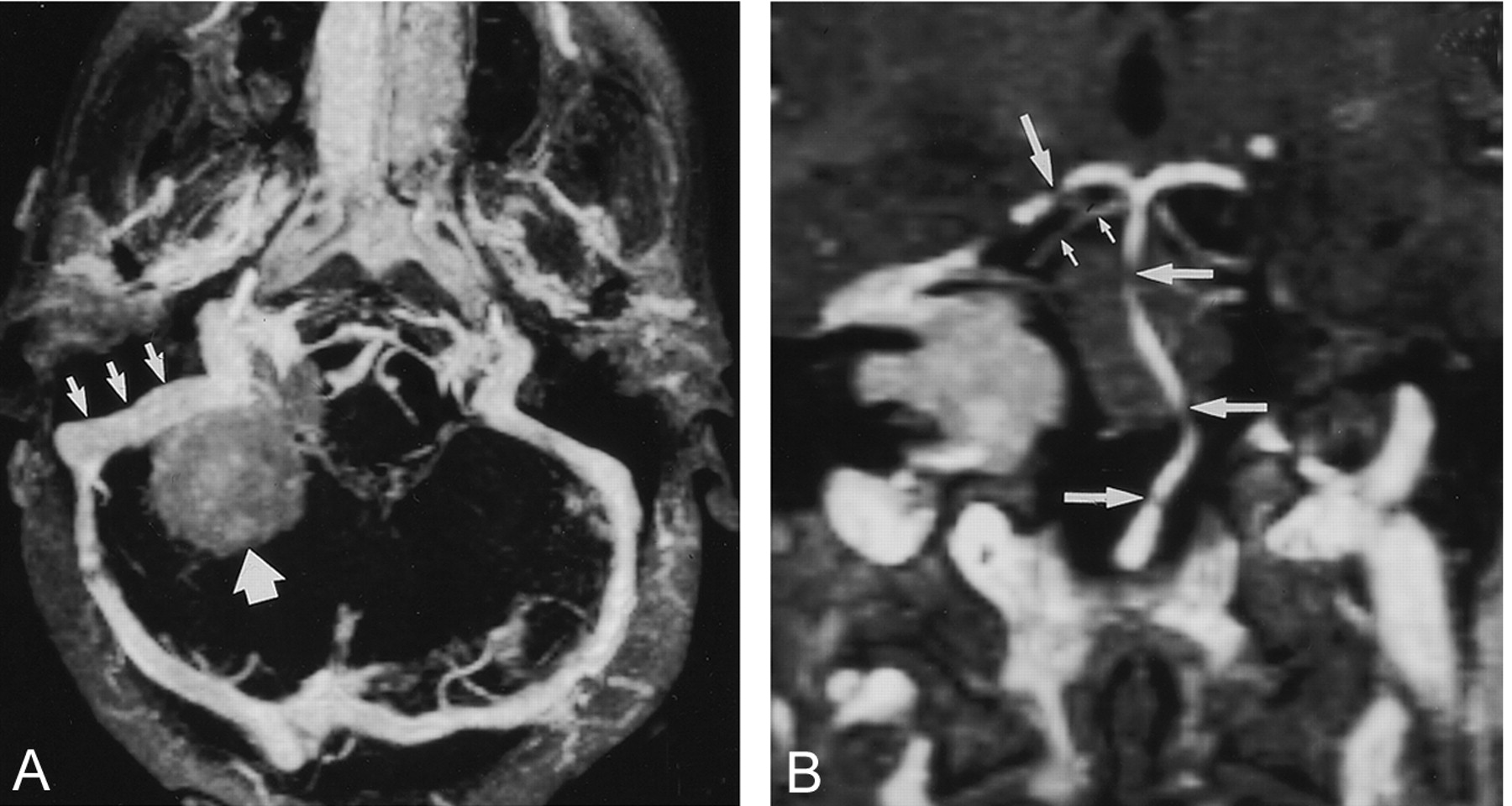
  • Fig 5.
    • Download figure
    • Open in new tab
    • Download powerpoint
    Fig 5.

    Images in a 42-year-old patient with meningioma in right cerebellopontine angle.

    A, Real-time para-axial MIP image with 15-mm thickness shows the sigmoid sinus (small arrows) in relation to a meningioma (large arrow), which has signal intensity lower than that of the sinus. No signs of tumor infiltration are depicted.

    B, Real-time paracoronal MIP image with 2-mm thickness shows the relation of the posterior fossa arteries to the tumor. Gaps in the basilar artery (large horizontal arrows) and in the right posterior cerebral artery (large vertical arrow) are due to partial volume effects. The proximal superior cerebellar artery is shown with great detail (small arrows).

Tables

  • Figures
    • View popup
    TABLE 1:

    Histopathology, contrast enhancement, and comparison of rCBV and vascularity score on DSA images of 22 tumors

    Patient No.HistopathologyContrast EnhancementrCBVVascularity on DSA Image
    1Mixed glioma (low grade)No1.00
    2Mixed glioma (low grade)No1.10
    3Leiomyosarcoma (metastasis)Yes1.81
    4Glioblastoma multiformeYes2.41
    5Anaplastic astrocytomaYes2.60
    6Mixed glioma (low grade)No2.70
    7MeningiomaYes4.12
    8Oligodendroglioma (anaplastic)Yes4.71
    9Mixed glioma (anaplastic)Minimal5.20
    10Anaplastic astrocytomaYes6.12
    11Glioblastoma multiformeYes6.22
    12Mixed glioma (anaplastic)Yes7.62*
    13Glioblastoma multiformeMinimal8.61
    14Adenocarcinoma (metastasis)Yes10.32
    15MeningiomaYes11.82
    16Glioblastoma multiformeYes12.62
    17Mixed neuroepithelial tumor (anaplastic)Minimal13.41
    18Glioblastoma multiformeYes14.22
    19Glioblastoma multiformeYes19.53*
    20Glioblastoma multiformeYes23.63*
    21Glioblastoma multiformeYes23.82
    22Glioblastoma multiformeYes23.92*
    • * Arteriovenous shunting was present.

    • View popup
    TABLE 2:

    Tumor location, surgical approach, and critical vessels evaluated in 22 tumors

    Patient No.Location and SideSurgical ApproachVessels Evaluated*Arterial Segments Evaluated
    1Temporal, LPterionalCortical veinsMCA (2–4)
    2Temporal, RTemporal craniotomyCortical veinsNone
    3TectalStereotactic biopsyCortical veins, ISS, deep venous systemPCA, SCA
    4Temporal, LStereotactic biopsyCortical veinsMCA (2–4), AchA, Pch
    5Parietal, LStereotactic biopsyCortical veins, deep venous systemNone
    6High parietal, LParietal craniotomyNone
    7Cerebellopontine, LTranspetrosalCortical veins, sigmoid sinus, SPSPCA, Pcom, SCA, AICA, PICA
    8Frontal, LFrontal craniotomyCortical veins, ISS, IV, deep venous systemACA (2–4)
    9Parietal, RPosterior frontal craniotomyCortical veinsMCA (2–4)
    10Frontal, RFrontal craniotomyCortical veins, SSSNone
    11Frontal, RFrontal craniotomyCortical veins, IVACA (2–4), MCA (2–4)
    12Insula, LPosterior frontal craniotomyCortical veinsMCA (4)
    13Frontal, RFrontal craniotomyCortical veinsACA (2–4)
    14High parietal, LFrontal craniotomyCortical veinsMCA (4)
    15Parasagittal, LFrontal craniotomyCortical veins, SSS, IVNone
    16Parietal, LParietal craniotomyCortical veins, IVNone
    17High parietal, LStereotactic biopsyCortical veins, ISS, IVACA (2–4)
    18High occipital, LSuperior parietal lobuleCortical veins, SSV, IVACA (2–4)
    19Parietotemporal, LPosterior temporal craniotomyCortical veinsNone
    20Thalamic, LTranscorticalCortical veins, deep venous systemAchA, PchA, thalamoperforators
    21Frontal, LFrontal craniotomyCortical veins, IVACA (3–4)
    22Occipital, LOccipital craniotomyCortical veins, SSS, IVNone
    • * ISS indicates inferior sagittal sinus; SPS, superior petrosal sinus; IV, interhemispheric veins; and SSS, superior sagittal sinus.

    • † ACA indicates anterior cerebral artery; Acha, anterior choroidal artery; AICA, anterior inferior cerebellar artery; MCA, middle cerebral artery; PCA, posterior cerebral artery; Pcha, posterior choroidal artery; Pcom, posterior communicating artery; PICA, posterior inferior cerebellar artery; and SCA, superior cerebellar artery.

    • View popup
    TABLE 3:

    Scoring table for vascular information provided on VIBE images with DSA as the standard of reference

    Vascular StructureNo. of PatientsScore*
    AllPartialNone
    Total veins221750
     Cortical veins221840
     Interhemispheric veins8710
     Venous sinuses8800
     Deep venous system4400
    Total arteries14077
    • * Scores were as follows: All indicated that VIBE imaging provided all of the relevant information; partial, VIBE imaging provided some but not all of the relevant information; and none, VIBE imaging provided none of the relevant information.

PreviousNext
Back to top

In this issue

American Journal of Neuroradiology: 23 (10)
American Journal of Neuroradiology
Vol. 23, Issue 10
1 Nov 2002
  • Table of Contents
  • Index by author
Advertisement
Print
Download PDF
Email Article

Thank you for your interest in spreading the word on American Journal of Neuroradiology.

NOTE: We only request your email address so that the person you are recommending the page to knows that you wanted them to see it, and that it is not junk mail. We do not capture any email address.

Enter multiple addresses on separate lines or separate them with commas.
Preoperative Assessment of Intracranial Tumors with Perfusion MR and a Volumetric Interpolated Examination: A Comparative Study with DSA
(Your Name) has sent you a message from American Journal of Neuroradiology
(Your Name) thought you would like to see the American Journal of Neuroradiology web site.
CAPTCHA
This question is for testing whether or not you are a human visitor and to prevent automated spam submissions.
Cite this article
Stephan G. Wetzel, Soonmee Cha, Meng Law, Glyn Johnson, John Golfinos, Peter Lee, Peter Kim Nelson
Preoperative Assessment of Intracranial Tumors with Perfusion MR and a Volumetric Interpolated Examination: A Comparative Study with DSA
American Journal of Neuroradiology Nov 2002, 23 (10) 1767-1774;

Citation Manager Formats

  • BibTeX
  • Bookends
  • EasyBib
  • EndNote (tagged)
  • EndNote 8 (xml)
  • Medlars
  • Mendeley
  • Papers
  • RefWorks Tagged
  • Ref Manager
  • RIS
  • Zotero
0 Responses
Respond to this article
Share
Bookmark this article
Preoperative Assessment of Intracranial Tumors with Perfusion MR and a Volumetric Interpolated Examination: A Comparative Study with DSA
Stephan G. Wetzel, Soonmee Cha, Meng Law, Glyn Johnson, John Golfinos, Peter Lee, Peter Kim Nelson
American Journal of Neuroradiology Nov 2002, 23 (10) 1767-1774;
del.icio.us logo Twitter logo Facebook logo Mendeley logo
  • Tweet Widget
  • Facebook Like
  • Google Plus One

Jump to section

  • Article
    • Abstract
    • Methods
    • Results
    • Discussion
    • Conclusion
    • Footnotes
    • References
  • Figures & Data
  • Info & Metrics
  • Responses
  • References
  • PDF

Related Articles

  • No related articles found.
  • PubMed
  • Google Scholar

Cited By...

  • No citing articles found.
  • Crossref
  • Google Scholar

This article has not yet been cited by articles in journals that are participating in Crossref Cited-by Linking.

More in this TOC Section

  • Progression of Microstructural Damage in Spinocerebellar Ataxia Type 2: A Longitudinal DTI Study
  • Usefulness of Quantitative Susceptibility Mapping for the Diagnosis of Parkinson Disease
  • Evaluating the Effects of White Matter Multiple Sclerosis Lesions on the Volume Estimation of 6 Brain Tissue Segmentation Methods
Show more Brain

Similar Articles

Advertisement

Indexed Content

  • Current Issue
  • Accepted Manuscripts
  • Article Preview
  • Past Issues
  • Editorials
  • Editors Choice
  • Fellow Journal Club
  • Letters to the Editor

Cases

  • Case Collection
  • Archive - Case of the Week
  • Archive - Case of the Month
  • Archive - Classic Case

Special Collections

  • Special Collections

Resources

  • News and Updates
  • Turn around Times
  • Submit a Manuscript
  • Author Policies
  • Manuscript Submission Guidelines
  • Evidence-Based Medicine Level Guide
  • Publishing Checklists
  • Graphical Abstract Preparation
  • Imaging Protocol Submission
  • Submit a Case
  • Become a Reviewer/Academy of Reviewers
  • Get Peer Review Credit from Publons

Multimedia

  • AJNR Podcast
  • AJNR SCANtastic
  • Video Articles

About Us

  • About AJNR
  • Editorial Board
  • Not an AJNR Subscriber? Join Now
  • Alerts
  • Feedback
  • Advertise with us
  • Librarian Resources
  • Permissions
  • Terms and Conditions

American Society of Neuroradiology

  • Not an ASNR Member? Join Now

© 2025 by the American Society of Neuroradiology All rights, including for text and data mining, AI training, and similar technologies, are reserved.
Print ISSN: 0195-6108 Online ISSN: 1936-959X

Powered by HighWire