
Article Figures & Data
Figures
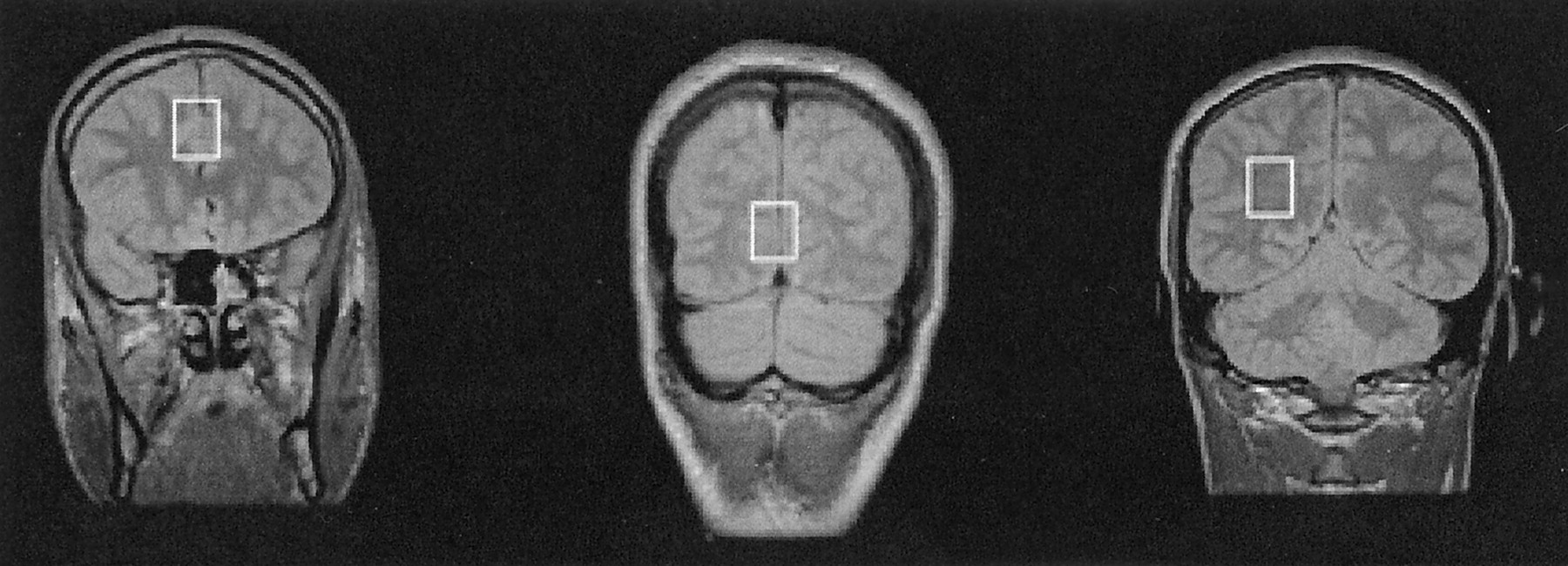
- Fig 1.
Proton density-weighted MR image (4000/22) shows the three voxel locations for the localized 1H MRS studies: midfrontal gray matter (left), mid-occipital gray matter (middle), and right parietal white matter (right).
- Fig 2.
Proton MRS spectra from the midfrontal gray matter region in a control subject (age, 23 years) and an MDMA user (cumulative lifetime exposure, 200 tablets; age, 26 years). The spectrum in the MDMA user is representative for the whole group and shows a reduction in NAA levels, with Cr levels similar to those in the control subject. Note that MI levels are elevated in this specific MDMA user, although for the group as a whole, mean MI measures in MDMA users did not statistically differ from those of control subjects.
- Fig 3.
Plot shows the correlation between the NAA/Cr ratio in the frontal cortex and the extent of previous MDMA use.


Tables
Characteristic Control Subjects(N = 12) MDMA Users(N = 15) Age (y) 27.0 ± 4.1 27.2 ± 5.3 MDMA use Duration (y) NA 5.6 (2.5–12.0) Usual dose (no. of tablets) NA 2.1 (1.5–3.5) Lifetime dose (no. of tablets) NA 723 (55–2176) Time since last tablet (wk) NA 12.0 (1–40) Other drugs Alcohol (units per wk) 13.4 ± 11.9 17.5 ± 13.8 Tobacco (no. of cigarettes per day) 10.8 ± 3.7 13.3 ± 14.9 Cannabis (no. of joints in the last 3 mo) 2.3 ± 0.5 158.3 ± 178.9* Amphetamine (no. of uses in the last 3 mo) NA 5.0 ± 8.7 Note.—Data are the means ± SDs or means (ranges). NA indicates not applicable.
* The difference between MDMA users and control subjects was statistically significant (P < .01, unpaired Student t test).
- TABLE 2:
1H MRS findings in gray and white matter regions in MDMA users and control subjects
Voxel Location Control Subjects* (N = 12) MDMA Users* (N = 15) Difference (%) P Value† Gray matter ratios‡ Midfrontal NAA/Cr 1.62 ± 0.20 1.43 ± 0.21 −11.7 .04 NAA/Cho 2.06 ± 0.23 1.78 ± 0.27 −14.0 .03 MI/Cr 0.65 ± 0.08 0.64 ± 0.06 −1.5 .62 Midoccipital NAA/Cr 1.56 ± 0.19 1.54 ± 0.21 −0.1 .72 NAA/Cho 3.04 ± 0.52 2.67 ± 0.61 −12.2 .12 MI/Cr 0.60 ± 0.07 0.57 ± 0.08 −5.0 .29 Right parietal white matter ratios§ NAA/Cr 1.90 ± 0.10 1.78 ± 0.19 −6.3 NA NAA/Cho 1.84 ± 0.16 1.96 ± 0.32 +6.5 NA MI/Cr 0.64 ± 0.06 0.62 ± 0.09 −3.1 NA * Data are the means ± SDs.
† NA indicates not applicable.
‡ At MANOVA, F = 2.82, df = 6.0, P = .045.
§ At MANOVA, F = 1.79, df = 3.0, P = .180.


