
Article Figures & Data
Figures
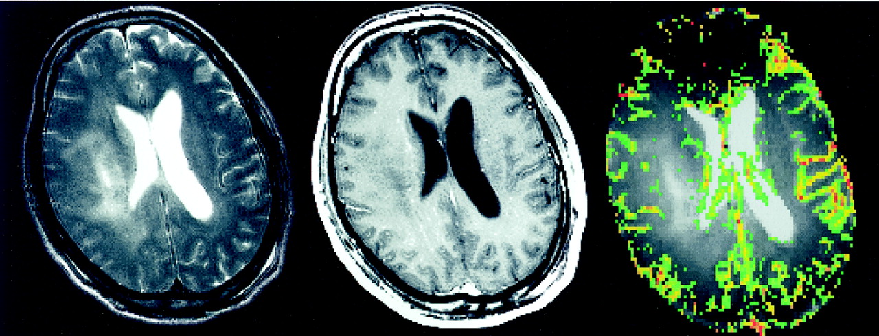
- Fig 1.
Images in a 35-year-old woman with pathologically confirmed gliomatosis cerebri.
Left, T2-weighted (3400/119) MR image demonstrates diffuse bilateral frontal signal intensity abnormality.
Middle, Contrast-enhanced axial T1-weighted (600/14) image demonstrates no evidence of contrast enhancement.
Right, Color overlay rCBV map demonstrates no evidence of increased perfusion in the region of signal intensity abnormality on the T2-weighted image.
- Fig 2.
Images in a 42-year-old man with pathologically confirmed gliomatosis cerebri.
Left, T2-weighted (3400/119) MR image demonstrates a region of signal intensity abnormality in the right periventricular white matter.
Middle, Contrast-enhanced axial T1-weighted (600/14) image demonstrates subtle evidence of contrast enhancement.
Right, Color overlay rCBV map reveals that the areas corresponding to signal intensity abnormality on the T2-weighted image do not demonstrate increased perfusion.
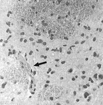
- Fig 3.
High-power view of histopathologic section of gliomatosis cerebri (hematoxylin-eosin with Luxol fast-blue stain, original magnification ×200). There are moderately pleomorphic glial cells in a diffusely infiltrating pattern with relative preservation of underlying cytoarchitecture. No evidence of vascular proliferation or necrosis is seen. A small collapsed vascular structure (arrow) is noted.
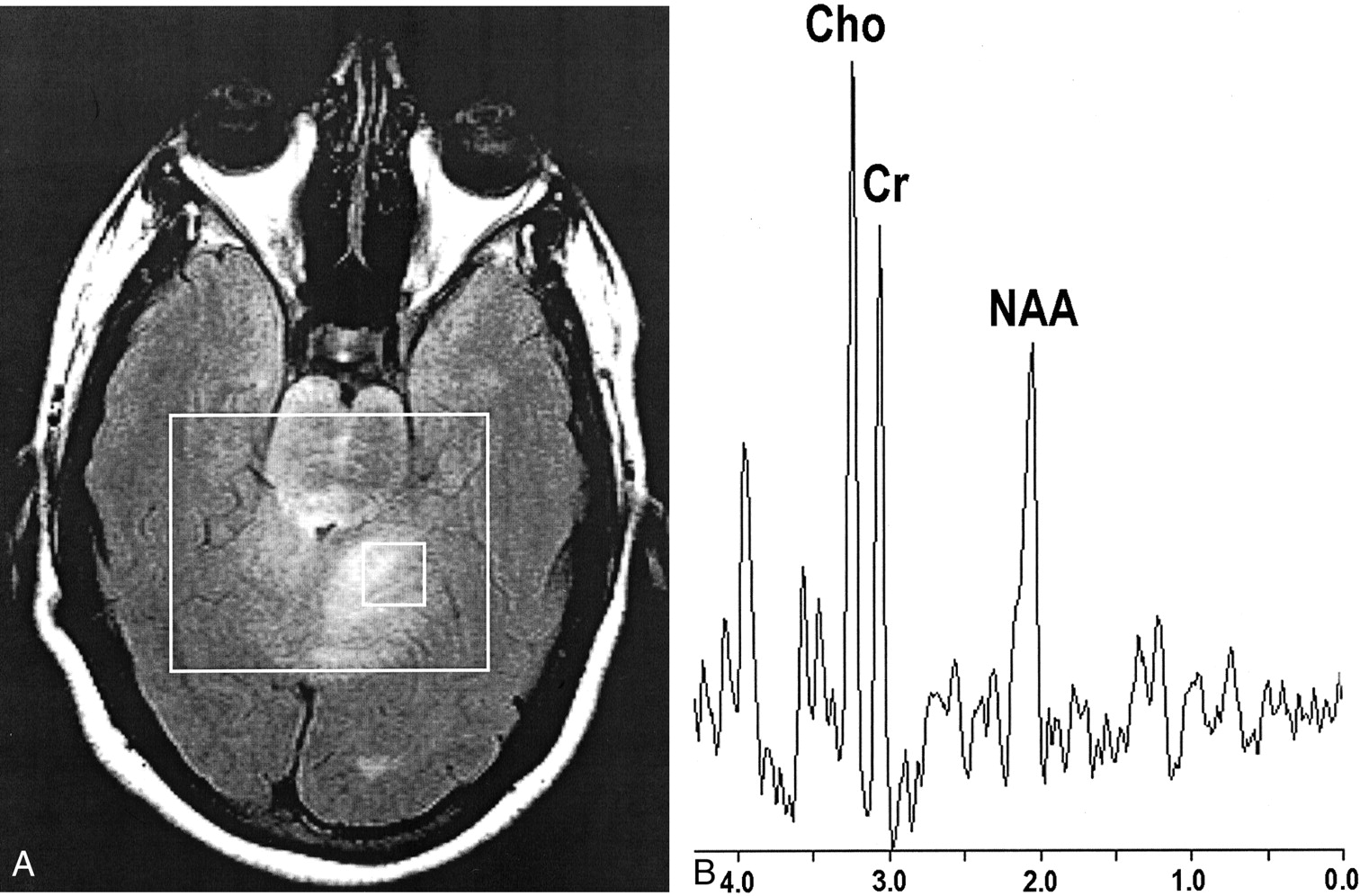
- Fig 4.
Findings in a 21-year-old woman with gliomatosis cerebri.
A, Axial fluid-attenuated inversion recovery (9000/110) image with localizer (box).
B, Corresponding spectrum (chemical shift imaging, PRESS, 1500/144) reveals elevated Cho/Cr and depressed NAA/Cr ratios.


Tables
Patient No./Age (y)/Sex rCBV* Location Clinical Presentation 1/11/M 0.92 ± 0.66 Left hemisphere, extension into right thalamus Headache 2/18/F 1.17 ± 0.43 Right temporoparietal lobes Headache 3/21/F 0.75 ± 0.15 Bilateral centrum semiovale and periventricular white matter; bilateral thalami; bilateral cerebellar hemispheres and brain stem Headache, ataxia, cranial nerve disturbances 4/27/M 1.26 ± 0.29 Left frontoparietal lobe and centrum semiovale Paresthesia 5/35/F 0.87 ± 0.37 Left frontal lobe, extension into corpus callosum and right frontal lobe Headache, memory deficits, visual disturbance 6/42/M 1.09 ± 0.39 Right hemisphere involving basal ganglia, frontal lobe and thalamus, extension into contralateral basal ganglia Fatigue, headache, memory deficits 7/75M 1.09 ± 0.35 Right hemisphere white matter, involvement of bilateral thalami, cerebral peduncles, pons, cerebellar peduncles Mood disturbance, gait disturbance * Date are the mean ± SD. Overall mean was 1.02 ± 0.42.
Patient No./Age(y)/Sex Abnormal Voxels Maximal Minimal Normal-Appearing Voxels Cho/Cr Cho/NAA NAA/Cr Cho/Cr Cho/NAA NAA/Cr 2/18/F 2.14 2.57 0.83 0.73 0.48 1.53 3/21/F 1.44 1.76 0.7 0.76 0.52 1.46 6/42/M 2.0 1.15 0.83 1.0 0.69 1.45 Mean ± SD 1.86 ± 0.37 1.83 ± 0.71 0.79 ± 0.08 0.83 ± 0.15 0.56 ± 0.11 1.48 ± 0.04
In this issue


Jump to section
Related Articles
Cited By...
- No citing articles found.