
Article Figures & Data
Figures
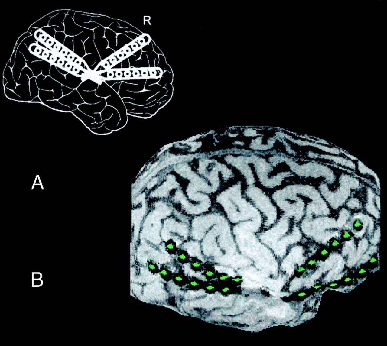
- Fig 1.
Positions of subdural strip electrodes in a 16-year-old male adolescent with pharmacorefractory focal epilepsy resulting from tuberous sclerosis. Invasive recordings were necessary to identify the electrophysiologically leading tuber of three tubers shown on MR images. Recordings showed a seizure-onset zone over the right temporal tuber with rapid spread to the frontal cortex; surgical excision of the temporal tuber based on the recordings rendered the patient seizure free.
A, Schematic displays the planned implantation of subdural strip electrodes. R indicates right.
B, Lateral curvilinear reformation shows the cortical surface with four overlying subdural strip electrodes consisting of six electrode contacts each (green). Whereas planar MR imaging sections (not shown) were difficult to interpret because of the underlying convexity of the cortical surface, the curvilinear reformation clearly depicts the spatial relationships between electrodes and the sylvian fissure and cortical gyrus.
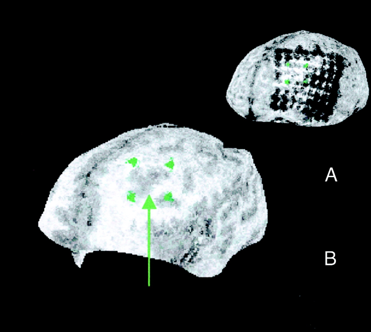
- Fig 2.
Curvilinear reformations show relative position of subdural electrode contacts to a cortical dysplasia located deep in the left frontal lobe. In this 23-year-old man with pharmacoresistant focal epilepsy with hypermotor and right tonic seizures from his first year of life, an 8 × 8 subdural grid electrode was implanted over the left frontal cortex for identification of the seizure-onset zone and mapping of language and motor cortex. As mapping of the speech area using electrical stimulation showed a wide extension of the language area, which corresponded to extended activated areas at functional MR imaging during language tasks, the relative position to an underlying suspected cortical dysplasia was crucial for the possibility of removing the lesion without language deficit. Identification of the electrode contacts overlying the dysplasia was based on the results of curvilinear reformatting of 3D MR imaging data. They were found to be part of both the seizure-onset zone and language area. Because of the possible risk of aphasia with a resection of the seizure-onset zone, surgery was deferred in this patient.
A, Projection of grid positions to the underlying cortical dysplasia at a depth of 20 mm.
B, Selected grid positions are indicated by green markers, and the dysplastic cortex showing blurred gray matter–white matter junction is indicated by a green arrow.
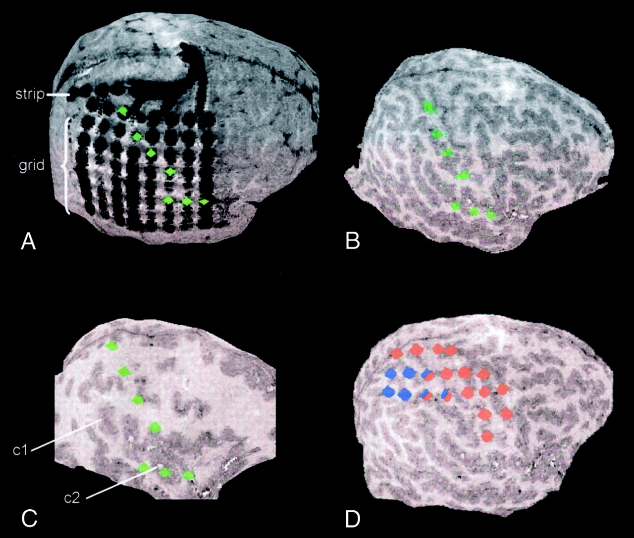
- Fig 3.
Curvilinear reformations show electrode positions of a 64-contact subdural grid and a parasagittally placed six-contact-strip electrode. Subdural recordings were performed in this 12-year-old patient with an extended cortical dysplasia including the right supramarginal gyrus to delineate the borders of the irritative zone and seizure-onset zone and for functional mapping of the sensory and motor strip. On planar MR imaging sections (not shown), the spatial relationship between electrode positions and the central sulcus could not be depicted clearly, and the relationship between the subdural strip electrode and the grid electrode remained uncertain. Curvilinear reformatting provided exact information about the relative positions of strip and grid electrodes, as well as the spatial relationship of grid electrode positions and the central sulcus, sylvian fissure, and dysplastic cortex. This was used to plan functional mapping of the somatosensory and somatomotor cortex by electrical stimulation and to plan the resection, which rendered the patient seizure free without persistent neurologic deficits.
A, At a depth of 1 mm, the relative positions of strip and grid electrodes are displayed. Green markers indicate the electrode contacts overlying the postcentral gyrus and the sylvian fissure.
B, At a depth of 10 mm, the relative position of the anatomically marked electrodes (green) and cortical gyration is shown.
C, At a depth of 22 mm, thickening of the cortex along the supramarginal gyrus (c1) and around the superior temporal sulcus (c2) is prominent. Again, the relative position of the dysplastic areas to the anatomically marked electrode contacts (green) is shown.
D, Markers display the results of electrocorticographical mapping of the motor (red) and sensory (blue) strip.


