
Article Figures & Data
Figures
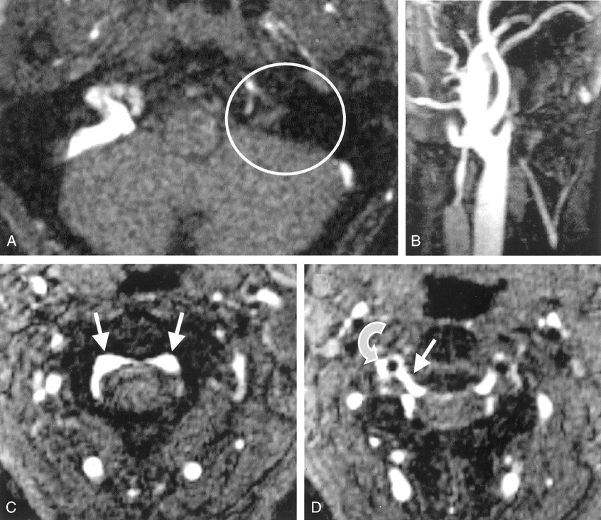
- Fig 1.
Cervical 2D TOF MR angiograms.
A, Axial source image depicts no contrast enhancement within the left sinus sigmoideus (circle).
B, Maximum intensity projection (MIP) reconstruction image of the left IJV depicts only a short proximal vessel segment with regular contrast enhancement.
C, Axial source image obtained at the level of C2 shows prominent epidural veins of the anterior intraspinal system (arrows).
D, Axial source image obtained at the level of the C2–3 intervertebral space shows that a radicular vein (straight arrow) connects the anterior intraspinal system to the right vertebral vein (curved arrow) next to the vertebral artery (flow void).
- Fig 2.
Duplex sonogram depicts the right vertebral vein (VV) and vertebral artery (VA) at the level of C4 and C5. Note the prominent segmental venous inflow (SV) with a high systolic flow velocity of 38 cm/s.
- Fig 3.
MIP of the contrast-enhanced T1-weighted gradient-echo MRV image of the left IJV. The vein has good contrast enhancement below the skull base (arrows).
- Fig 4.
Axial images from multisection CTA.
A, Bilateral asymmetrical venous contrast enhancement at the level of the jugular foramen is depicted (circles).
B, Prominent posterior condylar emissary vein is depicted on the right side (circle).


In this issue


Jump to section
Related Articles
Cited By...
- Positional Venous MR Angiography: An Operator-Independent Tool to Evaluate Cerebral Venous Outflow Hemodynamics
- Comparison of MR and Contrast Venography of the Cervical Venous System in Multiple Sclerosis
- Value of MR Venography for Detection of Internal Jugular Vein Anomalies in Multiple Sclerosis: A Pilot Longitudinal Study