
Article Figures & Data
Figures
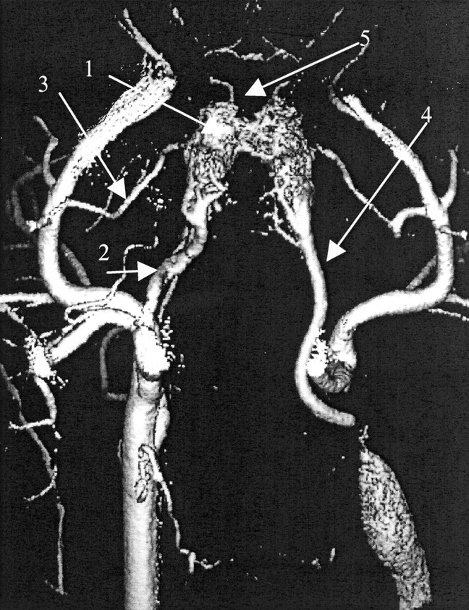
- Fig 1.
Three-dimensional rotation digital subtraction angiographic (DSA) image of the RM nidus (arrow 1) after surgical construction of the end-to-end anastomosis between the CCA and external jugular vein (EJV) on the left side. The left CCA distal to the fistula has been ligated. Note the main feeding ascending PA (arrow 2), RA of the meningeal branch of the right ECA (arrow 3) and left PA (arrow 4, draining vein). Also note that the anterior branch of the right internal carotid artery (ICA) originates from the RM (arrow 5).
- Fig 2.
Angiograms of the RA.
A, Superselective angiogram demonstrates collaterals between the RM and basilar system (arrow).
B, Angiogram obtained after surgical creation of the fistula between the CCA and EJV shows the extensive collateral network between the right VA (arrow 1) and ascending PA (arrow 2) on the left side. Note filling of the right ascending PA (arrow 3) and RM (arrow 4) by ipsilateral collaterals. The BA (arrow 5) has a small diameter.
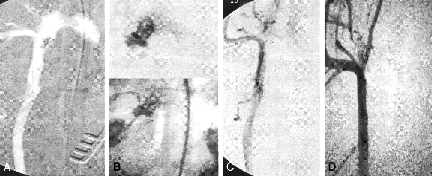
- Fig 3.
Angiograms obtained before and after embolization.
A, Oblique image obtained prior to embolization of the RM with roadmap technique shows that the microcatheter is positioned in the ascending PA close to the nidus and distal to the collaterals to the VA.
B, Oblique image obtained after embolization of the PA with pure 2-P-HEMA. The embolic agent completely fills the compartment. Note the intense radiopacity. Although the microcatheter is embedded in the 2-P-HEMA, it can be retracted without sticking. Top image shows embolization with the roadmap technique. Bottom image is an unsubtracted picture of the embolized nidus.
C, Oblique control angiogram of the right CCA obtained after embolization of the ascending PA. The microcatheter is in the same position as in A. Collateral filling of the RM by the RA and AA is preserved.
D, Control angiogram of the right CCA obtained 8 mo after embolization depicts long-term occlusion of the embolized feeder.
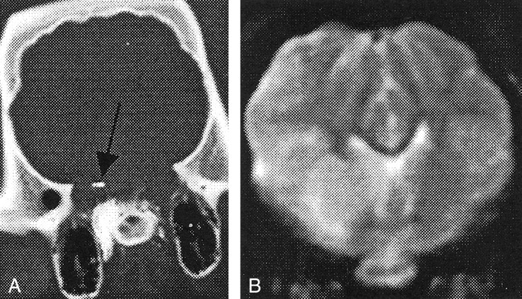
- Fig 4.
Image obtained after embolization with 50% 2-P-HEMA.
A, CT scan the of intracerebral vessels (arrow) shows intense contrast in the embolized RM.
B, Coronal T2-weighted MR image obtained after embolization with 50% 2-P-HEMA shows a large cerebral infarction on the right side; the animal died.
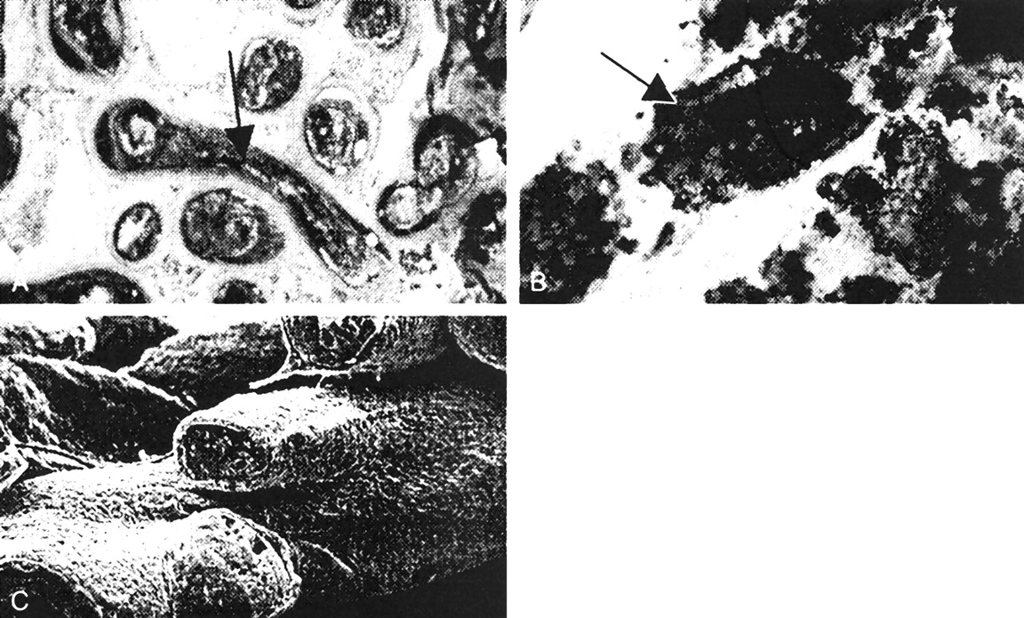
- Fig 5.
Histopathologic and electron microscopic findings after embolization.
A, Photomicrograph of an RM obtained 15 h after embolization with 50% 2-P-HEMA shows that the vessels (arrow) of the embolized RM are mostly filled with precipitated 2-P-HEMA, which is dark because of the presence of tungsten. As many as 90% of the vessels were completely patched with the embolic agent; others were partially occluded and thrombosed (original magnification ×25).
B, Photomicrograph obtained 6 mo after embolization with pure 2-P-HEMA shows that the tungsten has been replaced by amorphous material resembling a calcification (arrow) (original magnification ×50).
C, Electron micrograph of an AVM embolized with 50% 2-P-HEMA shows homogeneous casting of the vessel lumen due to precipitated 2-P-HEMA.


Tables
Animal Patent AVM 2-P-HEMA Concentration Injection Location and Volume (mL) Injection Speed (mL/mim) Occlusion of Embolized Nidus Compartment Complications Detected at Angiography Postembolization MR Imaging and CT Finding Time of Follow-Up Angiography and Nidus Occlusion 1 Yes Pure PA, 0.6; RA, 0.4 0.1 PA complete, RA complete None Partial infarction in right hemisphere, no 2-P-HEMA in brain vessels At 1 and 8 mo: PA complete, RA complete 2 Yes Pure PA, 0.4 0.4 PA complete Vasospasm Normal At 3 mo: complete; at 5 mo: partial recanalization 3 Yes Pure PA, 0.7; RA, 0.4 0.1 PA complete, RA complete None Normal At 2 and 7 mo: PA complete, RA complete 4 Yes Pure PA, 0.6 0.1 PA complete None Normal At 1 and 6 mo: complete 5 Yes Pure PA, 0.5 0.1 PA complete None Normal At 2 and 6 mo: complete 6 Yes Pure PA, 0.8 0.1 PA complete None Normal At 1 and 8 mo: complete 7 Yes 50% PA. 0.6 0.1 PA incomplete Embolic events Infarction in right hemisphere, 2-P-HEMA in brain vessels At 15 h: incomplete * To make pure 2-P-HEMA, 11% 2-P-HEMA by volume was dissolved in 89% ethenol by volume. To make 50% 2-P-HEMA, 5.5% 2-P-HEMA by volume was dissolved in 79.5% ethanbol boy volume. Sterilized 2-P-HEMA (1 mL) was mixed with tungsten (0.005 g).


