Advertisement

Main menu

  • Home
  • Content
    • Current Issue
    • Accepted Manuscripts
    • Article Preview
    • Past Issue Archive
    • AJNR Case Collection
    • Case of the Week Archive
    • Classic Case Archive
    • Case of the Month Archive
  • Special Collections
    • Spinal CSF Leak Articles (Jan 2020-June 2024)
    • 2024 AJNR Journal Awards
    • Most Impactful AJNR Articles
  • Multimedia
    • AJNR Podcast
    • AJNR Scantastics
    • Video Articles
  • For Authors
    • Submit a Manuscript
    • Author Policies
    • Fast publishing of Accepted Manuscripts
    • Graphical Abstract Preparation
    • Manuscript Submission Guidelines
    • Imaging Protocol Submission
    • Submit a Case for the Case Collection
  • About Us
    • About AJNR
    • Editorial Board
  • More
    • Become a Reviewer/Academy of Reviewers
    • Subscribers
    • Permissions
    • Alerts
    • Feedback
    • Advertisers
    • ASNR Home
  • Other Publications
    • ajnr

User menu

  • Alerts
  • Log in

Search

  • Advanced search
American Journal of Neuroradiology

American Journal of Neuroradiology

ASHNR American Society of Functional Neuroradiology ASHNR American Society of Pediatric Neuroradiology ASSR
  • Alerts
  • Log in

Advanced Search

  • Home
  • Content
    • Current Issue
    • Accepted Manuscripts
    • Article Preview
    • Past Issue Archive
    • AJNR Case Collection
    • Case of the Week Archive
    • Classic Case Archive
    • Case of the Month Archive
  • Special Collections
    • Spinal CSF Leak Articles (Jan 2020-June 2024)
    • 2024 AJNR Journal Awards
    • Most Impactful AJNR Articles
  • Multimedia
    • AJNR Podcast
    • AJNR Scantastics
    • Video Articles
  • For Authors
    • Submit a Manuscript
    • Author Policies
    • Fast publishing of Accepted Manuscripts
    • Graphical Abstract Preparation
    • Manuscript Submission Guidelines
    • Imaging Protocol Submission
    • Submit a Case for the Case Collection
  • About Us
    • About AJNR
    • Editorial Board
  • More
    • Become a Reviewer/Academy of Reviewers
    • Subscribers
    • Permissions
    • Alerts
    • Feedback
    • Advertisers
    • ASNR Home
  • Follow AJNR on Twitter
  • Visit AJNR on Facebook
  • Follow AJNR on Instagram
  • Join AJNR on LinkedIn
  • RSS Feeds

Welcome to the new AJNR, Updated Hall of Fame, and more. Read the full announcements.


AJNR is seeking candidates for the position of Associate Section Editor, AJNR Case Collection. Read the full announcement.


Getting new auth cookie, if you see this message a lot, tell someone!
OtherSPINE

Diffusion-Weighted Imaging in a Patient with Vertebral and Epidural Abscesses

James D. Eastwood, Robin T. Vollmer and James M. Provenzale
American Journal of Neuroradiology March 2002, 23 (3) 496-498;
James D. Eastwood
  • Find this author on Google Scholar
  • Find this author on PubMed
  • Search for this author on this site
Robin T. Vollmer
  • Find this author on Google Scholar
  • Find this author on PubMed
  • Search for this author on this site
James M. Provenzale
  • Find this author on Google Scholar
  • Find this author on PubMed
  • Search for this author on this site
  • Article
  • Figures & Data
  • Info & Metrics
  • Responses
  • References
  • PDF
Loading

Article Figures & Data

Figures

  • Fig 1.
    • Download figure
    • Open in new tab
    • Download powerpoint
    Fig 1.

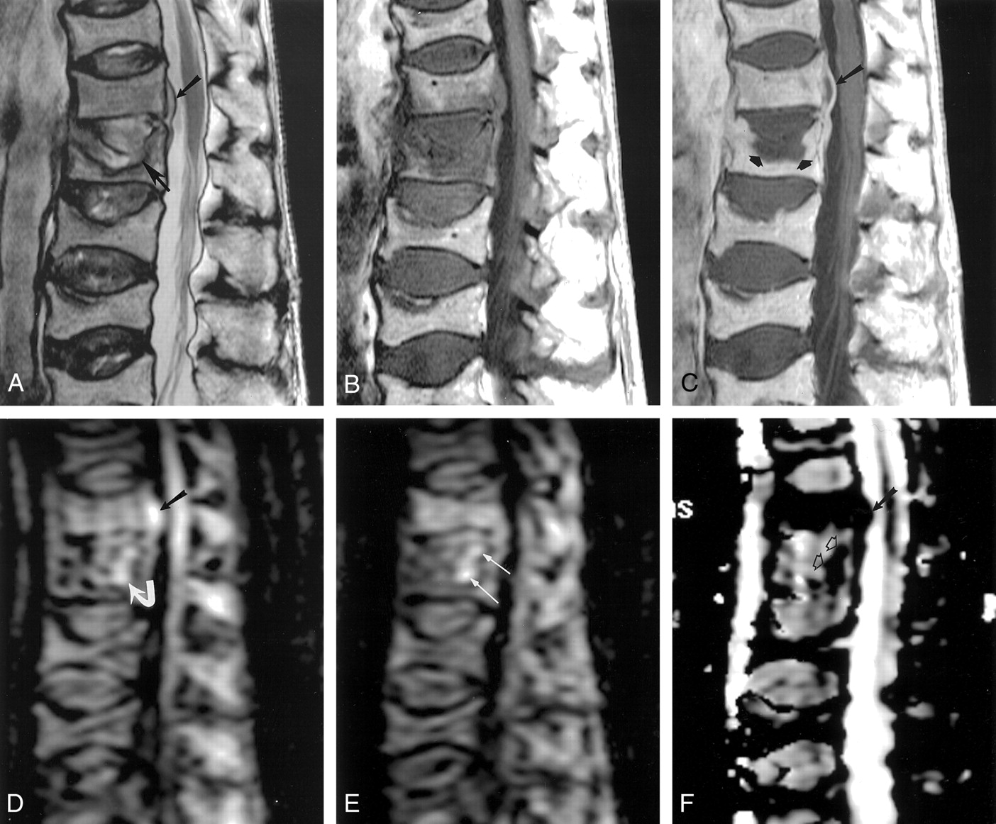
    Images obtained in an 83-year-old man with bacteremia and back pain.

    A, T2-weighted sagittal MR image (3000/103) shows a small ventral extradural mass (upper arrow) and a fracture and abnormal signal intensity adjacent to the superior endplate of the L1 vertebral body (lower arrow).

    B and C, T1-weighted sagittal MR images (815/30) obtained before (B) and after (C) IV administration of gadolinium-based contrast material show central nonenhancement in the extradural mass (long arrow in C) and in a substantial portion of the superior portion of the L1 vertebral body (short arrows in C). Both areas of nonenhancement were interpreted as abscesses.

    D, Sagittal diffusion-weighted MR image (1215/80/2, b = 500) obtained at the same location as in A–C shows very high signal intensity within the extradural abscess (straight arrow) and high signal intensity within the region of the vertebral abscess (curved arrow).

    E, Sagittal diffusion-weighted MR image (1215/80/2) obtained immediately to the left of D shows high signal intensity in the vertebral abscess (arrows).

    F, ADC map obtained at same location as in D shows the dark appearance of both epidural (solid arrow) and vertebral abscess (open arrows) regions.

  • Fig 2.
    • Download figure
    • Open in new tab
    • Download powerpoint
    Fig 2.

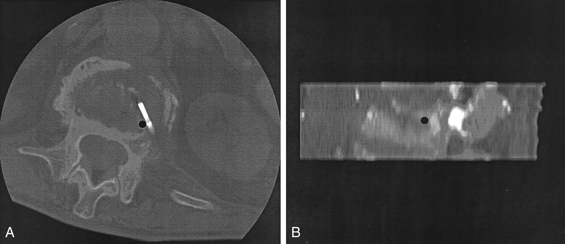
    Images obtained during biopsy of the vertebral body thought to contain an abscess.

    A, Transverse CT image obtained at the level of L1 during needle placement for biopsy shows the position of the biopsy needle with the inner stylet at the final position prior to the aspiration of fluid from the bone. The black dot marks the location of the tip of the needle after removal of the inner stylet.

    B, Sagittal reformatted image that was constructed from the prebiopsy CT scan. The black dot shows the position of the needle tip shown in A.

  • Fig 3.
    • Download figure
    • Open in new tab
    • Download powerpoint
    Fig 3.

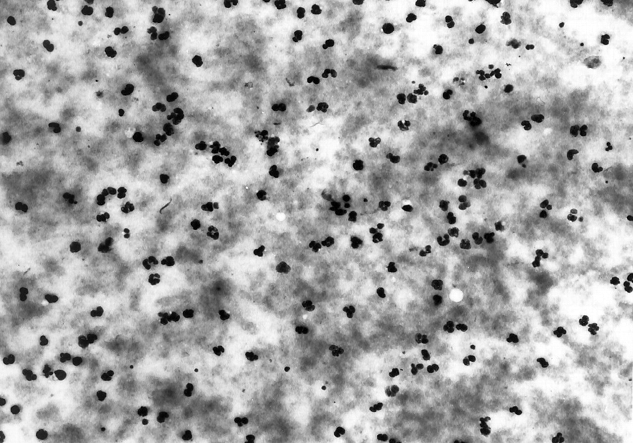
    Photomicrograph shows debris and multiple polymorphonuclear leukocytes. The findings were interpreted as being consistent with acute infection (hematoxylin-eosin, original magnification ×800).

PreviousNext
Back to top

In this issue

American Journal of Neuroradiology: 23 (3)
American Journal of Neuroradiology
Vol. 23, Issue 3
1 Mar 2002
  • Table of Contents
  • Index by author
Advertisement
Print
Download PDF
Email Article

Thank you for your interest in spreading the word on American Journal of Neuroradiology.

NOTE: We only request your email address so that the person you are recommending the page to knows that you wanted them to see it, and that it is not junk mail. We do not capture any email address.

Enter multiple addresses on separate lines or separate them with commas.
Diffusion-Weighted Imaging in a Patient with Vertebral and Epidural Abscesses
(Your Name) has sent you a message from American Journal of Neuroradiology
(Your Name) thought you would like to see the American Journal of Neuroradiology web site.
CAPTCHA
This question is for testing whether or not you are a human visitor and to prevent automated spam submissions.
Cite this article
James D. Eastwood, Robin T. Vollmer, James M. Provenzale
Diffusion-Weighted Imaging in a Patient with Vertebral and Epidural Abscesses
American Journal of Neuroradiology Mar 2002, 23 (3) 496-498;

Citation Manager Formats

  • BibTeX
  • Bookends
  • EasyBib
  • EndNote (tagged)
  • EndNote 8 (xml)
  • Medlars
  • Mendeley
  • Papers
  • RefWorks Tagged
  • Ref Manager
  • RIS
  • Zotero
0 Responses
Respond to this article
Share
Bookmark this article
Diffusion-Weighted Imaging in a Patient with Vertebral and Epidural Abscesses
James D. Eastwood, Robin T. Vollmer, James M. Provenzale
American Journal of Neuroradiology Mar 2002, 23 (3) 496-498;
del.icio.us logo Twitter logo Facebook logo Mendeley logo
  • Tweet Widget
  • Facebook Like
  • Google Plus One

Jump to section

  • Article
    • Abstract
    • Case Report
    • Discussion
    • References
  • Figures & Data
  • Info & Metrics
  • Responses
  • References
  • PDF

Related Articles

  • No related articles found.
  • PubMed
  • Google Scholar

Cited By...

  • Diffusion-Weighted MRI "Claw Sign" Improves Differentiation of Infectious from Degenerative Modic Type 1 Signal Changes of the Spine
  • Crossref
  • Google Scholar

This article has not yet been cited by articles in journals that are participating in Crossref Cited-by Linking.

More in this TOC Section

  • MP2RAGE 7T in MS Lesions of the Cervical Spine
  • Bern Score Validity for SIH
  • Deep Learning for STIR Spine MRI Quality
Show more Spine

Similar Articles

Advertisement

Indexed Content

  • Current Issue
  • Accepted Manuscripts
  • Article Preview
  • Past Issues
  • Editorials
  • Editors Choice
  • Fellow Journal Club
  • Letters to the Editor

Cases

  • Case Collection
  • Archive - Case of the Week
  • Archive - Case of the Month
  • Archive - Classic Case

Special Collections

  • Special Collections

Resources

  • News and Updates
  • Turn around Times
  • Submit a Manuscript
  • Author Policies
  • Manuscript Submission Guidelines
  • Evidence-Based Medicine Level Guide
  • Publishing Checklists
  • Graphical Abstract Preparation
  • Imaging Protocol Submission
  • Submit a Case
  • Become a Reviewer/Academy of Reviewers
  • Get Peer Review Credit from Publons

Multimedia

  • AJNR Podcast
  • AJNR SCANtastic
  • Video Articles

About Us

  • About AJNR
  • Editorial Board
  • Not an AJNR Subscriber? Join Now
  • Alerts
  • Feedback
  • Advertise with us
  • Librarian Resources
  • Permissions
  • Terms and Conditions

American Society of Neuroradiology

  • Not an ASNR Member? Join Now

© 2025 by the American Society of Neuroradiology All rights, including for text and data mining, AI training, and similar technologies, are reserved.
Print ISSN: 0195-6108 Online ISSN: 1936-959X

Powered by HighWire