
Article Figures & Data
Figures
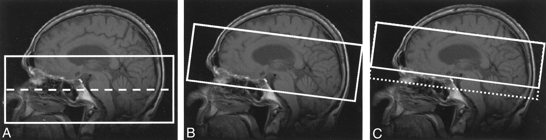
- Fig 1.
Acquisition geometry for precontrast (A) and postcontrast (B, C) 3D TOF MR angiography.
A, Two-slab acquisition used to cover the distal extracranial vessels as well as the intracranial circulation (dashed line indicates slab boundary).
B and C, Oblique-axial slabs used to image the intracranial circulation. Slab placement in C was optimal as it excludes the cavernous sinus and thus avoids venous enhancement (dotted line indicates slab position). Effective placement in C can be obtained by projection through only a subset of the sections acquired in B.
- Fig 2.
Schematic illustrates labeling of vessel segments and regions report on in this study. ACA indicates anterior cerebral artery region; BA, basilar artery; ICA, internal carotid artery; M1, M1 segment of the MCA; M2, M2 segment of the MCA; P1, P1 segment of the PCA; P2, P2 segment of the PCA; P3, P3 segment of the PCA. For grading purposes, ACA, M2, and P3 were segment regions, and the P1 and P2 segments were scored together and denoted by P1-P2.
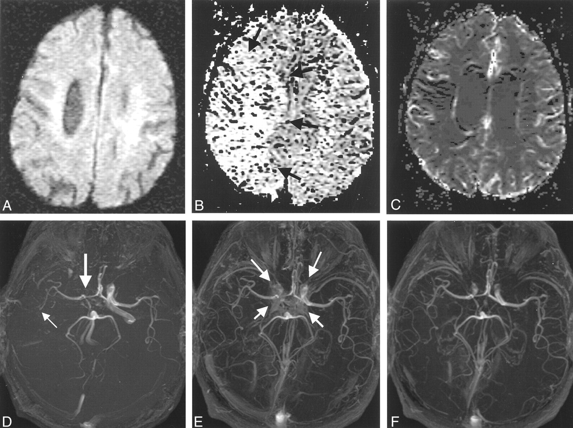
- Fig 3.
Images in a 78-year-old man with changes in the right anterior circulation.
A, Normal DW image.
B, Relative mean transit time map shows a region of delayed flow in the right hemisphere (arrows), which was consistent with the findings on the pre- and postconrast MR angiograms (D–F).
C, Normal relative cerebral blood volume map.
D, Precontrast 3D TOF MR angiogram suggests a diminished right ICA (thick arrow) and M1 and an occluded right M2 branch (thin arrow).
E and F, Postcontrast 3D TOF MR angiograms show improved depiction of the right ICA and the M1 and M2 MCA. Some contrast enhancement from the cavernous sinus is evident (arrows in E); however, this problem can be removed by changing the position of the slab. Slab positions for images D–F correspond to Fig 1B and C.
- Fig 4.
Images in a 75-year-old woman treated with intravenous tPA 2.7 hours after stroke onset. MR imaging commenced 4.1 hours after onset.
A–C, DW image (A) shows diffusion changes (arrow in A) and PW images (B and C) show relative mean-transit time defects (arrow in B) and relative cerebral blood volume defects (arrow in C).
D, Precontrast TOF MR angiogram suggests a diminished left MCA, which is consistent with the DW and PW imaging findings. Also, the right M1 and M2 appear diminished.
E, Postcontrast TOF MR angiogram confirms the left MCA changes, but the right M1 and M2 appear occluded (arrow). Postcontrast findings suggest that the right MCA territory is also at risk.
- Fig 5.
Images in a 91-year-old man treated with intravenous tPA at 2.9 hours after stroke onset. MR imaging commenced 5.5 hours after onset.
A, DW image shows an infarct (arrow).
B, Precontrast TOF MR angiogram appears to indicate a diminished left M1 and an occluded left M2 MCA (thick arrow). The ACA (thin arrow) appears occluded.
C, Postcontrast TOF MR angiogram shows the ACA segment is normal (thin arrow), the left M1 is occluded (thick arrow), and the left M2 is diminished.


Tables
Variable All Patients(n = 55) Treated Patients Only(n = 22) Age (y) Mean ± SD 66.4 ± 16.1 67.9 ± 17.9 Range 26–91 31–91 Sex (M:F) 41:15 14:8 No. treated with tPA 22 22 Onset-to-imaging time (h) Mean ± SD 4.2 ±2.1 3.7 ±1.3 Range 1.5–13.5 1.5–6.8 Pre- to postcontrast imaging interval (min) Mean ± SD 26.3 ±7.1 27.1 ±6.6 Range 8–53 18–44 NIHSS score at baseline Median (50% range) 9(4–15) 12.5(9–16) Range 0–28 2–28 NIHSS score at 24 hours Median (50% range) 4(2–9) 8(4.5–10.75) Range 0–28 0–26 Patient No. DW Changes PW Changes Precontrast TOF Changes Postcontrast TOF Changes 1 L ACA, R MCA R MCA R ICA, R M1, R M2 R ICA, R M1, R M2 2 BA L MCA L M1, L M2, L P3 L M1, L M2, L P1–P2 3 R MCA R MCA R M2 – 4 L MCA L MCA – – 5 L MCA NA L M2 L M1, L M2, L P1–P2 6 L VA – – – 7 L ICA L ICA L ICA L ICA, L M2 8 L MCA L MCA – – 9 L MCA L MCA ACA, L M1, L M2, R M2 L M1, L P1–P2 10 R MCA R MCA R M1, R M2, R P1–P2, R P3 – 11 R MCA R MCA R M1, R M2 R M1, R M2 12 L MCA L MCA – L M2, L P1–P2 13 R PCA, L MCA – RP3 – 14 R ACA – – – 15 R MCA – – R M2 16 L MCA L MCA L M2 L M2, L P1–P2 17 – – L M2 – 18 – R MCA R ICA, R M1, R M2 – 19 – – – – 20 L MCA – – – 21 L MCA L MCA L ICA, L M1, L M2 L ICA, L M1, L M2, L P1–P2 22 L MCA L MCA L ICA, L M1, L M2 L M1, L M2, L P1-P2 23 L MCA NA L ICA, L M1, L M2 L M2 24 L MCA L MCA – – 25 L MCA L MCA – – 26 L MCA NA – – 27 R MCA R MCA R ICA, R M1, R M2 R ICA, R M1, R M2 28 – – – – 29 R PCA NA R P1–P2, R P3 – 30 R MCA R MCA R M2 R M1, R M2 31 L MCA – – – 32 L MCA L MCA L M1, L M2 – 33 – – – – 34 R MCA R MCA R M1, R M2 R M1, R M2 35 R PCA R PCA – – 36 R PCA, R MCA NA R ICA, R M1, R M2, R P3 R M1, R M2 37 – – – – 38 – – – – 39 L MCA L MCA – – 40 L MCA NA L M1, L M2 L M1, L M2, L P1–LP2 Note.—L indicates left; NA, not available; R, right; VA, vertebral artery; -, no visible changes.
* Abnormalities related to acute ischemic stroke are listed by the vascular territories they affect (see Fig 2 for labeling convention).
- TABLE 3:
Agreement between combined DW and PW imaging and combined pre- and postcontrast 3D TOF MR angiography in identifying vascular territories with infarction
Agreement Left Right Both Anterior Posterior Both Anterior Posterior None Overall 1 19 1 1 10 3 5 DWI-PWI and TOF agree 1 10 1 1 10 2 5 DWI-PWI and TOF disagree 9 1 DWI-PWI changes but no TOF changes 8 1 No DWI-PWI changes but TOF changes 1 Note.—Anterior circulation infarcts include ICA, ACA, and MCA vessels; posterior circulation infarcts include vertebral artery, BA, and PCA vessels.
Pair-Wise κ Scores Observer 1 Observer 2 Observer 3 Observer 4 Observer 5 Vascular Signal: Precontrast Assessment Observer 1 1.00 0.44 0.36 0.46 0.33 Observer 2 1.00 0.42 0.61 0.49 Observer 3 1.00 0.54 0.44 Observer 4 1.00 0.57 Observer 5 1.00 Vascular Signal: Postcontrast Assessment Observer 1 1.00 0.39 0.44 0.48 0.44 Observer 2 1.00 0.30 0.35 0.41 Observer 3 1.00 0.41 0.39 Observer 4 1.00 0.54 Observer 5 1.00 Impact of Contrast Enhancement Observer 1 1.00 0.03 0.04 0.34 0.05 Observer 2 1.00 0.03 0.00 0.21 Observer 3 1.00 0.00 0.01 Observer 4 1.00 0.14 Observer 5 1.00 Note.—Overall agreement for vascular signal grade at precontrast assessment was 0.41, for vascular signal grade at postcontrast assessment was 0.48, and for impact of contrast enhancement grades was 0.08.
Grade Vascular Signal* Impact of Contrast Enhancement† Precontrast Images Postcontrast Images Postcontrast Images Frequency Percentage Frequency Percentage Frequency Percentage 0 430 89.6 436 90.8 455 94.8 1 23 4.8 32 6.7 16 3.3 2 26 5.4 12 2.5 9 1.9 NR 1 0.2 0 0.0 — — Total 480 100 480 100 480 100 Note.—Data are for median grades across all five observers.
* Vascular signal grade 0 indicates normal segment or region; 1, diminished flow; 2, absent flow; and NR, not readable.
† Impact of contrast enhancement grade 0 indicates no effect; 1, adverse effect but tolerable; 2, nondiagnostic.
- TABLE 6:
Change between postcontrast and precontrast vascular signal grades in all segments and regions
Vascular Signal Grade Change Frequency BA ACA ICA M1 M2 P1–P2 P3 Total All Vessels NR to readable 0 0 0 0 1 0 0 1 Two-grade increase 0 1 2 0 2 0 2 7 One-grade increase 0 0 6 4 10 2 3 25 No change 40 39 72 71 63 69 75 429 One-grade decrease 0 0 0 4 4 8 0 16 Two-grade decrease 0 0 0 1 0 1 0 2 Total 40 40 80 80 80 80 80 480 Only Abnormal Vessels on the Precontrast Angiograms NR to readable 0 0 0 0 1 0 0 1 Two-grade increase 0 1 2 0 2 0 2 7 One-grade increase 0 0 6 4 10 2 3 25 No change 0 0 0 8 7 0 0 15 One-grade decrease 0 0 0 2 0 0 0 2 Two-grade decrease 0 0 0 0 0 0 0 0 Total 0 1 8 14 20 2 5 50 - TABLE 7:
Impact of contrast enhancement in all segments and regions for all vessels and for only abnormal vessels
Impact of Contrast Enhancement Grade Change Frequency BA ACA ICA M1 M2 P1/P2 P3 Total All Vessels No effect 40 39 71 71 76 79 79 455 Adverse effect but tolerable 0 0 5 6 3 1 1 16 Nondiagnostic 0 1 4 3 1 0 0 9 Total 40 40 80 80 80 80 80 480 Only Abnormal Vessels on Precontrast MR Angiograms No effect 0 0 4 13 17 2 5 41 Adverse effect but tolerable 0 1 3 1 3 0 0 8 Nondiagnostic 0 0 1 0 0 0 0 1 Total 0 1 8 14 20 2 5 50
In this issue


Jump to section
Related Articles
Cited By...
- Diagnostic Performance of TOF, 4D MRA, Arterial Spin-Labeling, and Susceptibility-Weighted Angiography Sequences in the Post-Radiosurgery Monitoring of Brain AVMs
- Does contrast-enhancement improve visualisation of lenticulostriate arteries in cerebral small vessel disease using time-of-flight magnetic resonance angiography at 7 Tesla?
- Value of Contrast-Enhanced MRA versus Time-of-Flight MRA in Acute Ischemic Stroke MRI
- Optimal MRI Sequence for Identifying Occlusion Location in Acute Stroke: Which Value of Time-Resolved Contrast-Enhanced MRA?
- Prediction of Infarction and Reperfusion in Stroke by Flow- and Volume-Weighted Collateral Signal in MR Angiography
- Imaging in children presenting with acute neurological deficit: stroke
- Quality-Evaluation Scheme for Cerebral Time-Resolved 3D Contrast-Enhanced MR Angiography Techniques
- Fetal Origin of the Posterior Cerebral Artery Produces Left-Right Asymmetry on Perfusion Imaging
- Accuracy of Pre- and Postcontrast 3D Time-of-Flight MR Angiography in Patients with Acute Ischemic Stroke: Correlation with Catheter Angiography
- Postcontrast Time-of-Flight MR Angiography Demonstrating Collateral Flow in Acute Stroke
- Silent ischemia in minor stroke and TIA patients identified on MR imaging
- The probability of middle cerebral artery MRA flow signal abnormality with quantified CT ischaemic change: targets for future therapeutic studies
- Comparison of Preperfusion and Postperfusion Magnetic Resonance Angiography in Acute Stroke
- Mild Neurological Symptoms Despite Middle Cerebral Artery Occlusion
- Evaluation of Hyperintense Vessels on FLAIR MRI for the Diagnosis of Multiple Intracerebral Arterial Stenoses