
Article Figures & Data
Figures
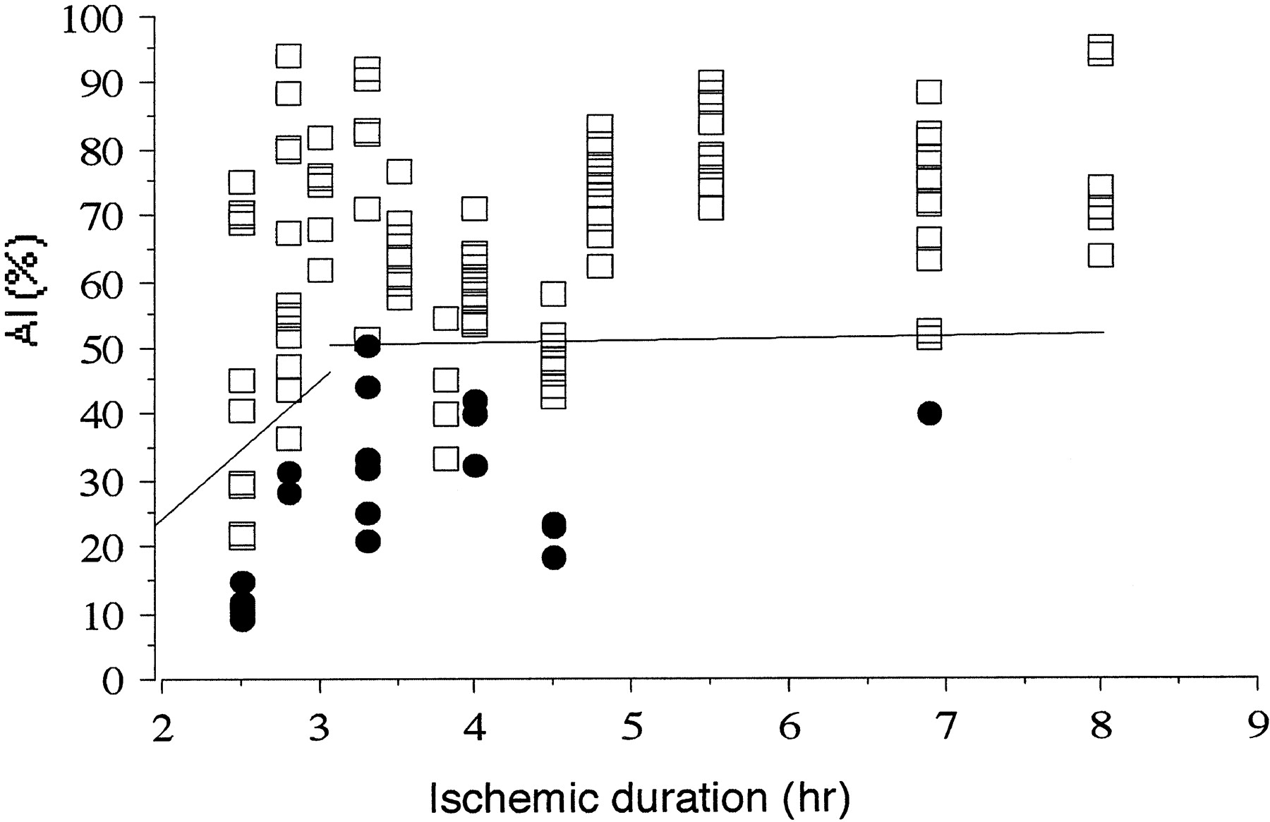
- Fig 1.
Graph shows relationship between outcome in tissue regions that were successfully reperfused and ischemic duration/severity. Plots of AI (%) of irreversible (•) and reversible (□) lesions versus ischemic duration are shown. A discriminant line of AI (%) = 21.53 time (h) − 19.15 (n = 35) within 3 hours of ischemic duration can be seen. There are six (17%) falsely discriminated ischemic lesions. A discriminant line of AI = 0.50 time − 48.27 (n = 92) after 3 hours’ ischemic duration can be seen. There are 10 (11%) falsely discriminated ischemic lesions.
- Fig 2.
Case 9.
A, Final CT scan shows that an infarct occupied approximately half the right MCA territory. Posterior half of the MCA territory escaped cerebral infarction.
B, SPECT scan shows severe hypoperfusion with an AI of 14.89 in the anterior half of the MCA territory, which experienced infarction, and mild hypoperfusion with an AI of 70.82 in the posterior half of the MCA territory, which escaped infarction.


Tables
Case Age (y)/Sex Occlusion Site Treatment Thrombolytic Agents Time to Reperfusion (h) Initial Symptoms Infarct on CT Outcome 1 58/F L M2 t-PA 7.2 IA 4.0 Hemiparesis, aphasia 3-cm frontal infarct E 2 71/M L M2 t-PA 7.2 IA 3.0 Hemiplegia, aphasia E 3 79/F R M1 PTA 3.3 Hemiplegia 2-cm basal ganglia infarct G 4 81/M R M1 PTA, t-PA 7.2 IA 4.8 Hemiparesis 2-cm basal ganglia infarct G 5 74/F R M2 t-PA 7.2 IA 2.5 Hemiplegia E 6 86M L M1 PTA 3.5 Hemiplegia, aphasia 4-cm basal ganglia infarct F 7 58/F L M2 t-PA 7.2 IA 3.3 Hemiplegia, aphasia 4-cm temporoparietal infarct G 8 78/M R M1 PTA, t-PA 7.2 IA 8.0 Hemiparesis 3-cm basal ganglia infarct G 9 65/M R M1 PTA 2.5 Hemiparesis 60% of the MCA territory F 10 76/M L M1 t-PA 7.2 IA 4.8 Hemiparesis, aphasia E 11 72/F L M1 PTA, t-PA 7.2 IA 5.5 Hemiplegia, aphasia 2-cm basal ganglia infarct E 12 74/F R M1 PTA, t-PA 7.2 IA 2.8 Hemiparesis 2-cm parietal infarct E 13 71/M L M1 PTA, t-PA 7.2 IA 2.8 Hemiplegia, aphasia E 14 67/M R M1 PTA, t-PA 7.2 IA 3.8 Hemiparesis 3-cm basal ganglia infarct G 15 80/M R M1 PTA, t-PA 7.2 IA 4.5 Hemiplegia 3-cm temporoparietal infarct G 16 68/M R M1 PTA, t-PA 7.2 IA 5.5 Hemiparesis 2-cm basal ganglia infarct E 17 60/F L M1 PTA, t-PA 7.2 IA 4.0 Hemiplegia, aphasia 3-cm basal ganglia infarct E 18 49/M R M1 PTA, t-PA 7.2 IA 6.9 Hemiparesis 2-cm temporoparietal infarct E 19 58/F L M2 t-PA 7.2 IA 3.3 Hemiplegia, aphasia 3-cm frontal infarct E Note.—L indicates left; R, right; t-PA, tissue plasminogen activator (mg); IA, intraarterial infusion; PTA, percutaneous transluminal angioplasty; MCA, middle cerebral artery; E, excellent (complete recovery); G, good (partially recovered with self-care); F, fair (no neurologic change).


