
Article Figures & Data
Figures
- Fig 1.
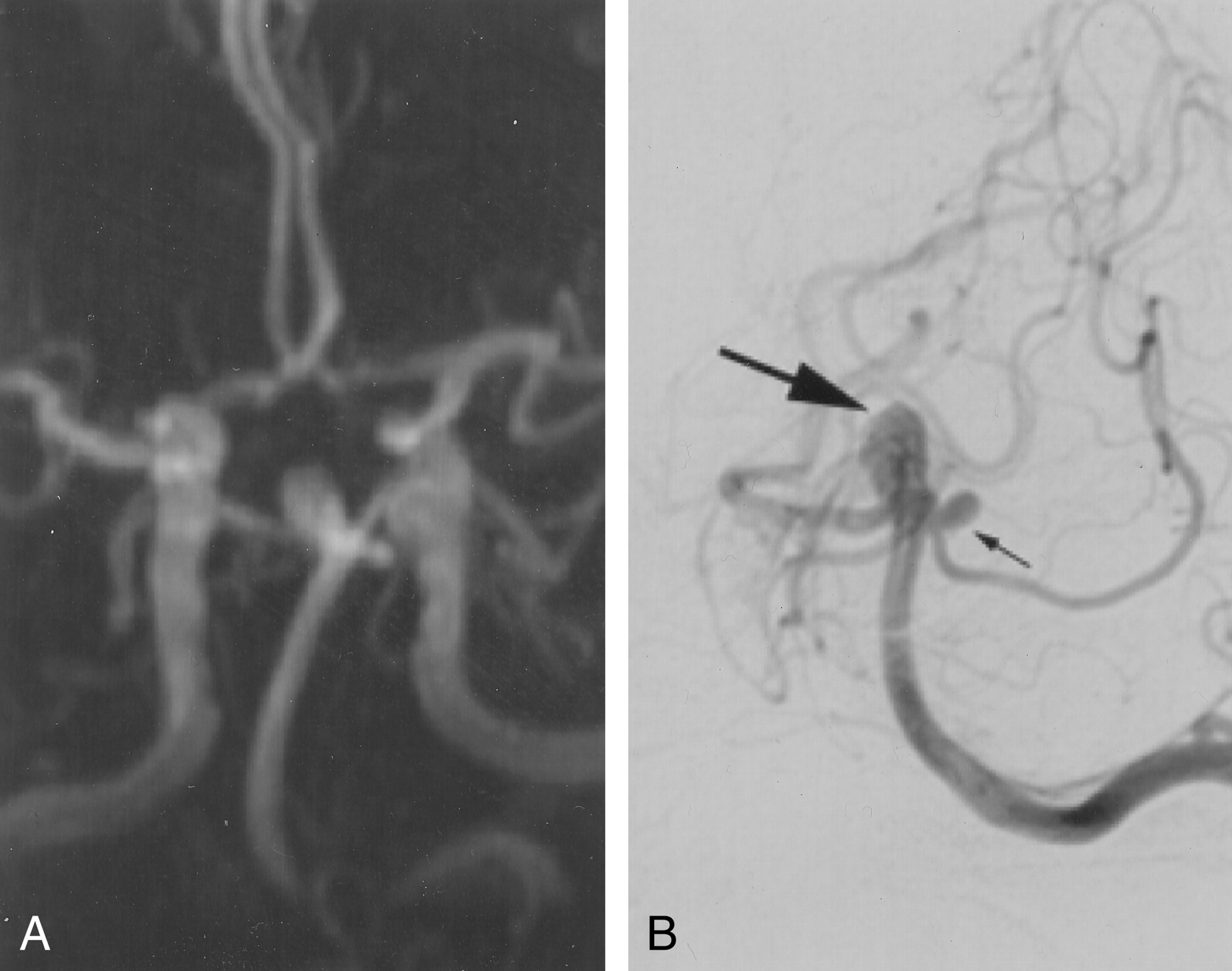
Angiograms obtained in a 55-year-old woman with worsening headaches.
A, Three-dimensional time-of-flight MR angiogram demonstrates an aneurysm in the basilar tip that measures approximately 8 × 6 mm.
B, Conventional digital subtraction angiogram obtained with a left vertebral artery injection better shows how the left superior cerebellar artery originates from the aneurysm neck.
- Fig 2.
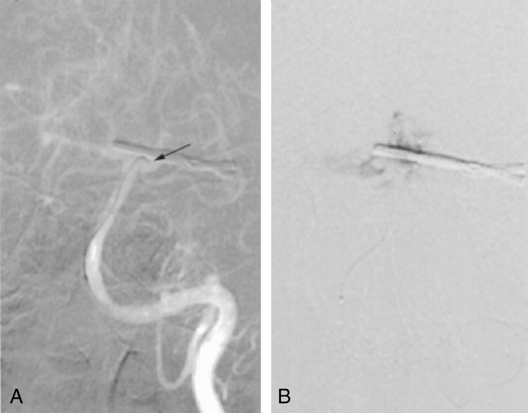
Roadmap images.
A, Anteroposterior (AP) oblique projection shows that, in this plane, the microcatheter tip (arrow) overlies the aneurysm.
B, However, gentle injection of the microcatheter in this location causes the contrast material to fill the subarachnoid space and not the aneurysm.
- Fig 3.
AP (left) and lateral (right) angiographic images obtained with an injection through the guiding catheter in the left vertebral artery demonstrate filling of the aneurysm with contrast material. No contrast agent extravasation is depicted; this result indicates that the microcatheter is sealing the hole in the aneurysm wall.
- Fig 4.
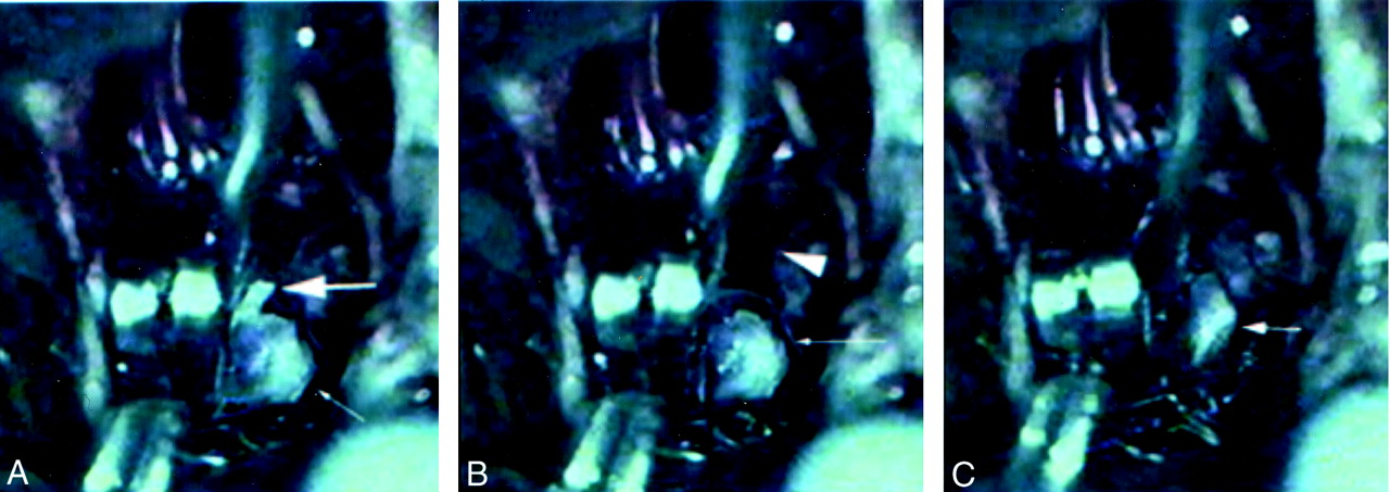
Sequence of pictures from an intraoperative videotape.
A, An aneurysm clip is held in the open position across the neck of the aneurysm (small arrow). The microcatheter protrudes through the aneurysm neck (large arrow).
B, The microcatheter is withdrawn from the aneurysm (arrow); this results in a brief spurt of hemorrhage from the hole in the aneurysm neck (arrowhead).
C, The aneurysm clip is released, with resultant decompression of the aneurysm (arrow).


