
Article Figures & Data
Figures
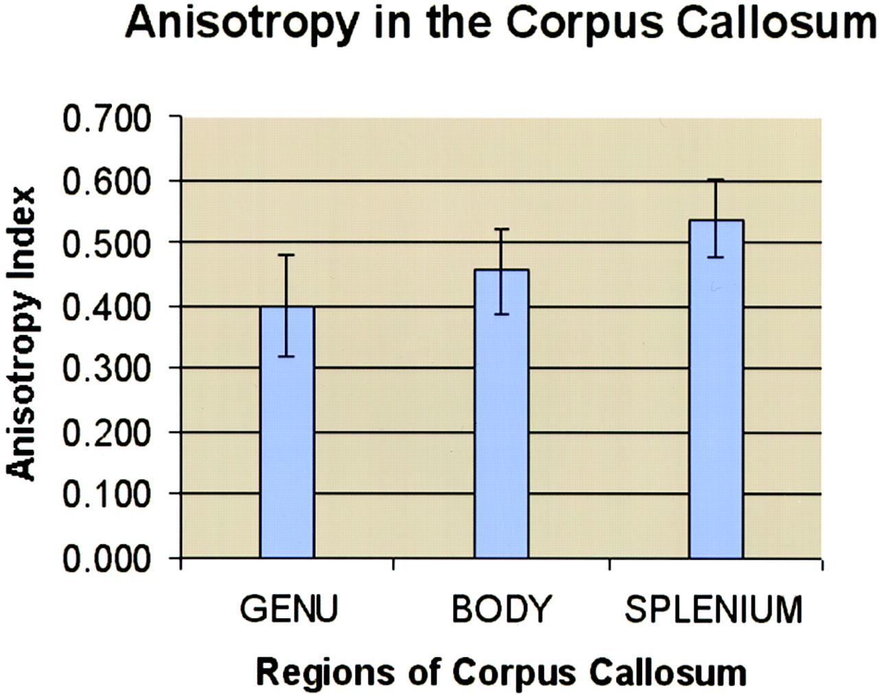
- Fig 1.
Graphical representation of anisotropy data obtained from different regions of the corpus callosum in all 42 patients. Anisotropy data were calculated by using the volume-ratio method as discussed in the Appendix. A value of 0 represents perfect isotropy; 1 represents perfect anisotropy.
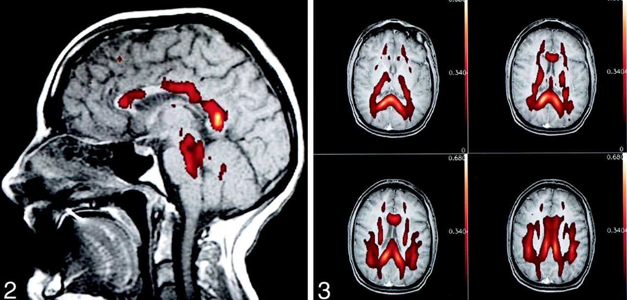
- Fig 2.
Typical appearance of anisotropy in healthy human adult brain. Anisotropy maps obtained with the volume-ratio method are represented by color overlay on a gray-scale sagittal spin-echo T1-weighted image. Note the increased anisotropy of the posterior aspect of the corpus callosum compared with that of the anterior portion.
- Fig 3.
Typical appearance of anisotropy in healthy human adult brain. The same anisotropy maps from Figure 2 were overlaid on four axial spin-echo T1-weighted images through the corpus callosum.
- Fig 4.
Histologic findings in cadaver corpora callosa
A, Perivascular fibrous alae on a sagittal celloidin 100-μm-thick section through the corpus callosum (alkaline phosphatase stain, cresyl violet acetate and light green counterstain). A large arteriole (black arrow) is obliquely sectioned, and the extent of the collagenous perivascular ala (white arrows) is seen in an artifactual gap in the tissue.
B, Axial celloidin 100-μm-thick section through the corpus callosum (alkaline phosphatase stain, trichrome counterstain). Collagenous (green) sheets (arrows) are contiguous with and extend laterally away from penetrating callosal arterioles. These alae descend through the corpus callosum in a curtain-like fashion. Note that the axons horizontally oriented in this section.
C, Sagittal paraffin 10-μm-thick section through the corpus callosum (Bielschowsky stain). Axons are stained black, and most are cut in cross-section and appear as black dots (as expected). Two small bundles of obliquely oriented axons (curved arrows) are depicted. Obliquely cut vessels and associated fibrous ala are seen in a tissue cleft (straight arrows).
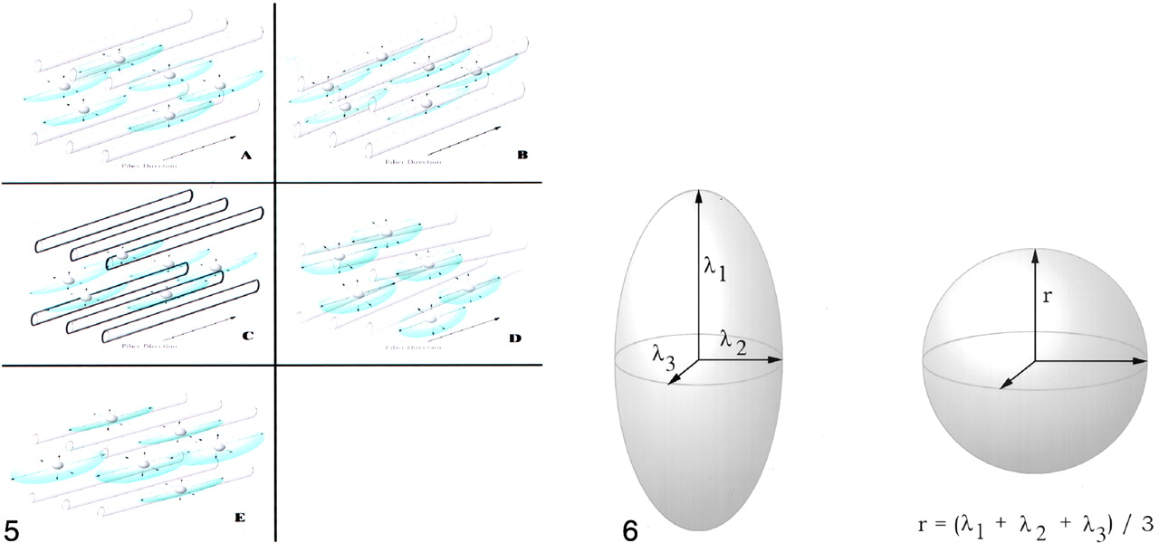
- Fig 5.
Schematic representation of diffusion anisotropy in brain white matter. Obliquely oriented cylinders represent axonal fibers and myelin sheaths, and small spheres represent water molecules. When the randomly chosen direction of diffusion is transverse to the fiber direction, the path of diffusion is blocked by hydrophobic myelin sheaths. When the randomly chosen direction of diffusion is parallel to the fiber direction, there are no barriers to free diffusion. Thus, the water molecules demonstrate a preference for diffusion along the direction of the fiber tract (12, 13, 16) (A). Schematic representations illustrate the situations in which axons are more tightly packed (B), the myelin sheaths are thicker (C), obliquely oriented axons are present (D), and the axonal diameter is reduced (E).
- Fig 6.
Schematic representation of the ellipsoid that describes the mean diffusion of a water molecule at the center of a voxel in all directions. The eigenvalues of the diagonalized diffusion tensor (λ1, λ2, λ3) are the axes of this ellipsoid. Also represented is a sphere with a radius that is the average value of the three axes of the corresponding ellipsoid. The volume ratio used is the ratio of the volume of the ellipsoid to the volume of the sphere, as discussed in the Appendix and in reference 13. The anisotropy index used in this investigation was as follows: 1 − volume ratio.


In this issue


Jump to section
Related Articles
Cited By...
- Altered White Matter Tracts in Bipolar Disorder: Insights from DTI Analysis
- Unifying Orientation-Dependent Relaxation and Diffusion Around Axonal Fibers from DTI: Characterizing Fiber-Tract-Specific Anisotropic R2 Profiles of the Corpus Callosum
- Diffusion Measures Indicate Fight Exposure-Related Damage to Cerebral White Matter in Boxers and Mixed Martial Arts Fighters
- Microstructural organization of corpus callosum projections to prefrontal cortex predicts bimanual motor learning
- Diffusion Tensor Group Tractography of the Corpus Callosum in Clinically Isolated Syndrome
- Callosal Contributions to Simultaneous Bimanual Finger Movements
- Structural Organization of the Corpus Callosum Predicts the Extent and Impact of Cortical Activity in the Nondominant Hemisphere
- Neuroradiological characterization of normal adult ageing
- Diffusion Tensor MR Imaging Visualizes the Altered Hemispheric Fiber Connection in Callosal Dysgenesis