
Article Figures & Data
Figures
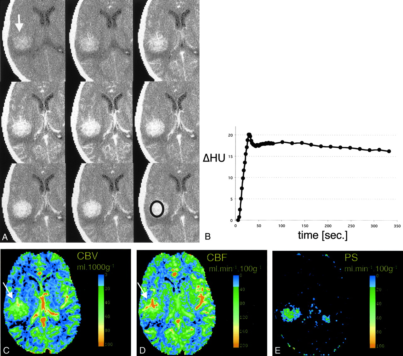
- Fig 1.
CT scans and data analysis obtained from patient 1.
A, Representative images from the dynamic contrast-enhanced CT study. Top row, Before the administration of contrast material, a subtle tumoral hyperattenuation indicates hemorrhage (arrow). Middle row, After the injection of contrast agent, the tumor is immediately and strongly enhancing. Bottom row, The tumor remains hyperattenuating even after the contrast agent washes out.
B, Time-attenuation curve of the tumor from the ROI in the bottom right image in A. Strong enhancement of approximately 15 HU is present during the first pass. A slight decrease in tumoral attenuation is followed by steady enhancement during the equilibrium phase. Contrast-agent washout is not observed during the studied period of approximately 5 minutes.
C–E, Pixel-by-pixel parameter maps of CBV (C), CBF (D) and PS area (E) in the section in A. CBV is slightly increased in the tumor and decreased in the surrounding edematous tissue. No increase in blood flow is seen. Tumoral conspicuity is highest on the PS map (arrow), against the near-zero permeability of the normal brain tissue.
- Fig 2.
CT scans and data analysis obtained from patient 2
A, Representative images from the dynamic contrast-enhanced CT study. Top row, Before contrast administration, hyperattenuation within the tumor indicates hemorrhage (arrow). Middle row, After the injection of contrast agent, the tumor is immediately and strongly enhancing. Bottom row, The tumor remains hyperattenuated even after the contrast agent has washed out.
B, Time-density curve of the tumor, from the ROI in the bottom right image in A. Strong enhancement of approximately 20 HU is present during first pass. A slight decrease in tumoral attenuation is followed by a steady enhancement during the equilibrium phase. Contrast-agent washout is not observed during the studied period.
C–E, Pixel-by-pixel parameter maps of CBV (C), CBF (D) and PS area (E) from the section in A. Both CBV and CBF are markedly increased in the tumor; this is most pronounced in the more anterior tumoral areas (arrows). In vascularized tumor tissue, CBF is as high as in the CBF in the contralateral insular vessels. Tumor conspicuity is highest on the PS map, with near-zero permeability of the normal brain tissue. (The contralateral area of high PS indicates the plexus, which does not have a blood-brain barrier and is inherently permeable to the contrast agent).
- Fig 3.
A, Representative images from the dynamic contrast-enhanced MR study in the patient in (Figure 2. As seen on the CT scans Fig 2), contrast enhancement is strong and immediate, and wash-out is slow. After the injection of contrast agent, the tumor is immediately and strongly enhancing (top row, middle and right images), and it remains hyperintense relative to the vascular signal intensity (bottom row).
B and C, Pixel-by-pixel parameter maps of CBV (B) and PS (C) from the section in A. As seen on the CT maps, blood volume is increased mostly in the more anterior tumor areas (B, arrow). Permeability is highest in the more anterior and peripheral tumor areas (C, arrow) as well.


In this issue


Jump to section
Related Articles
Cited By...
- Blood-Brain Barrier Compromise Does Not Predict Perihematoma Edema Growth in Intracerebral Hemorrhage
- Carotid Plaque Enhancement and Symptom Correlations: An Evaluation by Using Multidetector Row CT Angiography
- Increased Blood-Brain Barrier Permeability on Perfusion CT Might Predict Malignant Middle Cerebral Artery Infarction
- Neuroimaging applications of multislice CT perfusion