
Article Figures & Data
Figures
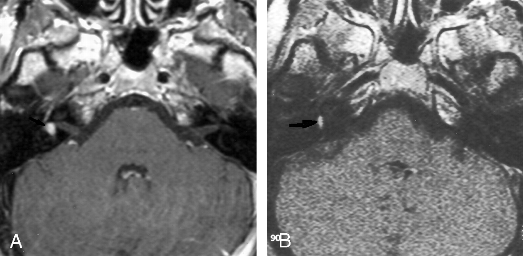
- Fig 1.
Axial contrast-enhanced T1-weight-ed MR images show a small vestibular schwannoma.
A, The 1.5-T image (550/20/3) obtained at the level of the IAC shows a small (2-mm) left vestibular schwannoma in the fundus of the IAC.
B, The 0.2-T image (650/15/3) obtained at the same level depicts the small vestibular schwannoma.
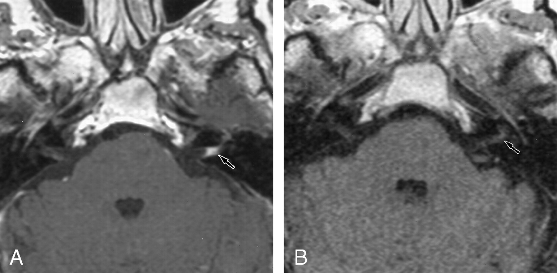
- Fig 2.
Axial contrast-enhanced T1-weight-ed MR images show an intra-labyrinthine schwannoma.
A, The 1.5-T image (550/20/3) shows a posterior enhancement of the right labyrinth (arrow), which corresponds to a schwannoma in the vestibule.
B, The 0.2-T image (650/15/3) obtained at the same level depicts this intravestibular schwannoma (arrow).
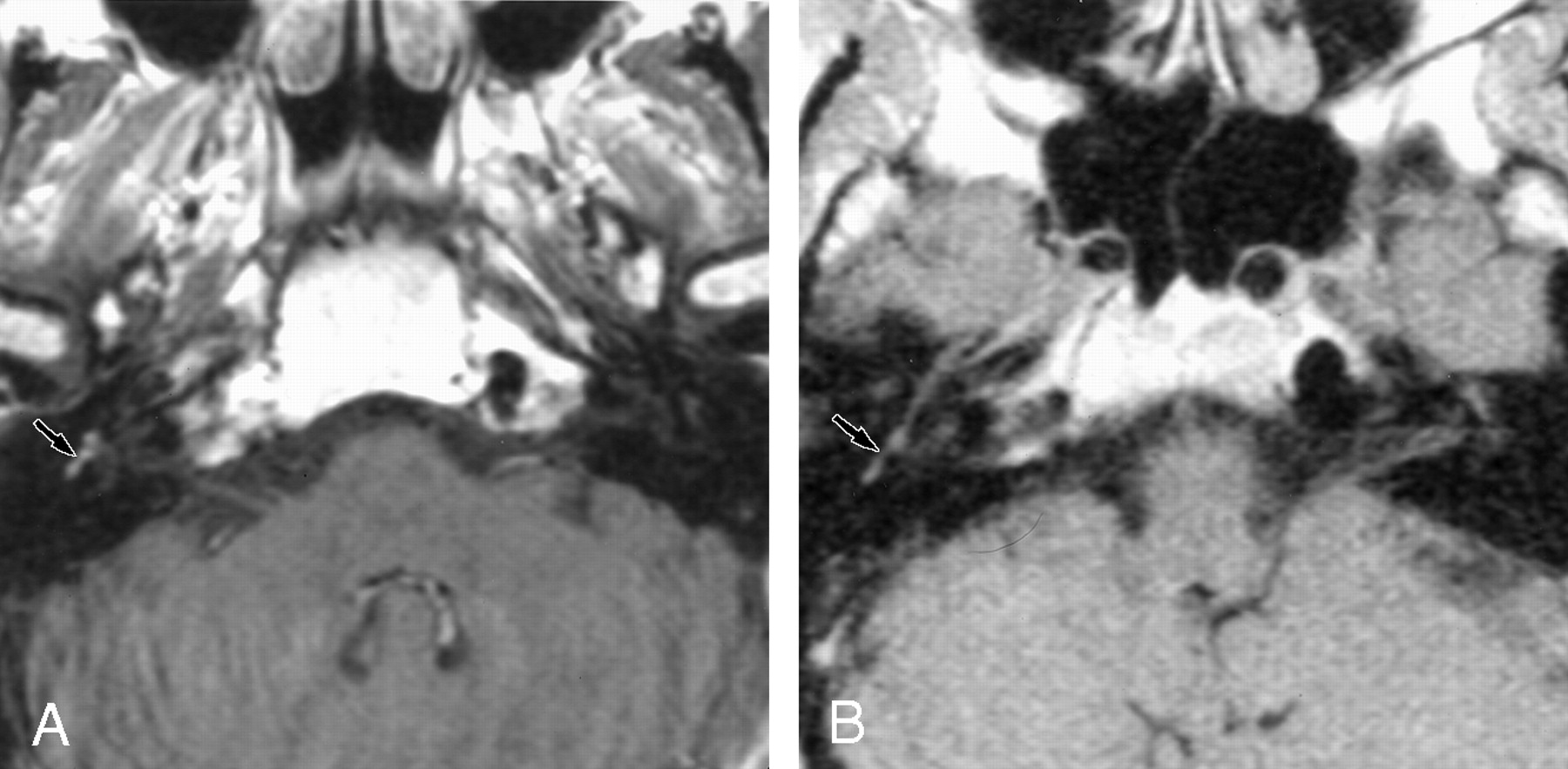
- Fig 3.
Axial contrast-enhanced T1-weight-ed MR images show meningeal enhancement in the IAC.
A, The 1.5-T image (550/20/3) obtained at the level of the IAC shows contrast enhancement in the left IAC, with concave limits corresponding to a meningeal enhancement (arrow).
B, The 0.2-T image (650/15/3) shows no contrast enhancement in the left IAC (arrow).
- Fig 4.
Axial contrast-enhanced T1-weight-ed MR images show enhancement of the facial nerve.
A, The 1.5-T image (550/20/3) shows contrast enhancement of the second portion of the right facial nerve.
B, The 0.2-T image (650/15/3) depicts no significant contrast enhancement at this level.


Tables
Result Group 1(n = 33) Group 2(n = 153) Group 3(n = 74) Group 4(n = 21) Findings on 1.5-T images Total disorders 21 (63.6) 27 (17.6) 3 (4.1) 12 (57.1) Tumoral disorders 20 (60.6) 27 (17.6) 3 (4.1) 6 (28.6) Nontumoral disorders 1 (3.0) 0 (0) 0 (0) 6 (28.6) Findings on 0.2-T images Total disorders 21 (63.6) 27 (17.6) 3 (4.1) 7 (33.3) Tumoral disorders 20 (60.6) 27 (17.6) 3 (4.1) 6 (28.6) Nontumoral disorders 1 (3.0) 0 (0) 0 1 (4.8) Note.—Data in parentheses are percentages.
In this issue


Jump to section
Related Articles
Cited By...
- No citing articles found.