Advertisement

Main menu

  • Home
  • Content
    • Current Issue
    • Accepted Manuscripts
    • Article Preview
    • Past Issue Archive
    • AJNR Case Collection
    • Case of the Week Archive
    • Classic Case Archive
    • Case of the Month Archive
  • Special Collections
    • Spinal CSF Leak Articles (Jan 2020-June 2024)
    • 2024 AJNR Journal Awards
    • Most Impactful AJNR Articles
  • Multimedia
    • AJNR Podcast
    • AJNR Scantastics
    • Video Articles
  • For Authors
    • Submit a Manuscript
    • Author Policies
    • Fast publishing of Accepted Manuscripts
    • Graphical Abstract Preparation
    • Manuscript Submission Guidelines
    • Imaging Protocol Submission
    • Submit a Case for the Case Collection
  • About Us
    • About AJNR
    • Editorial Board
  • More
    • Become a Reviewer/Academy of Reviewers
    • Subscribers
    • Permissions
    • Alerts
    • Feedback
    • Advertisers
    • ASNR Home
  • Other Publications
    • ajnr

User menu

  • Alerts
  • Log in

Search

  • Advanced search
American Journal of Neuroradiology

American Journal of Neuroradiology

ASHNR American Society of Functional Neuroradiology ASHNR American Society of Pediatric Neuroradiology ASSR
  • Alerts
  • Log in

Advanced Search

  • Home
  • Content
    • Current Issue
    • Accepted Manuscripts
    • Article Preview
    • Past Issue Archive
    • AJNR Case Collection
    • Case of the Week Archive
    • Classic Case Archive
    • Case of the Month Archive
  • Special Collections
    • Spinal CSF Leak Articles (Jan 2020-June 2024)
    • 2024 AJNR Journal Awards
    • Most Impactful AJNR Articles
  • Multimedia
    • AJNR Podcast
    • AJNR Scantastics
    • Video Articles
  • For Authors
    • Submit a Manuscript
    • Author Policies
    • Fast publishing of Accepted Manuscripts
    • Graphical Abstract Preparation
    • Manuscript Submission Guidelines
    • Imaging Protocol Submission
    • Submit a Case for the Case Collection
  • About Us
    • About AJNR
    • Editorial Board
  • More
    • Become a Reviewer/Academy of Reviewers
    • Subscribers
    • Permissions
    • Alerts
    • Feedback
    • Advertisers
    • ASNR Home
  • Follow AJNR on Twitter
  • Visit AJNR on Facebook
  • Follow AJNR on Instagram
  • Join AJNR on LinkedIn
  • RSS Feeds

Welcome to the new AJNR, Updated Hall of Fame, and more. Read the full announcements.


AJNR is seeking candidates for the position of Associate Section Editor, AJNR Case Collection. Read the full announcement.


Research ArticleHEAD AND NECK

Evaluation of Distal Extracranial Internal Carotid Artery by Transoral Carotid Ultrasonography in Patients with Severe Carotid Stenosis

Kazuhiro Kishikawa, Masahiro Kamouchi, Yasushi Okada, Tooru Inoue, Setsuro Ibayashi and Mitsuo Iida
American Journal of Neuroradiology June 2002, 23 (6) 924-928;
Kazuhiro Kishikawa
aDepartment of Cerebrovascular Disease, Graduate School of Medical Sciences, Kyushu University, Fukuoka, Japan
  • Find this author on Google Scholar
  • Find this author on PubMed
  • Search for this author on this site
Masahiro Kamouchi
aDepartment of Cerebrovascular Disease, Graduate School of Medical Sciences, Kyushu University, Fukuoka, Japan
  • Find this author on Google Scholar
  • Find this author on PubMed
  • Search for this author on this site
Yasushi Okada
aDepartment of Cerebrovascular Disease, Graduate School of Medical Sciences, Kyushu University, Fukuoka, Japan
  • Find this author on Google Scholar
  • Find this author on PubMed
  • Search for this author on this site
Tooru Inoue
bDepartment of Neurosurgery, Graduate School of Medical Sciences, Kyushu University, Fukuoka, Japan
  • Find this author on Google Scholar
  • Find this author on PubMed
  • Search for this author on this site
Setsuro Ibayashi
cDepartment of National Kyushu Medical Center, and the Department of Medicine and Clinical Science, Graduate School of Medical Sciences, Kyushu University, Fukuoka, Japan
  • Find this author on Google Scholar
  • Find this author on PubMed
  • Search for this author on this site
Mitsuo Iida
cDepartment of National Kyushu Medical Center, and the Department of Medicine and Clinical Science, Graduate School of Medical Sciences, Kyushu University, Fukuoka, Japan
  • Find this author on Google Scholar
  • Find this author on PubMed
  • Search for this author on this site
  • Article
  • Figures & Data
  • Info & Metrics
  • Responses
  • References
  • PDF
Loading

Article Figures & Data

Figures

  • Tables
  • Fig 1.
    • Download figure
    • Open in new tab
    • Download powerpoint
    Fig 1.

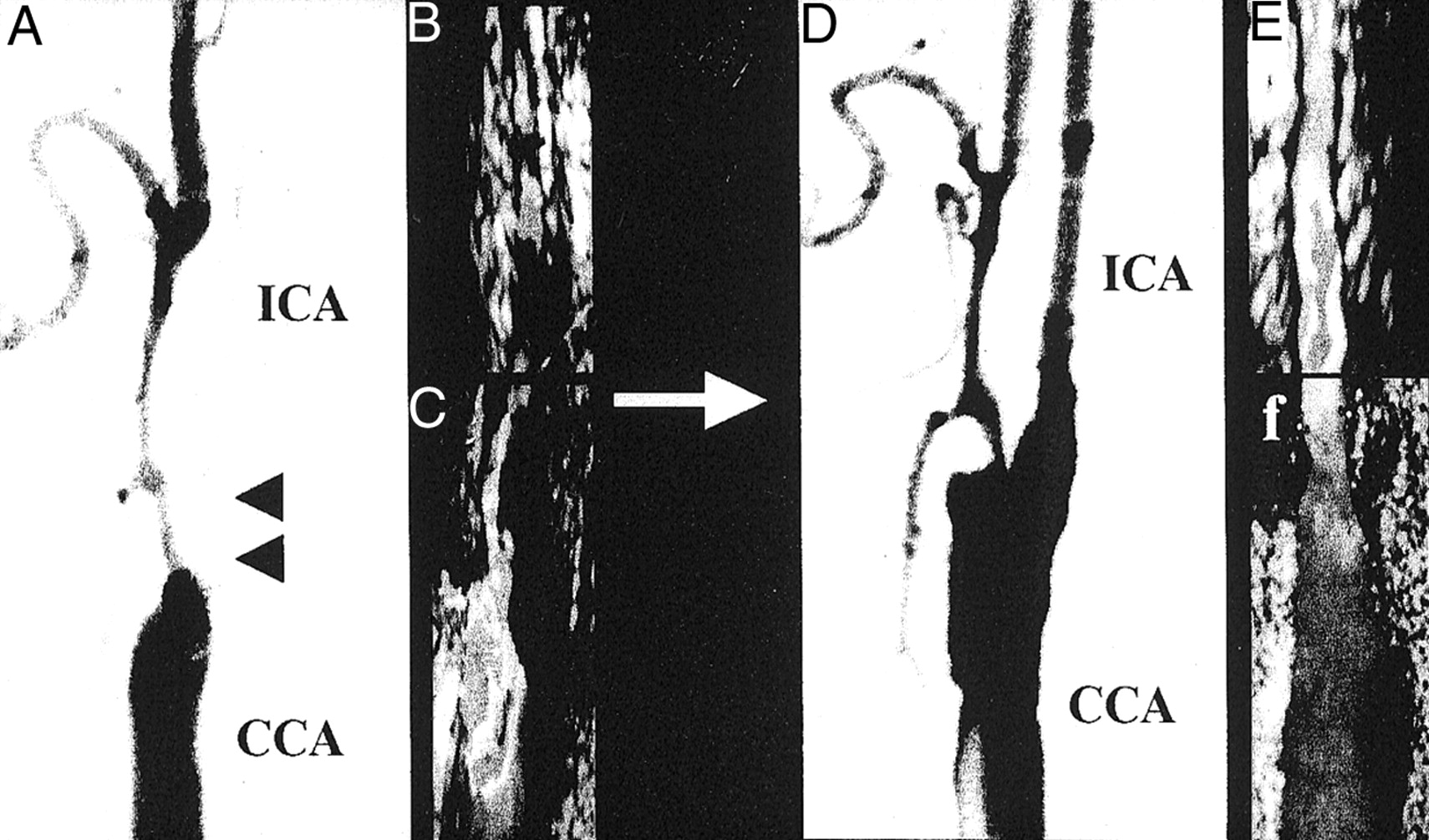
    Angiographic and ultrasonographic findings of carotid arteries before (left panels) and after (right panels) CEA. Arrowheads indicate severe stenosis in the origin of the ICA. Note that the distal ICA dilated after CEA.

    A, Angiographic findings before CEA.

    B, TOCU findings before CEA.

    C, Conventional carotid ultrasonographic findings before CEA.

    D, Angiographic findings after CEA.

    E, Transoral carotic ultrasonographic findings after CEA.

    F, Conventional carotd ultrasonographic findings after CEA.

  • Fig 2.
    • Download figure
    • Open in new tab
    • Download powerpoint
    Fig 2.

    Diameter of the distal ICA before and after CEA is plotted. Diameter was measured intraorally at the level of the post-pharyngeal portion by using TOCU. Closed circles and error bars indicate mean ± SD.

  • Fig 3.
    • Download figure
    • Open in new tab
    • Download powerpoint
    Fig 3.

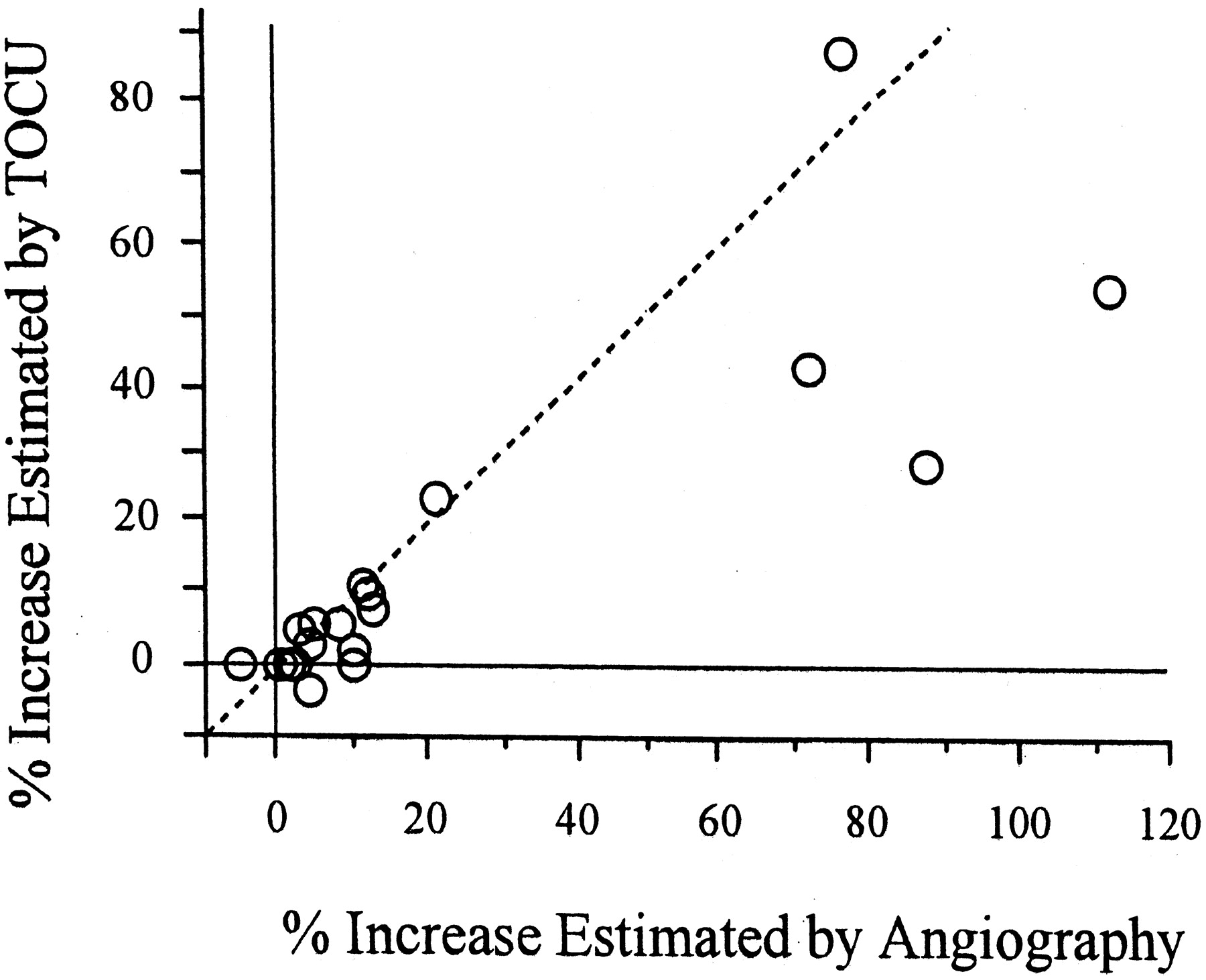
    Comparison of the percent increase in the diameter of the distal ICA as estimated by TOCU and cerebral angiography.

  • Fig 4.
    • Download figure
    • Open in new tab
    • Download powerpoint
    Fig 4.

    Relationship between degree of carotid stenosis and postoperative dilatation of the ICA. Percent increase in the diameter of the distal ICA was plotted against the grade of carotid stenosis. Grade of carotid stenosis in a cross-sectional area was estimated by conventional carotid ultrasonography. Diameter of the distal ICA was estimated by TOCU. Dotted line indicates 95% stenosis.

  • Fig 5.
    • Download figure
    • Open in new tab
    • Download powerpoint
    Fig 5.

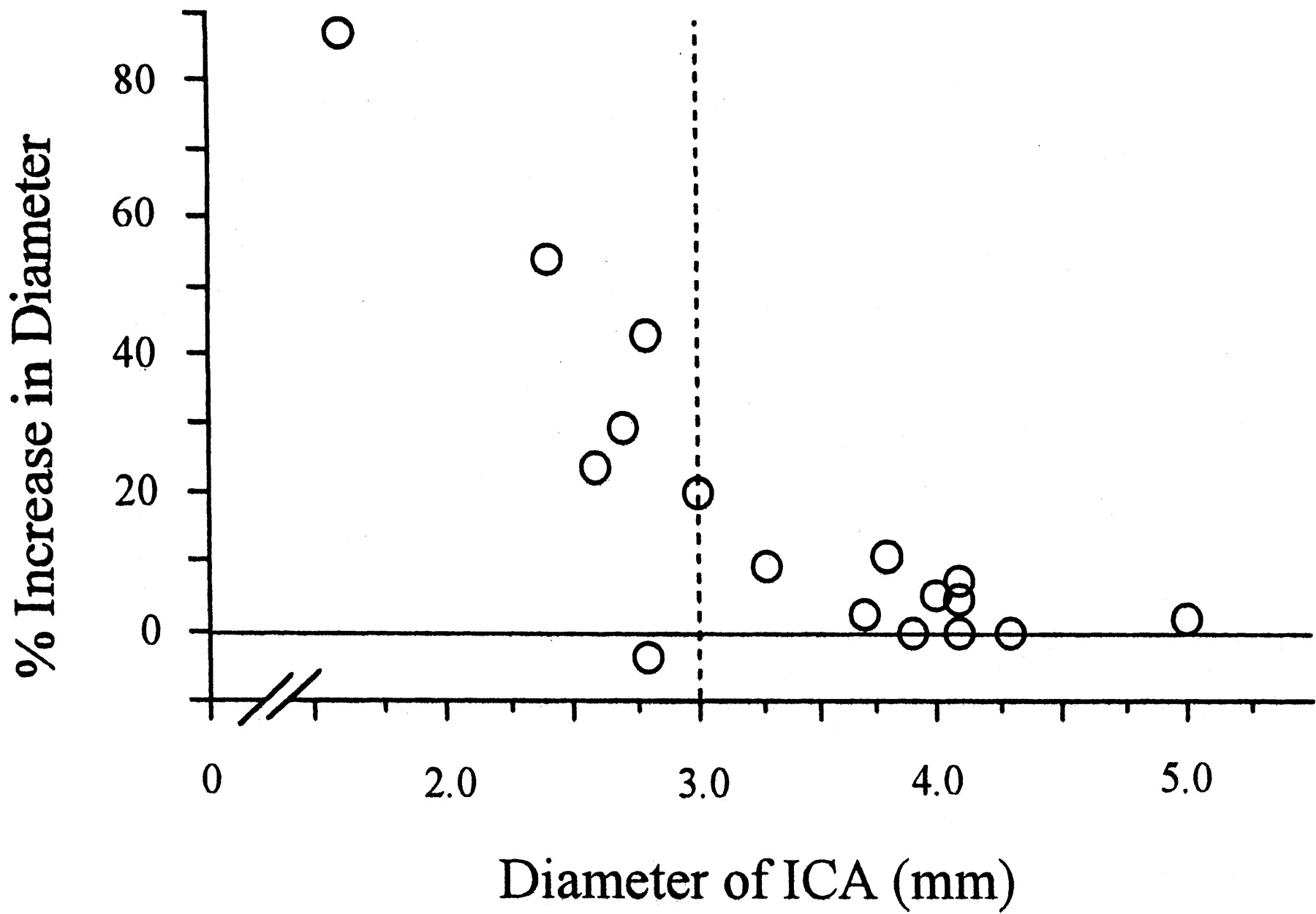
    Relationship between preoperative diameter and postoperative dilatation of the ICA. Percent increase in the diameter of the distal ICA was plotted against the preoperative diameter of the distal ICA. Diameter of the distal ICA was estimated by TOCU. Dotted line indicates diameter of 3.0 mm.

Tables

  • Figures
    • View popup
    TABLE 1:

    Demographic and clinical characteristics

    Age (years)67.6 ± 6.6 
    Male:Female (n)17:3
    Degree of stenosis (%)74.9 ± 14.6
    Symptomatic attack19/20 (95%)
     Minor stroke8/20 (40%)
     Transient ischemic attack11/20 (55%)
    Risk factors
     Hypertension16/20 (80%)
     Smoking15/20 (75%)
     Hyperlipidemia11/20 (55%)
     Diabetes mellitus7/20 (35%)
    Vascular complications
     Ischemic heart disease7/20 (35%)
     Arteriosclerosis obliterans2/20 (10%)
    • Note.—Values are expressed as mean ± SD. The degree of carotid stenosis was calculated using the method described by the North American Symptomatic Carotid Endarterectomy Trial group 1. Ischemic heart disease was diagnosed by myocardial scintigraphy, and arteriosclerosis obliterans was diagnosed by ankle pressure index, pulse wave, and clinical symptoms.

    • View popup
    TABLE 2:

    Change in parameters before and after carotid endarterectomy

    PreoperationPostoperationP
    Angiography
     ICA:CCA0.51 ± 0.160.59 ± 0.14<0.01
    TOCU
     B mode
      Diameter (mm)3.5 ± 0.83.9 ± 0.5<0.01
     Flow velocity (cm/s)
      Peak systolic velocity69.2 ± 25.671.5 ± 20.9NS
      End-diastolic velocity23.8 ± 7.124.2 ± 7.1NS
      Mean flow velocity38.8 ± 12.240.7 ± 11.8NS
    • Note.—ICA indicates internal carotid artery; CCA, common carotid artery; TOCU, transoral carotid ultrasonography; NS, not significant. Values are mean ± SD. P values exceeding .05 were considered to be significant. ICA:CCA indicates the ratio of the diameter of the internal carotid artery to that of the common carotid artery. The flow velocity of the distal internal carotid artery was obtained by transoral carotid ultrasonography.

PreviousNext
Back to top

In this issue

American Journal of Neuroradiology: 23 (6)
American Journal of Neuroradiology
Vol. 23, Issue 6
1 Jun 2002
  • Table of Contents
  • Index by author
Advertisement
Print
Download PDF
Email Article

Thank you for your interest in spreading the word on American Journal of Neuroradiology.

NOTE: We only request your email address so that the person you are recommending the page to knows that you wanted them to see it, and that it is not junk mail. We do not capture any email address.

Enter multiple addresses on separate lines or separate them with commas.
Evaluation of Distal Extracranial Internal Carotid Artery by Transoral Carotid Ultrasonography in Patients with Severe Carotid Stenosis
(Your Name) has sent you a message from American Journal of Neuroradiology
(Your Name) thought you would like to see the American Journal of Neuroradiology web site.
CAPTCHA
This question is for testing whether or not you are a human visitor and to prevent automated spam submissions.
Cite this article
Kazuhiro Kishikawa, Masahiro Kamouchi, Yasushi Okada, Tooru Inoue, Setsuro Ibayashi, Mitsuo Iida
Evaluation of Distal Extracranial Internal Carotid Artery by Transoral Carotid Ultrasonography in Patients with Severe Carotid Stenosis
American Journal of Neuroradiology Jun 2002, 23 (6) 924-928;

Citation Manager Formats

  • BibTeX
  • Bookends
  • EasyBib
  • EndNote (tagged)
  • EndNote 8 (xml)
  • Medlars
  • Mendeley
  • Papers
  • RefWorks Tagged
  • Ref Manager
  • RIS
  • Zotero
0 Responses
Respond to this article
Share
Bookmark this article
Evaluation of Distal Extracranial Internal Carotid Artery by Transoral Carotid Ultrasonography in Patients with Severe Carotid Stenosis
Kazuhiro Kishikawa, Masahiro Kamouchi, Yasushi Okada, Tooru Inoue, Setsuro Ibayashi, Mitsuo Iida
American Journal of Neuroradiology Jun 2002, 23 (6) 924-928;
del.icio.us logo Twitter logo Facebook logo Mendeley logo
  • Tweet Widget
  • Facebook Like
  • Google Plus One

Jump to section

  • Article
    • Abstract
    • Methods
    • Results
    • Discussion
    • Conclusion
    • Acknowledgments
    • Footnotes
    • References
  • Figures & Data
  • Info & Metrics
  • Responses
  • References
  • PDF

Related Articles

  • No related articles found.
  • PubMed
  • Google Scholar

Cited By...

  • Bottle Neck Sign of the Proximal Portion of the Internal Carotid Artery in Moyamoya Disease.
  • Internal Carotid Artery Stenosis Measurement: Comparison of 3D Computed Rotational Angiography and Conventional Digital Subtraction Angiography
  • Diagnostic Impact of Transcranial Color-Coded Real-Time Sonography With Echo Contrast Agents for Hyperperfusion Syndrome After Carotid Endarterectomy
  • Crossref
  • Google Scholar

This article has not yet been cited by articles in journals that are participating in Crossref Cited-by Linking.

More in this TOC Section

  • Correlation of Apparent Diffusion Coefficient at 3T with Prognostic Parameters of Retinoblastoma
  • Parathyroid Lesions: Characterization with Dual-Phase Arterial and Venous Enhanced CT of the Neck
  • Efficacy of Diffusion-Weighted Imaging for the Differentiation between Lymphomas and Carcinomas of the Nasopharynx and Oropharynx: Correlations of Apparent Diffusion Coefficients and Histologic Features
Show more Head and Neck

Similar Articles

Advertisement

Indexed Content

  • Current Issue
  • Accepted Manuscripts
  • Article Preview
  • Past Issues
  • Editorials
  • Editors Choice
  • Fellow Journal Club
  • Letters to the Editor

Cases

  • Case Collection
  • Archive - Case of the Week
  • Archive - Case of the Month
  • Archive - Classic Case

Special Collections

  • Special Collections

Resources

  • News and Updates
  • Turn around Times
  • Submit a Manuscript
  • Author Policies
  • Manuscript Submission Guidelines
  • Evidence-Based Medicine Level Guide
  • Publishing Checklists
  • Graphical Abstract Preparation
  • Imaging Protocol Submission
  • Submit a Case
  • Become a Reviewer/Academy of Reviewers
  • Get Peer Review Credit from Publons

Multimedia

  • AJNR Podcast
  • AJNR SCANtastic
  • Video Articles

About Us

  • About AJNR
  • Editorial Board
  • Not an AJNR Subscriber? Join Now
  • Alerts
  • Feedback
  • Advertise with us
  • Librarian Resources
  • Permissions
  • Terms and Conditions

American Society of Neuroradiology

  • Not an ASNR Member? Join Now

© 2025 by the American Society of Neuroradiology All rights, including for text and data mining, AI training, and similar technologies, are reserved.
Print ISSN: 0195-6108 Online ISSN: 1936-959X

Powered by HighWire